
Maxillary Obturator Prosthesis for a Hemimaxillectomy Patient-a Case Report
Department Of Prosthodontics And Crown & Bridge, Seema Dental College and Hospital, Rishikesh
ABSTRACT
Malignant tumours of the upper gums and hard palate account for 1-5% of malignant neoplasms of the oral cavity. Most of such carcinomas are diagnosed late, when they have already invaded the underlying bone. They are usually treated by surgical excision, the procedures of choice for removal may be: alveolectomy, palatectomy, maxillectomy, which can be total or partial. To prevent reoccurrence extensive surgical intervention would leave the patient with anatomical defects. Rehabilitation of such patients improves facial form, function and social acceptance. Rehabilitation aims on: restoring the separation between the oral and nasal cavities, enables the patient to swallow, maintain or provide mastication, sufficient occlusion, supports the soft facial tissues, re-establishes speech and restores an aesthetically pleasing smile. It is crucial to work in collaboration with the doctor who makes the prosthesis and who evaluates the case when the surgery is being planned.
KEY WORDS: Oral cavity, Malignant tumors, Maxillectomy, Jaw Resection, Palatal Resection, Obturator Prosthesis.
INTRODUCTION
Morphology of the maxilla has a functional and aesthetic role. The oral cavity is separated from the nasal fossa by the palate. Occlusion between the dental arches provides the mandible with stability which thus enables the pharyngeal muscles to initiate swallowing. The maxillary bone is also responsible for the projection of the hemi-face, nose and cheeks which makes it aesthetically important too.
A maxillary defect will have serious consequences between form and function i.e. the inability to chew and swallow, disorders in phonation and important psychological and social implications which follow.1
The procedures preferred for removal of carcinomas of limited size (T1N0) of the upper maxilla and hard palate are: alveolectomy and palatectomy which lead to a small deficit which can be reconstructed through simple repair surgeries with the use of a local pedicle flap, stage T2-T4 carcinomas require a more extensive surgery, such as2:
- TYPE I PARTIAL MAXILLECTOMY (resection of a single wall),
- TYPE II SUBTOTAL MAXILLECTOMY (resection of ≤ 5 walls),
- TYPE IIIA TOTAL MAXILLECTOMY (resection of all 6 walls with preservation of the orbital contents),
- TYPE IIIB (resection of all 6 walls with orbital exenteration),
- TYPE IV ORBITO-MAXILLECTOMY (resection of 5 walls, orbital exenteration with the preservation of the hard palate).
The defect can be repaired using free microvasularized flaps3 or by leaving a wide breach which will heal spontaneously, over which a stomatologic prosthesis can be given which may be mobile like palatal obturators or permanent like osteo-integrated implants.
Rogers et al. have reported that there is no difference in the quality of life of patients rehabilitated by Mobile Palatal Obturators or by Permanently retained Osseointegrated implants but, if insufficient bone tissue is present after palatectomy or total maxillectomy, then osteointegrated implants cannot be used.4
In maxillofacial rehabilitation cases the lesser the remaining teeth in the arch, the more complicated becomes the closure of the defects through flaps. In particular, the separation between the oral and nasal cavity with a muscular diaphragm which is not resting upon hard tissue leads to a mobile support, for which it is impossible to make adequate impressions.5
MATERIALS & METHODS
At Seema Dental College & Hospital the use of palatal obturators is generally preferred as they offer a safe oncological follow-up in patients with high risk of local recurrences and are a fast, inexpensive mode of rehabilitation in terms of aesthetic appearance, phonation and mastication.
CASE REPORT
A 37 year old male patient reported to the Department Of Prosthodontics, Seema Dental College & Hospital Rishikesh in November 2019 with an ill fitting obturator prosthesis, with which he complained of problems in speech and eating, he also said that he found no space for his tongue and wanted to get another prosthesis fabricated. He had undergone maxillectomy for Mucoepidermoid Carcinoma in April 2019[ Figure 1] and was trying to use his prosthesis since June 2019.

Figure 1: INTA ORAL PRE PROSTHODONTIC REHABILITATIVE PICTURE
PROCEDURE
Primary impression was made using alginate [Figure 2]. Primary cast was made [Figure 3] and special tray was fabricated using cold cure acrylic material [Figure 4]. It was adjusted according to the patients mouth and Border molding was done using green stick compound[Figure 5]. Then the final impression was made using elastomeric impression material [Figure 6]. Master cast was obtained over which a denture base was fabricated[Figure 7]. Jaw relation was recorded[Figure 8.a,b.]. Teeth arrangement and try in was done.
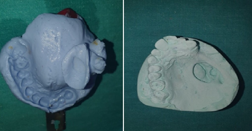
Figure 2: PRIMARY IMPRESSION & Figure 3: PRIMARY CAST

Figure 4: SPECIAL TRAY & Figure 5: BORDER MOLDING DONE USING GREEN STICK AND HOLES DRILLED FOR SECONDARY IMPRESSION

Figure 6: SECONDARY IMPRESSION MADE USING ELASTOMERIC IMPRESSION MATERIAL & Figure 7: MASTER CAST POURED USING DIE STONE AND BLOCK OUT DONE

Figure 8a: BITE RIM FABRICATED & Figure 8b: BITE REGISTRATION DONE
Wax-up of the trial obturator prosthesis was done. It was flasked, dewaxed, packed and processed. The obturator was finished and polished.[Figure 9]

Figure 9: FINAL PROSTHESIS FINISHED AND POLISHED
With the final adjustments it was inserted and delivered to the patient.[ Figure 10] Patient examination was performed after specific time intervals. The patient reported good aesthetics and improved functioning with the prosthesis without any discomfort.
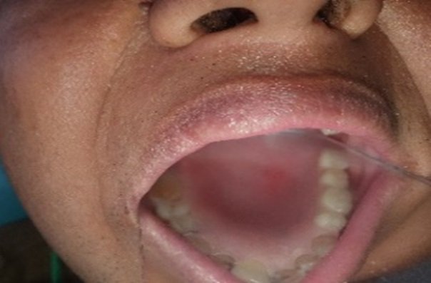
Figure 10: INTRA ORAL PICTURE POST INSERTION OF THE PROSTHESIS

Figure 11: Intraoral Pre Prosthodontic Rehabilitative Picture & Figure 12: Intraoral Post Prosthodontic Rehabilitative Picture
DISCUSSION
An obturator prosthesis can play a very crucial role in the rehabilitation of post surgical maxillectomy patients.6 The design for obturators may vary based on the classification system of the defect.7 All the removable obturator prosthesis should be made with the basic prosthodontic principles which include broad stress distribution, cross arch stabilization.8 The Support for the prosthesis is provided by the remaining teeth, palate. In patients with remaining dentition, teeth play an important role in providing retention, support, and stability for the prosthesis. Retention can be achieved from the remaining teeth or ridge, lateral part of the defect, soft tissue undercut, scar band and clasps.9
CONCLUSION
The biggest challenge we face while rehabilitating patients post maxillectomy is of adequate retention, stability and support. Good knowledge and skills coupled with better understanding of the patients needs can enable a successful rehabilitation for such patients which would include better chewing, swallowing, comfort, esthetics, phonetics and an improved quality of life all together.
REFERENCES
- Genden EM, Okay D, Stepp MT, et al. Comparison of functional and quality-of-life outcomes in patients with and without palatomaxillary reconstruction. Arch Otolaryngol Head Neck Surg 2003;129:775-80.
- Cordeiro PG, Santamaria E. A classification system and algorithm for reconstruction of maxillectomy and midfacial defects. Plast Reconstr Surg 2000;105:2331-46.
- Chiapasco M, Biglioli F, Auteliano L, et al. Clinical outcoobturator prosthesisme of dental implants placed in fibula-free flaps used for the reconstruction of maxillo-mandibular defects following ablation for tumors or osteoradionecrosis. Clin Oral Impl Res 2006;17:220-8.
- Rogers SN, Lowe D, McNally D, et al. Health-related quality of life after maxillectomy: a comparison between prosthetic obturation and free flap. J Oral Maxillofac Surg 2003;61:174-81.
- G Tirelli, R Rizzo,1 M Biasotto,1 R Di Lenarda,1 B Argenti,1 A Gatto, and F Bullo. Obturator prostheses following palatal resection: clinical cases. Acta Otorhinolaryngol Ital. 2010 Feb; 30(1): 33–39.
- F. Keyf, “Obturator prostheses for hemimaxillectomy patients,” Journal of Oral Rehabilitation, vol. 28, no. 9, pp. 821–829, 2001.
- M. A. Aramany, “Basic principles of obturator design for partially edentulous patients. Part I: classification,” Journal of Prosthetic Dentistry, vol. 40, no. 5, pp. 554–557, 1978.
- M. A. Aramany, “Basic principles of obturator design for partially edentulous patients. Part II: design principles. 1978,” Journal of Prosthetic Dentistry, vol. 86, no. 6, pp. 562–568, 2001.
- R. Gurjar, S. Kumar, H. Rao, A. Sharma, and S. Bhansali, “Retentive aids in maxillofacial prosthodontics—a review,” International Journal of Contemporary Dentistry, vol. 2, no. 3, pp. 84–88, 2011.
Disclaimer from Guident
The views and opinions expressed in articles published in Guident are solely those of the authors and do not necessarily reflect the views of the editors, the editorial board or the publisher, Ivory India. Guident does not endorse any product, technique or treatment described. Clinical content is intended for qualified dental and medical professionals and is not a substitute for individual clinical judgement. Guident, its editors and the publisher accept no liability for any consequence arising from the use of information published in the journal.
How to cite this article
Dr. Yogeshwari Krishnan, Dr. Varun Kumar, Dr. Himanshu Aeran. Maxillary Obturator Prosthesis for a Hemimaxillectomy Patient-a Case Report. Guident. 2021 Mar;14(4). Available from: https://guident.in/articles/prosthodontics/maxillary-obturator-prosthesis-for-a-hemimaxillectomy-patient-a-case-report.html


