Dental Complications After COVID-19 Mucormycosis-Understanding the Pathophysiology-Case Series Part-1
Jaipur, India
ABSTRACT
The corona virus disease outbreaked in 2019 (COVID -19) became pandemic, which cause severe respiratory syndrome (SARS-CoV -2).COVID -19 is associated with inflammation that even resulted in fatal outcomes. Association with dental conditions have been found with COVID -19 complications. This case report discuss common dental complications in previously covid-19 infected individuals, thus considering such complications to be made in consideration by healthcare workers while treating their patients.
Key words : Covid-19,covid complications, dental conditions, inflammation
INTRODUCTION
Corona virus (COVID-19) a pandemic disease affecting respiratory system, still dissolute to impact a major portion of world’s population frightening the health care workers displaying its new strains and pathogenicity.1 Systemic inflammation is one of the important characteristic of dental condition.2 This emerging respiratory disease has resulted in over 75 million confirmed positive cases with 1.6 million death by Dec 22nd,2020.3
While few patients of covid-19 present with mild symptoms4,14% cases may develop severe condition. Covid-19 severe cases with ARDS5 generally show exacerbated immune response, presenting excessive levels of cytokines and widespread area of tissue damage. Covid-19 cases has been seen associated with increased levels of C-Reactive Protein(CRP), D-dimer, interleukin-6(IL-6) and ferritin,6 suggesting a link between severity and virality of non resolving inflammation.
It is still controversial to point out the role of oral cavity in covid-19,but few evidence based studies suggest role of oral cavity mucosa in transmission and pathogenicity of covid-19.7 Demonstrating oral diseases as a marker for increased severity of covid-19. In this we discuss about the case which was first looked upon as a regular dental infective case but later was co-related with covid-19 complication.
CASE REPORT-1
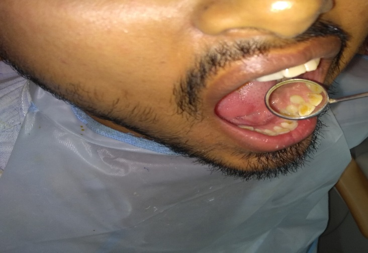
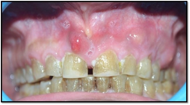
A 35year old male patient reported to our clinic with chief complaint of pain along with pus discharge from upper left tooth region upon clinical examination it was found that patient had multiple small abscess with suppuration ,gingiva was found to be inflammatory and oedematous.(Figure 1.a)Patient responded upon palpitation.
Figure 1a: edematous gingiva with Small abscess
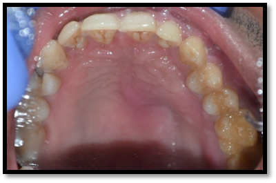
Figure 1b: Reflection of palatal mucosa
Intra oral examination revealed diffuse swelling in palatal mucosa of affected side.(Figure 1.b)Patient also presented with history of covid-19 infection.(Figure 2)
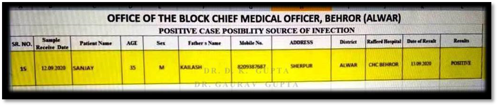
Figure 2. Covid-19 positive report
In the starting patient was assumed to be of regular dental case but later was thought it might be a complication of post covid -19 infection. So to rule out we referred the patient to undergo for haematology analysis and CT scan.
CBC findings of patient showed increased level of platelet count, low haemoglobin level.(Figure 3)
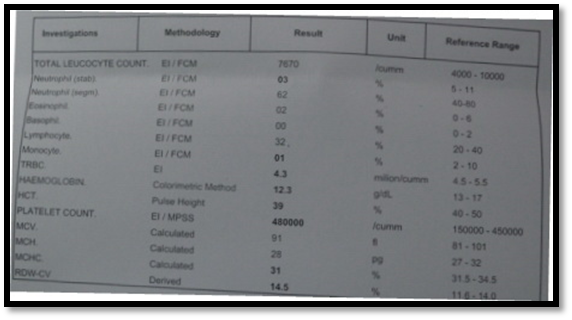
Figure 3. CBC report showing increased levels of platelet count and low haemoglobin
BT and CT were in normal ranges, while random blood glucose(321mg/dl),SGPT (192 U/L)) and SGOT (161 U/L) showed a peak rise in their values.(Figure 4a,b,c)
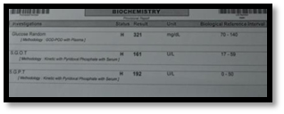


Figure 4a,b,c. Report showing values of BT,CT,SGOT,SGPT and random glucose
To rule out further damage and analyse the case CT scan with 3D reconstruction findings were suggestive of:
- ill marginatedpermeative lytic lesion in left upper maxillary alveolus and hard palate with extent, morphology and dimensions were suggestive of likely maxillary osteomyelitis.
- mucosal thickening with non enhancing secretions causing complete opacification of left maxillary sinus and blocked osteometal complex.
- mucosal thickening and retained secretions also in bilateral ethmoid ,sphenoid and right maxillary sinus with opacification of bilateral concha bullosa suggestive of panrhinosinusitis.(Figure 5 a, b and c)
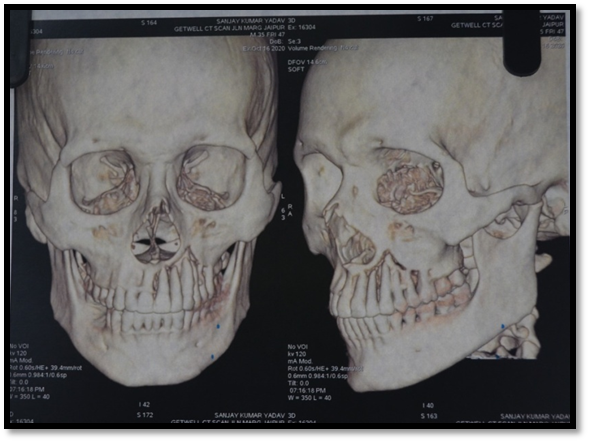
Figure 5a. 3D reconstruction of face
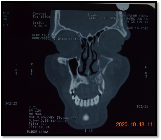
Figure 5b. CT scan of face

Figure 5c. CT scan findings
After going through the test reports ,the case as based upon clinical as well as pathological and radiographic investigations was suggestive to be of maxillary necrosis.
Treatment given : surgery was carried out by raising the flap and extraction of all the teeth except 3rd molar in affected area along with local debridement of necrosed tissue was done.(Figure 6 a,b,c)(Figure 7). 3rd molar was left to provide further support for prosthesis fabrication.

Figure 6a. Surgery of affected area
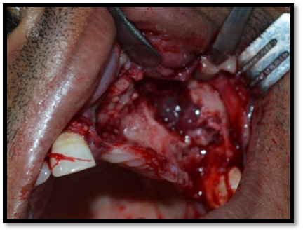

Figure 6b & c. Extraction of affected teeth except 3rd molar
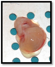
Figure 7. Removal of necrosed tissue
Patient was followed with a course of antibiotics and analgesics.to check for recovery patient was recalled with one month follow up and was delivered with removal prosthesis with clasps.(Figure 8.a,b)
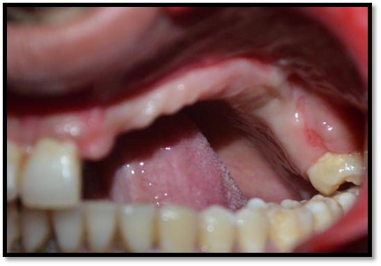
Figure 8a. Post operative image of affected side

Figure 8b. RPD with clasps delivered to improve esthetic
Post operative one month follow up OPG was taken to look for the bone healing.(Figure 9)
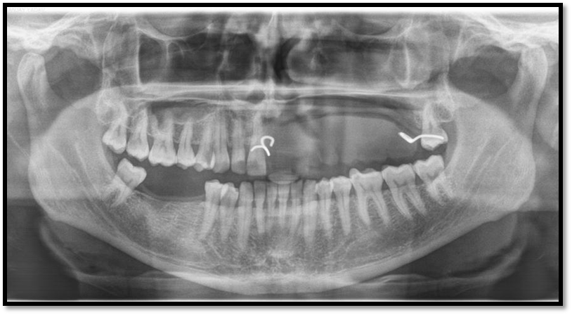
Figure 9. Post operative OPG depicting healing
DISCUSSION
Covid-19 a pandemic disease started to spread in December 2019 causing pneumonia of unknown etiology originated from seafood market of Wuhan, China.8 Before it was identified as Novel corona virus, later known to be severe acute respiratory syndrome coronavirus-2 (SARS-CoV-2) and disease called to be covid-19.8
Osteomyelitis means bone marrow inflammation, this term was coined by Nelaton in 1844.9
Osteomyelitis has more incidence in males (80.36 %) then females (19.64 %)10
Osteomyelitis is an opportunistic infection which may emerge as a complication of some underlining condition.11
However, specific mechanism and pathological pathway have been noticed linking periodontal diseases with systemic diseases, showing translocation of bacteria to blood, induced auto immune damage a systemic inflammation.12
Here in this case report we discuss a case of male patient as a regular dental infection which later on was co-related with covid-19 complication based upon systemic and radiographic findings presenting extra pulmonary manifestations of covid -19 depicting necrosis of maxilla due to some underlining systemic condition which may be a covid-19 post infection complication.
CONCLUSION
Osteomyelitis is mostly seen in patients with diabetes and immuno-suppression. In covid-19 individual have low immune response which may give rise to secondary infections other than respiratory distress. Care should be taken in diagnosis of such cases through radiographic investigations and other histopathological findings while treating patients in clinics. Clinicians should be aware of such pathologies which may be due to complication of some underlining infection.
REFERENCES
- Harshil bhatt and Sandeep singh.Venous thromboembolism and COVID-19: a case report and review of the literature.Journal of medical case reports 14,article no 188(2020) https://doi.org/10.1186/s13256-020-02516-4
- NadyaMarouf,WenjiCai,Khalid N. Said,HaninDaas,HananDiab, Venkateswara Rao Chinta,Ali Ait Hssain,BelindaNicolau,MarianoSanz,FalehTamimi . Association between periodontitis and severity of COVIDâ€Â19 infection: A case–control study.Journal of clinical periodontology 1stfeb 2021.https://doi.org/10.1111/jcpe.13435
- WHO (2020b). COVIDâ€19 weekly epidemiological update 20201222 https://www.who.int/publications/m/item/weekly-epidemiological-update-22-december-2020. Google Scholar
- Huang, C,Wang, Y, Li, X., Ren, L, Zhao, J, Hu, Y. I, Zhang, L. I, Fan, G, Xu, J, Gu, X, Cheng, Z, Yu, T, Xia, J, Wei, Y, Wu, W, Xie, X, Yin, W, Li, H, Liu, M, Cao, B (2020).Clinical features of patients infected with 2019 novel coronavirus in Wuhan, China. The Lancet, 395(10223), 497– 506. https://doi.org/10.1016/s0140-6736(20)30183-5. Crossref |CAS |PubMed |Web of Science®|Google Scholar
- Mehta, P, McAuley, D. F, Brown, M, Sanchez, E,Tattersall, R. S, & Manson, J. J (2020). COVIDâ€Â19: Consider cytokine storm syndromes and immunosuppression. The Lancet, 395(10229), 1033– 1034. https://doi.org/10.1016/s0140-6736(20)30628-0. PubMed|Google Scholar
- Chen, N, Zhou, M, Dong, X, Qu, J, Gong, F, Han, Y, Qiu, Y, Wang, J, Liu, Y, Wei, Y, Xia, J, Yu, T, Zhang, X, & Zhang, L. I (2020). Epidemiological and clinical characteristics of 99 cases of 2019 novel coronavirus pneumonia in Wuhan, China: A descriptive study. The Lancet, 395(10223), 507– 513. https://doi.org/10.1016/s0140-6736(20)30211-7. Crossref |CAS |PubMed |Web of Science®|Google Scholar
- Xu,H, Zhong, L, Deng, J, Peng, J, Dan, H, Zeng, X, Li, T, & Chen, Q(2020). High expression of ACE2 receptor of 2019â€ÂnCoV on the epithelial cells of oral mucosa. International Journal of Oral Science, 12(1), 8. https://doi.org/10.1038/s41368-020-0074-x. Crossref| CAS| PubMed| Web of Science®|Google Scholar
- Zhu N, Zhang D, Wang W, Li X, Yang B, Song J, Zhao X, Huang B, Shi W, Lu R, Niu P, Zhan F, Ma X, Wang D, Xu W, Wu G, Gao GF, Tan W, China Novel Coronavirus Investigating and Research Team. A novel coronavirus from patients with pneumonia in China, 2019. N Engl J Med. 2020;382(8):727-33 https://doi.org/10.1056/NEJMoa2001017.
- L. Klenerman, “A history of osteomyelitis from the journal of bone and joint Surgery,” Journal of Bone and Joint Surgery, vol. 89-B, no. 5, pp. 667–670, 2007.View at: Publisher Site | Google Scholar
- R. A. Mohsien, M. C. Al Mohammedawi, and A. G. Yahya, “Virulance factors enhancing microbial infection in chronic osteomylitis and antibiotic susceptability pattern,” American Journal of Medical Case Reports, vol. 2, no. 6, pp. 126–132, 2014.View at: Google Scholar
- Rajesh arani,S.N.H AfsarShareef and H. M. KhuthijaKhanam.Mucormycotic osteomyelitis involving the maxilla: A rare case report and review of the literature.Case Reports in Infectious Diseases / 2019 / Article.Volume 2019 |Article ID 8459296 https://doi.org/10.1155/2019/8459296
- Schenkein, H. A, Papapanou, P. N, Genco, R, & Sanz, M (2020). Mechanisms underlying the association between periodontitis and atherosclerotic disease. Periodontology 2000, 83(1), 90– 106. https://doi.org/10.1111/prd.12304. Wiley Online Library PubMed Web of Science®Google Scholar
Disclaimer from Guident
The views and opinions expressed in articles published in Guident are solely those of the authors and do not necessarily reflect the views of the editors, the editorial board or the publisher, Ivory India. Guident does not endorse any product, technique or treatment described. Clinical content is intended for qualified dental and medical professionals and is not a substitute for individual clinical judgement. Guident, its editors and the publisher accept no liability for any consequence arising from the use of information published in the journal.
How to cite this article
Dr. Gaurav Gupta, Dr. D. K. Gupta, Dr. Neelja Gupta Wisdom Dental Clinics. Dental Complications After COVID-19 Mucormycosis-Understanding the Pathophysiology-Case Series Part-1. Guident. 2021 Mar;14(4). Available from: https://guident.in/articles/general/dental-complications-after-covid-19-mucormycosis-understanding-the-pathophysiology-case-series-part-1.html