
Making Planes Out of Skulls- the Role of Cephalometrics in Prosthodontics
SS Dynamic Prosthetic Solutions, Chennai, India, Royal Dental College, Kerala, India, Department of Prosthodics
Abstract
Two skulls are not the same. The role of cephalometrics has been considered relavent only in the field of orthodontics and its importance in prosthodontics has remained very limited. But the use of cephalometrics for prosthodontic reconstruction dates back to 1931, when it was first used by Hofrath for prosthodontic reconstruction. Though the values obtained from this method cannot be used accurately without deviation, it can definitely help to lay down guidelines in individualized treatment planning.
INTRODUCTION
Some complete denture wearers suffer facial collapse as a result of severe occlusal wear of the artificial teeth, decreased occlusal vertical dimension, deterioration of the dentures, and residual ridge resorption. The stomatognathic system adapts to the new maxillomandibular and occlusal relationships and continues to function. The facial rehabilitation is usually using anatomical landmarks, both intraoral and extra oral. Facebow transfer is the most commonly employed clinical method of transferring these landmarks to the articulator.
The basic transfer with the Face-bow is the transfer of reference planes of the head into the articulator system. The use of different reference planes does not change the centric relation of the plaster cast. These reference landmarks are used as it undergoes very little change or no change at all. These landmarks can be clearly studied by using a radiographic technique called as CEPHALOMETRICS.
Cephalometrics have been in use since 1989. Chaconas expresses his belief in the use of cephalometrics in prosthodontic diagnosis and treatment planning for better understanding of the patient and to get a complete picture.
THE BASICS OF CEPHALOMETRICS
The basic knowledge of cephalometrics pertains to the landmarks, which must be clearly defined. Figure 1 describes the various anatomical landmarks that are commonly used in cephalometrics. They are2:
- Xi – Geographic centre of mandible
- M – Menton (Inferior point on symphysis )
- N - Nasion (Junction of frontal and nasal bone)
- O - Orbitale (Inferior most point on the orbit)
- P – Porion (Superiormost point of auditory meatud )
- ANS – Anterior nasal spine( Anterior point on spiny process of maxilla )
- A – Point A( Innermost curvature point of maxilla from ANS to Maxillary alveolar process)
- Po – Pogonion ( Anterior point on symphysis of Mandible )
- PM – Protruberance menti ( Point where curvature Turns from convex to concave on symphysis of Mandible )
- Po1/Pog – Soft tissue pogonion
- PN – Pronasale ( Anteriormost point on nose )
- Go – Gonion ( Inferior point on angle of mandible )
- PNS – Posterior nasal spine
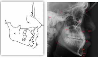
Figure 1
By connecting these landmarks, various cephalometric planes are obtained. The planes obtained are1: FH - Frankfurt horizontal - the line traced from O to P;
CL - Camper's line - the line connecting ANS and P; PL - Palatal line - the line connecting ANS and PNS;
OL - Occlusal line - line traced between points IS and M; and
ML- Mandibular line - the tangent line of the lower border of the mandible connecting points T1 and T2.
These planes will be seen in the following analytic criteria:
- Skeletal criteria
- Dental criteria
- Profile criteria
SKELETAL CRITERIA
1) Maxillary and mandibular depth
Skeletal relation of maxilla to the reference plane can be studied by orienting N – A line to the Frankfurt horizontal plane (O – P plane). The angle is found to average 90 degress. Any increase or decrease in the angle can be correlated clinically as maxillary excess or deficiency respectively3.
When the Frankfurt horizontal plane is oriented with the N – Po plane, the mandibular depth can be studied with an average angle of 90 degrees. Any increase or decrease in this angle can be correlated as Prognathism or Retrognathism (figure 2).
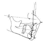
Figure 2
These criteria seem to be simple enough to be clinically appreciable but further analysis applying different facial types according to Simon’s classification (figure 3), it is clear that a cephalometric analysis would give better understanding.
- Protraction can be of 3 types – Maxillary, Mandibular and bimaxillary
- Retraction can be of 3 types – Maxillary, Mandibular and bimaxillary
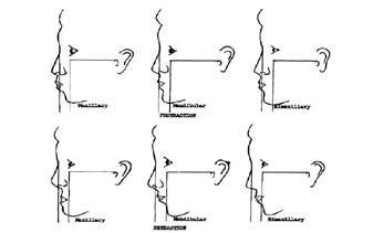
Figure 3.
This maxillomandibular relationship can be studied by connecting N – A plane to N – Po plane. In a straight profile patient, it should form an average of 0 degrees. Deviation of this will help us to determine the deficiency or excess is maxillary or mandibular.
1) Frankfurt – Mandibular plane angle
The relationship of Frankfurt horizontal plane to mandibular plane helps in understanding the skeletal open bite and closed bite. It has no relation with dental patterns. This angle is found to be 25 degrees on an average. But the variations in relation to age, sex and racial characteristics should be kept in mind. With a standard deviation of 5 degrees, the following is derived
- Low FMA – 20 degrees or less ( skeletal closed bite )
- High FMA – 30 degrees or more (skeletal open bite )
Girard DiPierto studied this angle extensively and said that a high or a low FMA is been correlated with the muscle potential. The bite forces generated by the elevator muscles (Temporalis, Masseter and Medial Pterygoid ) is high in low FMA and less in high FMA patients. The relationship between osseous tissue and functional requirements is evident. In low FMA patients,
- In natural teeth, the teeth are seen as small crowns not completely erupted or intruded which is susceptible to attrition due to high bite forces. This leads to a decrease in the vertical dimension
- This decrease in the vertical dimension can be thought to be treated with FDP by increasing the vertical dimension. But poor tolerance by the tissues are seen and this increase in vertical is reverted back to original by the process of intrusion with contribution of resorption of alveolar process. Also, cantilevered and long span bridges are poorly tolerated by these patients
- In edentulous patients, it has to be kept in mind that the high forces are generated and residual ridge is under stress. The available interridge distance is low. There is increased tendency for fracture of denture base.
In high FMA patients,
The opposite of the effects seen above in low FMA patients can be generally expected. There is an increase in lower facial height. This can be attributed to the physiologic freedom given by the muscles to growth of alveolar process. But it is not conclusive that decreased bite force leads to increased eruption and greater bite force causes decreased eruption. Again, it is interesting to note that
- The palatal contour also shows relations in these patients. Low FMA patients have flat, broad palate while it is V shaped in high FMA patients.
- The attached gingival level is more in high FMA patients than low FMA patients
- The muscle attachment is in basal bone in high FMA patients whereas it is near to crest of ridge in low FMA patients
- The muscle direction in high FMA patients give exaggeration to the anterior component of force causing susceptibility to mesial drifting of posterior teeth into edentulous space.
- The pharyngeal space is constricted causing tongue protrusion, mouth breathing. The tongue can interfere with stability and retention of complete denture in high FMA patients due to the tongue protrusion tendency.
2) Maxillomandibular dimension
- Measurement of the length is done by extending the occlusal plane( formed by connecting the Incisal edge of maxillary central incisor to distobuccal cusp of 1st maxillary molar) to the shadow of the posterior pharyngeal wall on lateral cephalograph.
- Measurement of height is done by connecting maxillary plane ( ANS – PNS plane ) vertically to Menton
These dimensions have found be on an average,
Maxillomandibular Length – 80 mm (>80 mm – Long, < 80 mm – Short )
Maxillomandibular height – 70 mm (>70 mm – High, < 70 mm – Low )
The average maxillomandibular relations are long and High, Short and Low
3) Lower facial height:
The angle formed by joining of ANS – Xi plane to Xi – PM plane gives the lower facial height angle which is averaging around 47 degrees. Increase or decrease in the angle can help in understanding skeletal patterns like ‘open bite’ and ‘deep bite’.
DENTAL CRITERIA
1. Occlusal plane:
It has been been extensively studied for more than 150 years starting from well known scientists like Bonwill, Brommell, Cummer, Gysi, Prothero, Wadsworth, Sears, Boucher.
Montieth explained that the anteroposterior dimension of maxillary base governs the plane angulation of occlusion. He gave his “Denture glass effect” (figure 4) model to demonstrate his theory. He also studied the relationship of N – Po – ANS angle and found that for every degree increase in the angle, there is a 0.9098 degree decrease in occlusal plane.

Figure 4: Denture-glass effect. Anteroposterior dimension of maxillary base governs angulation of occlusal plane in same way as diameter of glass of water determines tilt that upper denture immersed therein assumes. A, Glass of greater diameter permits flatter angle than that permitted by B, narrower glass
Montieth’s formula: Y = 77.3484 – (0.9098 x X ) where X is N-Po-ANS angle
Though it seems interesting to derive the occlusal plane from a formula, Karkazis studied the value of this formula in deriving the occlusal plane in dentulous subjects and found variability to be high and could only find close correlation to the clinically determined occlusal plane in edentulous patients.
Maxillomandibular dimensions as explained earlier was studied by L’estrange in order to see its relation to the occlusal plane and found variability from the average dimensions like in the case of LONG AND LOW and SHORT AND HIGH will see the occlusal plane varying from their average angle of 7.5 degrees5. This finding can be correlated to the “Denture glass effect” of Montieth6. Ow studied the occlusal plane in Chinese population and found SHORT and HIGH maxillomandibular relation in these population.
Different reference planes were tried out to find a relation to the occlusal plane like Sloane and Cook studied ANS – Hamular notch (Cook’s angle) 7 and also maxillary plane was tried out in other studies but it did not find any conclusive results.
Scientists like Lundquist has tried to orient the plane to clinical landmarks such as retromolar pad, Buccinator groove and corner of the lip and found high significance in its relation. Hartano studied the variability of the plane in different facial types as seen in simon’s classification.
The occlusal plane showed variable angles for the different facial types and a point to be noted is the incorporation of Gysi’s prosthetic plane which shows much closer parallelism to the occlusal plane in dentulous patients. Camper has not exactly mentioned which part of the ear to use as a landmark and some studies use the superior border of the tragus and some others use the middle border of the tragus when studying the relationsip with this plane.
Practically speaking, the facebow is oriented to the articulator by relating Orbital pointer to the auditory meatus. The condylar axis is incorporated in the average value semiadjustable articulators like the Hanau H2 model which is commonly used by using Beyron’s theory of 13 mm average distance from the auditory meatus. But keeping in mind, Weinstein’s statement that there is no need to locate the original hinge axis.
An within a radius of 5 mm will only create an error of 0.2mm in the vertical height at the second molar. This might seem like a small value but it has been determined that edentulous complete denture wearers can perceive an occlusal discrepancy of even 0.06mm. So keeping in mind that the facebow transfer made already includes error within the 5mm radius, the axis orbital plane is transferred to the articulator by its relationship to the Frankfurt horizontal plane already incorporated into the articulator.
Gonzales studied the relationship of the the two planes and said that the angular difference is compensated by placing the orbital indicator 7mm above the condylar axis. This correction is been incorporated in Hanau H2 and Dentatus articulator. In other articulators like the Whipmix, Panadent, TMJ and older Hanau articulators, the earlier concept of parallelism of these reference planes is still being used.
The most commonly used occlusal plane indicator is the ala tragal line and it was shown in a study that the most reliable posterior reference point of the ala tragal line was the midpoint of the tragus when cephalometrically compared with the occlusal plane and Frankfurt horizontal plane in dentulous patients4.
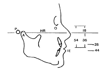
Figure 5
Pitchford further complicates the matter by stating the the occlusal plane is still too steep after this correction and explained this error as due to the failure to incorporate the reference esthetic position into the articulator. This position which is important to determine esthetics shows a relationship when a plane is drawn from condylar axis and extended into the horizon horizontally and measurement is done between this plane and the incisal edge of the maxillary central incisor.
In the reference esthetic position (figure 5), the average distance is found to be 36 mm where as a facebow transfer with Frankfurt horizontal plane is averaging 54mm. Potential esthetic errors could be expected due to this difference. Interestingly, the incisive notch in the Hanau H2 model averages a distance of 36mm from the condylar axis and the manual explains that this point could also be used to orient the occlusal plane. Pitchford says that correction could be done by measuring the orbital pointer to be placed 11 mm above the orbital indicator in the articulators with corrections done for the condylar axis – orbital plane and in the other articulators, he suggests 18 mm correction above the indicator8.
Franfurt horizontal plane has also been studied for its relationship with the occlusal plane. Ow expressed his concern that the position the orbitale when studied in different races and found the superioinferior variation in this position when he studied Chinese and Swedish population8.
Clincally speaking, Swenson says that the occlusal plane should be in relation to the area of the residual ridge available. If its unfavourable, the placement should be close to the ridge to decrease the leverage action on stability and retention. Wolff’s law of transformation says that the functional stress govers the shape of the bone. The occlusal plane should be at right angles the occlusal force, so variability with the facial types should be taken into consideration before deciding on the occlusal plane.
PROFILE CRITERIA
In cephalometric analysis, several reference lines have been introduced to assess anteroposterior position of the upper and lower lips. Ricketts’ ‘E’ line, Steiner’s S1 line, Holdaway’s ‘H’ line , Burstone’s ‘B’ line , and Sushner’s ‘S2’ line ( figure 6) are common lip assessment lines. 10 the most significant of these are E line and S1 line
1. E line
The patient’s soft tissue landmarks are taken into consideration by joining PN – Po1. The measurement is done in its relation to the lower lip. It is found that the esthetics is found to be favorable when the lower lip is 2mm behind this plane. Though a significant relationship has not been found for this relation, it is used for getting esthetics. Hence, the name ‘Esthetic line’ or ‘E line’.9
2. S1 line
S-Line is drawn from midpoint between subnasale (Sn) and Pronasale (Pn) to soft tissue pogonion (Pog) and lip prominence with reference to this line is assessed. Its cephalometric norms are as follows: Upper lip to S Line (0±2mm), Lower lip to S Line (0±2mm). Steiner used S-Line with the idea that E-Line is affected by nose length.11
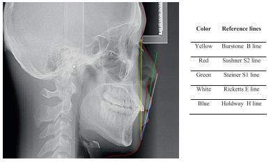
Figure 6
REFERENCES
- Seifert D, Jerolimov V. Relations Of Reference Planes For Orientation Of The Prosthetic Plane. Acta Stomat Croat. 2000;34(4):413-416.
- Majd M, Shoeleh F. A Novel Hybrid Approach For Cephalometric Landmark Detection.
- Dawson P. Functional Occlusion. St. Louis, Mo.: Mosby; 2007.
- Krishna V, Reddy V, Sankar V. Cephalometric Comparison Of Frankforts Horizontal Plane And Occlusal Plane With Variations In Ala-Tragus Line. Annals And Essences Of Dentistry. 2013;5(4).
- L'estrange P, Vig P. A Comparative Study Of The Occlusal Plane In Dentulous And Edentulous Subjects. The Journal Of Prosthetic Dentistry. 1975;33(5):495-503.
- Monteith Bd. A Cephalometric Method To Determine The Angulation Of The Occlusal Plane In Edentulous Patients. J Prosthet Dent 1985;54:81-7.
- Sloane Rh, Cook J. A Guide To Orientation Of The Occlusal Plane. J Prosthet Dent 1953;3:33
- Shetty S, Zargar N, Shenoy K, Rekha V. Occlusal Plane Location In Edentulous Patients: A Review. The Journal Of Indian Prosthodontic Society. 2013
- Ricketts R. Esthetics, Environment, And The Law Of Lip Relation. American Journal Of Orthodontics. 1968;54(4):272-289.
- Asad S. Assessment Of Antero-Posterior Position Of Lips: E-Line- S-Line. Pakistan Oral & Dental Journal. 2011;31(1):84-87.
More references are availabe on request.
Disclaimer from Guident
The views and opinions expressed in articles published in Guident are solely those of the authors and do not necessarily reflect the views of the editors, the editorial board or the publisher, Ivory India. Guident does not endorse any product, technique or treatment described. Clinical content is intended for qualified dental and medical professionals and is not a substitute for individual clinical judgement. Guident, its editors and the publisher accept no liability for any consequence arising from the use of information published in the journal.
How to cite this article
Dr. Sathya Shankar, Dr. Shreshta Sathishc. Making Planes Out of Skulls- the Role of Cephalometrics in Prosthodontics. Guident. 2015 Dec;9(1). Available from: https://guident.in/articles/prosthodontics/making-planes-out-of-skulls-the-role-of-cephalometrics-in-prosthodontics.html


