Regeneration Will Overcome Repairs..... Platelet Rich Fibrin (PRF) Forgotten Tool in Regenerative Dentistry
India. The Dental Clinic, Dehradun, India.
Authors : Dr. Suganya. B , Dr. Priyanka Mittal .
Abstract
Regenerative dental procedures are biologically based procedures with regeneration of defective oral tissues. In the past two decades, an increased understanding of the physiological roles of platelets in wound healing has led to ideas of using platelets as therapeutic tool in the field of regenerative dentistry.
Platelet rich fibrin or autologous platelet gel or platelet rich plasma is the component derived from whole blood, when used in the wound site especially in case of periodontal regenerative procedure; it accelerates the regeneration of the periodontal tissues because of its growth factors. One of the main advantages of autologous PRP is that as it is derived from patients own blood, transmission of diseases of immunogenic reactions can be avoided.
INTRODUCTION
Platelets are the smallest corpuscular components of the blood (diameter – 2-4 microns), number varies from 1,50,000 to 3,00,000 per/mm3 of blood. The origin of platelets is bone marrow, where megakaryocytes, as the result of mitotic proliferation of a progenitor cell-liberates platelets as the end product of protrusions of their membrane and cytoplasm. They lack nucleus for replication and die in 8-9 days. The platelets contain alpha-granules which is the reservoir for the growth factors or cytokines, nothing but small proteins each of 25,000 Daltons molecular weight.
We are behind these growth factors which enhances the tissue healing and regeneration.
GF’s secreted by platelets includes,
- PGDF: Platelet Derived Growth Factor.
- EGF: Epidermal Growth Factor.
- IGF-1: Insulin like Growth Factor.
- TGFβ1: Transforming Growth Factor.
- VEGP: Vascular endothelial Growth Factor.
- HGF: Hepatocyte Growth Factor.
- bFGF: Basic Fibroblast Growth Factor.
GROWTH FACTORS: MECHANISM OF ACTIONS.
TGF-β:
- Mesenchymal proliferation and differentiation.
- Cell mitogenesis.
- Collagen II, Proteoglycan and ectomycinchymal synthesis.
- Endothelial chemotaxis and angiogenesis.
- Macrophages and lymphocyte proliferation, inhibition and chondrocyte differentiation.
PGDF α & β:
- Osteoblast and mesenchymal mitogenesis.
- Macrophages, neutrophils and other cell chemotaxis, collagen I synthesis.
bFGF:
- Chondrocyte and osteoblast differentiation.
- Mesenchymal, chondrocytes and osteoblast mitogenesis.
EGF:
- Endothelial chemotaxis and angiogenesis.
- Collagen synthesis.
- Mesenchymal and epithelial cell mitogenesis.
CTGF:
- Angiogenesis.
- Cartilage regeneration.
- Fibrosis.
- Platelet adhesion.
VEGF:
- Angiogenesis.
- Endothelial cell mitogenesis.
IGF:
- Cell Proliferation.
- Collagen synthesis.
- Myoblast proliferation and differentiation.
FIBRIN IN PLASMA:
Fibrin is derived from the plasma and provides cell mobility in the wound. The role of fibrin which is crosslinked protein derived from fibrinogen in plasma is not only to serve as a scaffold or surface for cellular migration, but to entrap platelets. This will lead to the random distribution of platelets and growth factors throughout the wounds.
Platelet rich plasma is an autologous source for growth factors obtained from the sample of a patient’s blood collected in a dental office setting via centrifugation process. The PRP thus obtained has 2.16 times increase in the concentration of platelets when compared to normal range of platelets in Human blood.
PLATELET RICH FIBRIN PROCUREMENT PROCEDURE:
The classical preparation was done by Dr. CHOUKROUN in 2000. This technique does not contain anticoagulant.
The protocol for PRF procedure is very simple and simulates that of PRP.
REQUIREMENTS:
- Centrifugal table.

Fig: Centrifugal Machine.
- Blood collection Kit with 2 sterile vacutainer tubes of 6 ml each.
- 24 gauge butterfly needle.
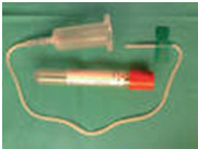
Fig: Vacutainer Tubes & Butterfly needle.
- Special kits for PRF membrane preparation are available where the collected PRF can be made as a membrane by squeezing out the fluid present in the fibrin clot.
Procedure includes collection of whole venous blood from the peripheral veins(around 5 ml) in each of the two sterile vacutainer tubes without anticoagulant and the vacutainer tubes are then placed in a centrifugal machine at 3000 revolution per minute for 10 mins, after which it settles into the following 3 layers.
Upper straw colored acellular plasma or PPP (Platelet Poor Plasma).
Middle PRF or fibrin clot rich in platelets.
Lower fraction containing Red blood corpuscles.
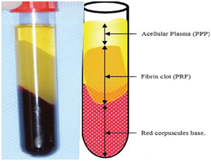
Fig:Centrifuged Tube - Three layers.
Then the middle layer is collected 2 mm below the lower dividing line, which is the PRF.
The mechanism involved in this is, the fibrinogen concentrated in upper part of the tube, combines with circulating thrombin due to centrifugation to form fibrin. A fibrin clot is then formed in the middle between the RBCs at the bottom and acellular plasma at the top. The middle part is platelets trapped massively in fibrin meshes.

Fig:PRF with 2mm of RBC.

Fig:PRF membrane prep.
CONTRAINDICATIONS
Absolute:
- Platelet dysfunction syndrome.
- Critical thrombocytopenia.
- Hemodynamic instability.
- Septicemia.
- Local infection at the site of procedure.
- Patient unwilling to accept risks.
Relative
- Consistent use of NSA IDs within 48 hours of procedure.
- Corticosteroid injection at treatment site within one month.
- Systemic use of corticosteroids within 2 weeks.
- Tobacco use.
- Recent fever or illness.
- Cancer – especially hematopoietic or of bone.
- HGB < 10 gram/dl.
- Platelet count <105 per µl.
Clinical Applications of PRF in Dentistry:
- Periodontal Intra bony defects repair.
- Apexification in endodontics.
- Inlay and onlay grafts.
- Sinus lift procedures.
- Closure of cleft lip and palate defects.
- Repair of bone defects created by removal of teeth and small cysts.
- Repair of fistulas between the sinus cavity and mouth.
PRF - REGENERATIVE AID:
When the PRF membrane is applied to the graft site and sutured, the higher concentration of platelets (average increase of 33.8%) Jump starts the cascade of regenerative events leading to the formation of a mature graft site. This is achieved mainly by the secretion of growth factor mainly PGDF and TGF beta.
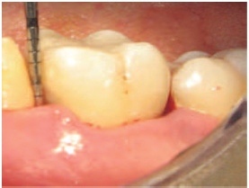
Fig 1: Initial clinical view of the intrabonydefect and probing depth
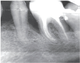
Fig 2: Initial radiographic view of theintrabony defect
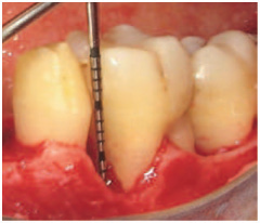
Fig 3: Intrasurgical measurement of theintrabony defect
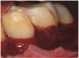
Fig 4: Application of PRP alone

Fig 5: 12-months clinical view of theintrabony defect and probing depth

Fig 6: 12-months radiographic view ofthe intrabony defect
CONCLUSION
In lieu of advances in science and technology, it’s important to adapt to the newer exciting possibilities of regeneration of tissues. The fact that PRP is an autologous preparation introduced at the time of surgery eliminates the concerns about disease transmission and immunogenic reactions, which are associated with allogenic or xenogenic preparation, and about the possibility of mislabeling a sample, which might occur through a laboratory system.
It is also evident that PRP is easy to prepare in a dental office setting. The growth factor enhancement can be used in case of healing in elderly where healing is impaired. The amplification of PDGF and TGF beta through the technique of platelet sequestration and concentration into platelet rich plasma is seen to be a practical tool for enhancing the rate of bone formation in regenerative process. In this review we present the preparation of PRP and its application in the dentistry and its shortcomings which need to be overcome for broader acceptance by clinicians for regenerative procedures in dental clinics and general dentistry.
REFERENCES
- W.V.Giannobile “the Potential role of growth and differentiation factors in periodontal regeneration” J.Periodontol.volume 67, 1996,P.545e53
- W.V.Giannobile “Periodontal tissue engineering by growth factors” Bone, volume 19, issue suppl.1,1996,pp.23s-37s.
- D.M.Dohan, J.Choukroun ,A.Diss et.al “Platelet-rich fibrin(PRF): a second –generation platelet concentrate, Part1:technological concept and evolution” oral surgoralmedoralpath oral radiolendod, volume 101,2006,pp.E37-E44.
- J.Choukroun, F.Adda, C.Schoeffer, A.vervelle “PRO: an opportunity in perio-implantology” implantodontie, volume 42,2000, pp.55-62
- V.Gupta, V.K.Bains, G.P.Singh, A.Mathur and R.Bains “Regenerative potential of platelet rich fibrin in dentistry: literature review”, Asian journal of oral health and allied sciences, volume 1, no:1, pp.22-28,2011.
- V.H.Kang, S.H.Jeon, J.Y.Park et.al., “Platelet rich fibrin is a bioscaffold and reservoir of growth factors for tissue regeneration”, Tissue engineering A.vol.17, no:3-4, pp.349-359, 2011.
- Hannink M, Donoghue DJ (1989). “Structure and function of platelet derived growth factor(PDGF) and related proteins”. Biochim.Biophys.Acta 989(1):1-10.
- Robert.E.Marx, Eric.R.Carlson oral surg oral med oral path oral radiolEndod June 1998,vol.85(6):638-646 “Growth factor enhancement for bone grafts”.
- Alan.R.Hartman, Dennis K.Galanakis, Mark.P.Honig, Frank.C.Seifert, Constantine “Autologous whole plasma fibrin gel” Arch surg .1992; 127(3):357-359.
- El-Sharkawy H, Kantarci A, Deady J, Hasturk H, Liu H,Alshahat M, et al. Platelet-rich plasma: growth factors and pro- andanti-inflammatory properties. J Periodontol 2007;78:661-9.
More references are availabe on request.
Disclaimer from Guident
The views and opinions expressed in articles published in Guident are solely those of the authors and do not necessarily reflect the views of the editors, the editorial board or the publisher, Ivory India. Guident does not endorse any product, technique or treatment described. Clinical content is intended for qualified dental and medical professionals and is not a substitute for individual clinical judgement. Guident, its editors and the publisher accept no liability for any consequence arising from the use of information published in the journal.
How to cite this article
Dr. Suganya. B, Dr. Priyanka Mittal Academy of Advanced Endodontics Bangalore. Regeneration Will Overcome Repairs..... Platelet Rich Fibrin (PRF) Forgotten Tool in Regenerative Dentistry. Guident. 2015 Dec;9(1). Available from: https://guident.in/articles/general/regeneration-will-overcome-repairs-platelet-rich-fibrin-prf-forgotten-tool-in-regenerative-dentistry.html