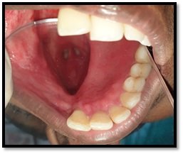
Interaction Between Occlusion and Human Brain Activity Exploring the Brain
Department of Prosthodontics, K. D. Dental College & Hospital, Mathura
ABSTRACT
In the area of human research that focuses on the interactions between occlusion and brain function this review discusses the relationship between the brain activity observed in the motor and sensory cortices and movements of the oral and maxillofacial area, such as gum chewing, tapping and clenching. It has been found that the sensorimotor cortex is also affected by slight changes in occlusion, such as those caused by occlusal interference devices, splints and implant prosthesis. However, findings such as these must be verified by evidence gathered from more subjects. Furthermore, individual variation is an important factor although it is assumed that any sensory movements in the oral and maxillofacial area are related to most regions of the cerebral cortex. In-depth research is necessary to clarify how changes in movement and sensation affect functional localisation in the brain.
Keywords: Tooth loss, Chewing, Brain activity, MRI, Dementia, Prosthetic rehabilitation.
INTRODUCTION
Does tooth loss increase the risk of dementia? Can improvements in chewing ability prevent cognitive impairment or ameliorate cognitive decline? The association between the brain and the stomatognathic system, which plays a key role in chewing and swallowing, has recently been hotly debated in the media. The world population is ageing and is estimated to increase from 264 million in 2009 to 416 million in 2050. Given the fact that ageing is one of the risk factors for developing dementia, an increase in patients suffering from dementia is to be anticipated. There are several risk factors for developing dementias like Alzheimer’s disease (AD), which is one of the most common subtypes of dementia. These risk factors include ageing, illiteracy, a lower level of education, lower socioeconomic status, head trauma, genetic factors such as the apolipoprotein E4 (apo E4) allele, cardiovascular risk factors such as being overweight, smoking, hypertension and diabetes mellitus, an inactive lifestyle and, perhaps surprisingly, loss of teeth. Loss of teeth has also been associated with malnutrition, mortality and disability, loss of cognitive function and prevalence of dementia. This review will focus on the relationship between masticatory and cognitive function in ageing and dementia.1
The literature on the relationship of mastication and cognition that will be addressed in this review will be subdivided into several categories: animal experimental studies, usually with a strong neuroanatomical focus; human experimental studies, with healthy (and typically young) subjects; and clinical studies, either cross-sectional or longitudinal, with a patient population suffering from either age-related or pathological loss of the ability to perform activities of daily living.2
OUTLINE 3
The mechanism describing this concept is as follows: a) Mastication increases cortical blood flow and widely activates various cortical areas of the somatosensory, supplementary motor and insular cortices. b) Mastication increases blood oxygen levels in the prefrontal cortex and the hippocampus which may be essential for learning and memory processes. c) Mastication prevents degradation of senile brain function and stress-related disorders without medication.
SYSTEMATIC LITERATURE REVIEWS SUPPORTING THE EVIDENCE
Evidence From Clinical And Epidemiological Research 2,4
When describing results, the following definitions for age groups will be used: “young” adults are individuals with an age under or equal to 30 years; “middle-aged” adults are individuals with an age equal to or higher than 30 years, but lower than 50 years; “senior” adults are individuals with an age equal to or higher than 50 years, but lower than 70 years and “aged” adults are individuals with an age equal to or higher than 70 years. When a study did not specify ages, but did include only senior and/or aged subjects, they are referred to as “old” or “older”. When considered of particular relevance, specific ages are mentioned.

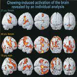
Chewing induced brain activation
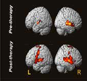
Increased brain activity pre & post prosthetic therapy
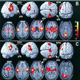
B)masticatory function of adults
shows greater brain activity (C)
than those of during chewing
older subject

Brain activation & broadmann area
during chewing gum in a healthy
subject
*brighten area indicates activated regions of brain
Evidence From Animal Research 2,4,5
Compared to clinical and epidemiological studies, animal research provides benefits in examining the relevant mechanistic cause-effect relationships. These studies have demonstrated that cognitive decline is related to cellular and neurochemical change in the hippocampus, including decreased cellular proliferation decreased levels of brain-derived neurotrophic factor as well as increased nitrous oxide levels and extracellular dopamine levels. The most common experimental animals accepted are Senescence Accelerated Mouse (SAM) P8 strain, wistar rat.

BRIDGING THE GAP
The role of brain neuroimaging in the investigation of the brain stomatognathic axis 6
fMRI-most common approach, EEG, NIRS (near-infraredspectrophotometry, PET, xenon tomography to locate cerebral blood flow changes).
Identifying the brain network associated with mastication (fig 1) 7,8,9,10
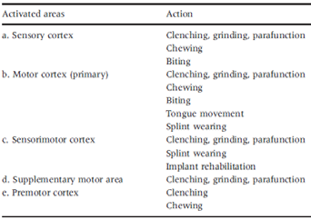
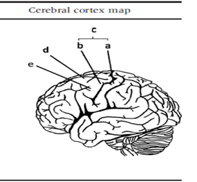
Fig: 1
Using brain imaging markers to explore the mechanisms underlying long-term changes in masticatory functions
Jaw-clenching task, wearing of an implant-supported overdenture (IOD),wearing of a complete denture (CD)11,conventional removable partial denture12, insertion of palatal plate13.it was found that there was a consistent increase in the activation of sensorimotor regions as a result of use of following prosthesis. These findings showed that differences in brain mechanisms may play critical roles in the individual variation in long-term adaptation to treatment, particularly for psychological adaptation (i.e., incorporating the denture as ‘part of the body’.
Exploring the evidence from experimental studies
Evidence from behavioral research has revealed that chewing is associated with changes in sustained attention and helps relieve stress. Evidence from animal research has consistently revealed that mastication attenuates activity in the hypothalamic-pituitary-adrenal axis and reduces the effect of chronic stress.
HYPOTHESES REGARDING THE NEURAL MECHANISMS UNDERLYING THE BRAIN-STOMATOGNATHIC AXIS AND RECENT SUPPORTING EVIDENCE
The Sesnsory Feedback Hypothesis 14 (Fig: 2)
According to this hypothesis there is consistent increase in activation of the sensorimotor area during a chewing task. Increased brain activation in these regions is associated with the use of dentures, as shown by cross-sectional and longitudinal research.

Fig: 2
The Compensation Hypothesis 15 (Fig: 3)
According to this hypothesis masticatory dysfunction as a result of ageing is associated with compensatory motor control processing esp. pre-frontal cortex area. Such compensatory mechanisms are closely related to the brain regions that are associated with cognitive control and sensory integration, including the PFC, the parietal lobe, the insula, and the cingulate cortex.
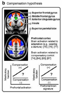
Fig: 3
The Cerebellum Hypothesis 16 (Fig: 4)
According to this hypothesis cerebellar atrophy or decreased cellular function is associated with both cognitive decline & masticatory dysfunction. Cerebellar atrophy is associated with poor cognitive performance and predictive of the differentiation of Alzheimer’s and Parkinson’s dementia.
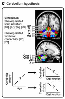
Fig: 4
CONSIDERATIONS FOR FUTURE RESEARCH 4 Refinement of behavioral assessments 17
A more comprehensive assessment of specific elements of stomatognathic function (eg: masticatory performance, biting force, oral stereognosis & masticatory muscles) and their interaction should be considered. The sustantial gap of findings in animal research compared to human studies should be overcomed.
Inclusion of baseline changes in mental & physical condition 18
Some of animal research has concluded that nutritional deficiency are associated with cognitive decline. Thus, these factors should be considered.

Experimental design that include prospective, longitudinal observation
The effect of long term alterations, such as neuroplasticity effects in the brain, cannot be inferred from cross-sectional findings. The difference between normal and pathological ageing should be clear.
A precise estimation of the effect size
A proper prognosis and diagnosis by clinicians must be done to know” to what extent does the change in a factor say X contribute to the change in other factor say Y.
CONCLUSIONS
The mechanism concluding the cause-and-effect relationship between cognitive decline and masticatory dysfunction requires more investigation. One cannot rely on cognitively impaired older persons to recognize the need for oral care, indicating the necessity for professional dental health care to be available and to be administered on a regular basis, regardless of demand from the patient. The clinical relevance of several evidences is compelling and suggest that oral care should be actively provided to older persons in nursing homes and should furthermore not be limited to individuals retaining some teeth but extended to edentate persons as well. The World Health Organization (WHO) recognized the importance of oral health care in 2006 and indicates a stringent need for research, training of care givers, and development of policy regarding oral health care (World Health Organization, 2006). There should be general awareness of the importance and value of good oral health, not only in the scientific community, but in the general and clinical population as well.
“TEETH FOR A BRIGHTER BRAIN”
REFERENCES
- Kalaria, R.N., Maestre, G.E., Arizaga, R., Friedland, R.P., Galasko, D., Hall, K., Luchsinger, J.A., Ogunniyi, A., Perry, E.K., Potocnik, F., Prince, M., Stewart, R., Wimo, A., Zhang, Z.X., Antuono, P., 2008. Alzheimer’s disease and vascular dementia in developing countries: prevalence, management, and risk factors. Lancet Neurol. 7, 812–826.
- R.A.F. Weijenberga etal. Mastication for the mind—The relationship between mastication and cognition in ageing and dementia. Neuroscience & Biobehavioral Reviews 2011.
- C. OHKUBO et al. Interactions between occlusion and human brain function activities. Journal of Oral Rehabilitation 2012.
- Chia-shu Lin. Revisiting the link between cognitive decline and masticatory dysfunction. Lin BMC Geriatrics (2018) 18:5
- Takeshi kato et al. The effect of the loss of molar teeth on spatial memory and acetylcholine release from the parietal cortex in aged rats. behavioural brain research 83 (1997).
- Bishop NA, Lu T, Yankner BA. Neural mechanisms of ageing and cognitive decline. Nature. 2010; 464 (7288):529–35.
- Onozuka M, Fujita M, Watanabe K, Hirano Y, Niwa M, Nishiyama K, Saito S. Age-related changes in brain regional activity during chewing: a functional magnetic resonance imaging study. J Dent Res. 2003; 82 (8): 657–60.
- Viggiano A, Manara R, Conforti R, Paccone A, Secondulfo C, Lorusso L, Sbordone L, Di Salle F, Monda M, Tedeschi G, et al. Mastication induces long-term increases in blood perfusion of the trigeminal principal nucleus. Neuroscience. 2015; 311: 75–80.
- Lin CS, SY W, CY W, Ko HW. Gray matter volume and resting-state functional connectivity of the motor cortex-cerebellum network reflect the individual variation in masticatory performance in healthy elderly people. Front Aging Neurosci. 2015; 7:247.
- Quintero A, Ichesco E, Schutt R, Myers C, Peltier S, Gerstner GE. Functional connectivity of human chewing: an fcMRI study. J Dent Res. 2013; 92(3): 272–8.
- Ohkubo C, Morokuma M, Yoneyama Y, Matsuda R, Lee JS. Interactions between occlusion and human brain function activities. J Oral Rehabil. 2013; 40(2): 119–29.
- Yan C, Ye L, Zhen J, Ke L, Gang L. Neuroplasticity of edentulous patients with implant-supported full dentures. Eur J Oral Sci. 2008;116(5): 387–93.
- Kimoto K, Ono Y, Tachibana A, Hirano Y, Otsuka T, Ohno A, Yamaya K, Obata T, Onozuka M. Chewing-induced regional brain activity in edentulous patients who received mandibular implant-supported overdentures: a preliminary report. Journal of prosthodontic research. 2011; 55(2): 89–97.
- Jiang H, Liu H, Liu G, Jin Z, Wang L, Ma J, Li H. Analysis of brain activity involved in chewing-side preference during chewing: an fMRI study. J Oral Rehabil. 2015; 42(1): 27–33.
- Ward NS. Compensatory mechanisms in the aging motor system. Ageing Res Rev. 2006; 5(3): 239–54.
- Thomann PA, Schlafer C, Seidl U, Santos VD, Essig M, Schroder J. The cerebellum in mild cognitive impairment and Alzheimer’s disease – a structural MRI study. J Psychiatr Res. 2008; 42(14): 1198–202.
- Onozuka M, Fujita M, Watanabe K, Hirano Y, Niwa M,Nishiyama K et al. Mapping brain region activity during chewing: a functional magnetic resonance imaging study. J Dent Res. 2002; 81: 743–746
- Shimazaki, Y., Soh, I., Saito, T., Yamashita, Y., Koga, T., Miyazaki, H., Takehara, T. 2001. Influence of dentition status on physical disability, mental impairment, and mortality in institutionalized elderly people. J. Dent. Res. 80, 340–345.
Disclaimer from Guident
The views and opinions expressed in articles published in Guident are solely those of the authors and do not necessarily reflect the views of the editors, the editorial board or the publisher, Ivory India. Guident does not endorse any product, technique or treatment described. Clinical content is intended for qualified dental and medical professionals and is not a substitute for individual clinical judgement. Guident, its editors and the publisher accept no liability for any consequence arising from the use of information published in the journal.
How to cite this article
Dr. Preeti Sagar, Dr. Sumver Jain, Dr. Manesh Lahori. Interaction Between Occlusion and Human Brain Activity Exploring the Brain. Guident. 2020 Apr;13(5). Available from: https://guident.in/articles/prosthodontics/interaction-between-occlusion-and-human-brain-activity-exploring-the-brain.html