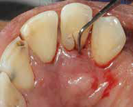
Leukemia Causing Gingival Enlargement a Case Report
Jaipur, India
ABSTRACT
For variety of systemic diseases, oral cavity functions as an early indicator. Thorough examination of the oral mucosa, gingiva, teeth, tongue and other oral tissues gives early and accurate diagnosis of these underlying systemic disorders. Systemic conditions such as blood dyscrasias, endocrinal imbalance, and nutritional deficiencies may lead to gingival changes which is related as local factors in the oral cavity. Leukemia is a dreadful disease and also malignancy of white blood cells, if not diagnosed properly and treated early may result in significant morbidity and mortality. In leukemic patients, oral changes may be the first and only presenting signs. The aim of this case report aims is to throw light on acute leukemia diagnosed on the basis of oral signs and emphasizes the importance of thorough oral examination to identify the threatening condition.
Keywords: Gingival enlargement, leukemia, oral manifestations
INTRODUCTION
Increase in size of the gingiva is “Gingival overgrowth” or “gingival enlargement” which accounts to one of the many types of periodontal diseases. Based on etiologic factors and pathologic changes, gingival enlargement may be classified as - (1) inflammatory enlargement: Acute and chronic; (2) drug induced enlargement: Anticonvulsants - phenytoin, calcium channel blockers - verapamil, nifedipine, immunosuppressant’s - cyclosporine; (3) Overgrowth associated with systemic diseases: Conditioned overgrowth – due to pregnancy, puberty, Vitamin C deficiency, plasma cell gingivitis, nonspecific conditioned overgrowth (granuloma pyogenicum), and systemic diseases causing gingival overgrowth – due to leukemia, granulomatous diseases (sarcoidosis, Crohn’s disease, Wegenersgranulomatosis); (4) neoplastic overgrowth (gingival tumors): Benign tumors, malignant tumors; (5) false enlargements.1
The clinical manifestation of gingival enlargement differs based on its etiology. Genetically induced gingival enlargement appears pink in color with minimal signs of inflammation, has a slow growth rate, and is firm in consistency. Soft, edematous, tender gingiva with ease to bleeding is a sign in blood disorders.2 Gingival enlargement in leukemia occurs due to infiltration of premature leukocytes. This is a common symptom that aids in the diagnosis of leukemia and warrants dental consultation.1 Many of the life-threatening diseases may present with oral lesions as the initial manifestation, thus, necessitating the familiarization of dental professionals with the manifestations of systemic diseases.2,3
CASE REPORT
An 68-year-old male patient from low socioeconomic status reported, with a complaint of swollen and bleeding gums for the past 1-month. Gingival swelling progressed slowly within the last 20 days and was associated with bleeding and tenderness. Medical history revealed that the patient experienced lethargy, slight weight loss and loss of appetite in the last few months. However, he denied the intake of any medications for chronic illness. Physical examination revealed that the patient was poorly built and nourished with petechial spots on the skin in the forearm and legs.
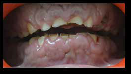
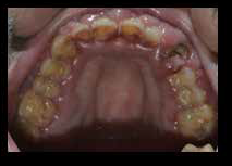
Bilateral submandibular and deep cervical lymphadenopathy was appreciated. Lymph nodes were indurated and tender on palpation. Intraoral examination revealed generalized gingival enlargement on the buccal, labial, palatal and lingual aspect of marginal and attached gingiva of the maxillary and mandibular arches [Figure. 1a, b, c, d].
(a)

(b)
(c)

(d)
Figure: 1a, b, c, d - Intra-oral oral pictures of patient
Gingiva was swollen and glazed. Gingival enlargement extended on to one-third to one-half of the crown portion of teeth. The patient had a foul odor and fair oral hygiene. On palpation, gingiva was soft, edematous, devoid of stippling and tender on palpation. Gingival bleeding on slight provocation was also appreciated. Orthopantomogram did not revealed bony involvement.
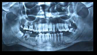
Figure. 2 - OPG of the patient
Inflammatory gingival enlargement, drug induced enlargement, conditioned gingival enlargement, and systemic gingival enlargement were considered in the differential diagnosis. Inflammatory enlargement was ruled out as the inflammatory component (plaque and calculus) was not significant to the severity of the presented gingival enlargement. Drug-induced enlargement was ruled out as thorough history taking did not reveal the intake of any drug significant for gingival enlargement. However, features of lethargy, weight loss and loss of appetite along with the rapidly progressive gingival overgrowth and presence of ecchymosis in the floor of the mouth substantiated an underlying systemic disease. Complete blood count was performed, and it revealed features of leukocytosis, anemia and thrombocytopenia [Figure. 3].

Figure: 3 - CBC Report
Peripheral blood smear reveals numerous monoblasts (60%) and promonocytes. Monoblasts are having moderate to severe basophilic cytoplasm with large, round nuclei and lacy chromatin. Scarcity of platelets can be appreciated with a nucleated red blood cell (RBC) in the background. Based on the blood parameters and peripheral blood smear, a final diagnosis of gingival enlargement due to acute myeloid leukemia (AML) was made. The patient was advised to maintain proper oral hygiene by use of soft bristle toothbrush and 0.2% oral chlorhexidine rinses. The patient was referred to oncology center for bone marrow aspiration and chemotherapy. However, the patient could not respond to the treatment and died later due to multi organ failure.
DISCUSSION
Leukemia is a malignancy of white blood cells (WBCs) and occurs either due to uncoordinated proliferation or increased viability of white cells (now called blasts). Based on the cell lineage and evolution of the disease, leukemia’s are broadly classified as lymphoid or myeloid, and acute or chronic respectively.4 In case of chronic leukemia, there is slow onset which runs an indolent course of months to years and involve relatively well-differentiated leukocytes. In acute leukemia’s, there is disorganized proliferation of poorly differentiated blast cells which are abrupt in onset, and if left untreated, it shows aggressive and rapidly fatal nature.5 Chronic and acute leukemia’s are classified according to the type of WBC involved, that is, lymphoid and myeloid. French - American - British classification further subdivides based on the degree of differentiation along cell lines and the extent of cell maturation into acute lymphoid leukemia and AML.5
Although various predisposing factors have been attributed, yet the exact etiology of leukemia remains obscure. Predisposing factors include: Exposure to ionizing radiation or electromagnetic fields, cytotoxic therapy, and viral infections.6
According to Stafford et al., oral lesions are more frequently seen in patients with acute leukemia.7 Oral manifestations may either be the result of direct infiltration of leukemic cells (primary) or secondary to underlying thrombocytopenia, neutropenia, or impaired granulocyte function.6 5% of frequency is seen in case of gingival infiltration as the initial presenting complication of AML.8 Dreizen et al. suggested that patients with acute monocytic leukemia had the greatest incidence of gingival infiltrates (M5) (66.7%) followed by acute myelomonocytic leukemia (M4) (18.5%) and acute myeloblastic leukemia (M1, M2) (3.7%).9
Common oral manifestations of acute leukemia’s include swollen gingiva, oral ulcers, spontaneous gingival bleeding, petechiae, pale mucosa, viral and fungal infections (herpes and candidosis).7,10 Some of the uncommon oral signs are Cracked lips, hemorrhagic bullae on the anterior dorsum of the tongue, buccal and labial mucosa, tooth pain and mobility, and petechiae.11 From minimal to complete tooth coverage and function and aesthetics which get hinders depend on gingival overgrowth severity.12 In case of edentulous individuals, leukemic gingival infiltration is not seen ,thus suggesting a potential role of tooth-associated local factors in its pathogenesis.9 Caries, calculus, and poor oral hygiene may exacerbate gingival signs and symptoms and predisposes the patient for oral pain, bleeding, super infection, and tissue necrosis.12
The diagnosis is suggested by a complete blood cell count showing pancytopenia and blast cells and is confirmed by examination of the bone marrow. Investigation of the specific type of leukemia simplifies the best treatment and most accurate prognosis.
Treatment options in acute leukemia’s include aggressive multidrug chemotherapy and allogenic bone marrow transplantation. Periodontal and dental treatment for leukemic patients should always be planned after medical evaluation and physicians consent. Periodontal intervention (scaling and root planning) should be done under prophylactic antibiotics. Patients are advised 0.12% chlorhexidine mouth rinses postoperative oral hygiene procedures. However, periodontal surgeries are postponed until complete remission of the underlying disease occurs.1
To maintain good oral hygiene , patient was advised to use soft bristle tooth brush and 0.2% chlorhexidine mouth rinses.
CONCLUSION
Nowadays its important that dentists and physicians should be aware of the importance of recognizing oral manifestations of systemic diseases. Although physicians most commonly diagnose leukemia, dentists can also diagnose patients with acute myelogenous leukemia. Proper awareness of diagnostic signs and complications associated with leukemia should be known by dentist so to better aid in diagnosis and subsequent treatment and management. Hence medical consultation must be essential in dental therapy in such cases.
REFERENCES
- Carranza FA, Hogan EL. Gingival enlargement. In: Newman MG, Takei HH, Carranza FA, editors. Clinical Periodontology. 10th ed. Philadelphia: WB Saunders Co; 2002. pp. 279–96. [Google Scholar]
- Demirer S, Ozdemir H, Sencan M, Marakoglu I. Gingival hyperplasia as an early diagnostic oral manifestation in acute monocytic leukemia: A case report. Eur J Dent. 2007;1:111–4. [PMC free article] [PubMed] [Google Scholar]
- Chavan M, Subramaniam A, Jhaveri H, Khedkar S, Durkar S, Agarwal A. Acute myeloid leukemia: A case report with palatal and lingual gingival alterations. Braz J Oral Sci. 2010;9:67–9. [Google Scholar]
- Love AA. Manifestations of leukemia encountered in otolaryngologic and stomatologic practices. Arch Otolaryngol. 1936;23:173–221. [Google Scholar]
- Dean AK, Ferguson JW, Marvan ES. Acute leukaemia presenting as oral ulceration to a dental emergency service. Aust Dent J. 2003;48:195–7. [PubMed] [Google Scholar]
- Benson RE, Rodd HD, North S, Loescher AR, Farthing PM, Payne M. Leukaemic infiltration of the mandible in a young girl. Int J Paediatr Dent. 2007;17:145–50. [PubMed] [Google Scholar]
- Stafford R, Sonis S, Lockhart P, Sonis A. Oral pathoses as diagnostic indicators in leukemia. Oral Surg Oral Med Oral Pathol. 1980;50:134–9. [PubMed] [Google Scholar]
- Williams WJ, Beutler E, Erslev AJ, Lichtman MA. 4th ed. New York: McGraw Hill; 1990. Hematology; pp. 243–4. [Google Scholar]
- Dreizen S, McCredie KB, Keating MJ, Luna MA. Malignant gingival and skin “infiltrates” in adult leukemia. Oral Surg Oral Med Oral Pathol. 1983;55:572–9. [PubMed] [Google Scholar]
- Bressman E, Decter JA, Chasens AI, Sackler RS. Acute myeloblastic leukemia with oral manifestations.Report of a case. Oral Surg Oral Med Oral Pathol. 1982;54:401–3. [PubMed] [Google Scholar]
- Gleeson P. Spontaneous gingival haemorrhage: Case report. Aust Dent J. 2002;47:174–5. [PubMed] [Google Scholar]
- Cooper CL, Loewen R, Shore T. Gingival hyperplasia complicating acute myelomonocytic leukemia. J Can Dent Assoc. 2000;66:78–9. [PubMed] [Google Scholar]
Disclaimer from Guident
The views and opinions expressed in articles published in Guident are solely those of the authors and do not necessarily reflect the views of the editors, the editorial board or the publisher, Ivory India. Guident does not endorse any product, technique or treatment described. Clinical content is intended for qualified dental and medical professionals and is not a substitute for individual clinical judgement. Guident, its editors and the publisher accept no liability for any consequence arising from the use of information published in the journal.
How to cite this article
Dr. Gaurav Gupta, Dr. D. K. Gupta, Dr. Neelja Gupta Wisdom Dental Clinics. Leukemia Causing Gingival Enlargement a Case Report. Guident. 2020 Apr;13(5). Available from: https://guident.in/articles/periodontics/leukemia-causing-gingival-enlargement-a-case-report.html