Esthetic and Functional Rehabilitation of Broad Anterior Saddles with Diastema Maintenance: A Case Series Utilizing Fixed Partial Dentures with Palatal Loop Connectors
Department of Prosthodontics, Sree Mookambika Institute of Dental Sciences, Kulasekharam, Tamil Nadu, India
ABSTRACT
Background: The management of broad edentulous saddles and the concomitant requirement to preserve or create interdental diastema in the anterior maxilla presents a significant esthetic and biomechanical challenge. This article aims to explore the rationale, design principles, and clinical outcomes of utilizing the specialized palatal loop connector Fixed Partial Denture (FPD) as a viable restorative modality in these complex cases.
Methods: This report details the successful prosthetic management of three distinct clinical scenarios involving anterior space management, where conventional treatment options (implants or standard FPDs) were either unsuitable or did not meet the patient's demand for diastema maintenance. The technical aspects, including abutment preparation, material selection (Metal-Ceramic, Zirconia, and Fiber-Reinforced Composite), and critical connector design parameters (e.g., cross-sectional area and contour), were rigorously applied, informed by biomechanical principles derived from modern restorative analysis.
Results: The loop connector FPD successfully restored the continuity of the dental arch while achieving the primary objective of esthetic rehabilitation and diastema preservation. The non-rigid nature of the connector helped distribute occlusal forces, and the refined design minimized phonetic interference. Long-term functional success was directly linked to the careful execution of the mandatory post-treatment oral hygiene protocol.
Conclusion: The loop connector FPD is a specialized, highly effective solution when confronted with broad anterior saddles requiring diastema maintenance. Its successful application is critically dependent on patient selection, precise biomechanical adherence (particularly the single-tooth span rule), and unwavering patient commitment to rigorous oral hygiene maintenance, addressing the inherent design vulnerability of the loop itself.
Keywords : Palatal Loop Connector, Fixed Partial Denture (FPD),Anterior Maxilla, Diastema Preservation/Maintenance, Edentulous Saddle, Biomechanical Adherence, Oral Hygiene Protocol.
INTRODUCTION: THE PROSTHODONTIC DILEMMA OF ANTERIOR SPACING
Fixed partial dentures for replacing teeth in the anterior region is always a challenge to a prosthodontist if a long edentulous span exists anteriorly.1 Congenitally missing maxillary anteriors, particularly lateral incisors, and generalized spacing (diastema) are common esthetic concerns in the population.2 Esthetic replacement of a single or multiple anterior teeth can be achieved through conventional fixed partial dentures, resin-bonded bridges, or dental implants.3 However, when the clinical situation involves a broad saddle—an edentulous space greater than the typical size of the tooth being replaced—and the patient insists on maintaining an existing, desired diastema, conventional rigid restorations often fall short.1
Standard FPDs, designed to rigidly connect abutments and close the entire space, may result in pontics or retainer crowns with unnaturally large mesio-distal dimensions or compromised emergence profiles, leading to poor esthetics and patient dissatisfaction.1 Dental implants, while providing excellent long-term bone preservation and hygiene3, present a complex path to prosthetic diastema maintenance, often requiring demanding soft tissue procedures to sculpt the desired interdental space.
The loop connector fixed partial denture (FPD) offers a unique and simple solution to this dilemma. It involves fabricating an FPD with a connector that loops on the palatal aspect, effectively maintaining the anterior edentulous space and the desired diastema without compromising the alignment of adjacent teeth.1 This allows for the creation of teeth with ideal proportions and a natural appearance while maintaining stable, fixed retention.
BIOMECHANICAL PRINCIPLES OF LOOP CONNECTORS
Connectors are those parts of a fixed partial denture or splint that join the individual retainers and pontics together. These are broadly classified as rigid or non-rigid.1
RIGID VS. NON-RIGID SYSTEMS
Usually, restoration fabrication is accomplished with rigid connectors, which fuse the components into a single, monolithic unit. Non-rigid connectors, such as the loop design or intracoronal attachments, are used occasionally when a common path of insertion for the abutment preparations is impossible, or when specific biomechanical management of occlusal forces is required.4
The physiological mobility of teeth is a complex, dynamic process involving the periodontal ligament (PDL).5 Teeth exhibit both horizontal (lateral) and vertical (intrasocket) mobility. For single-rooted teeth, the upper limit of horizontal mobility is approximately 0.15 mm.5 When multiple teeth are rigidly splinted, their independent physiological movements are restricted. The loop connector, by nature of its increased length and decreased cross-sectional area compared to a conventional rigid connector, is more flexible.1 This flexibility allows the adjacent abutment teeth a controlled degree of independent physiological movement, effectively distributing occlusal stresses away from the most vulnerable abutments, preventing stress concentration, and thus acting as a form of non-rigid coupling.5
INDICATIONS AND CONTRAINDICATIONS FOR NON-RIGID CONNECTORS
The loop connector FPD is indicated in specific scenarios where rigid coupling is contraindicated or undesirable1:
- Diastema Maintenance: The patient explicitly wishes to maintain the diastema.1
- Excessive Pontic Space: The available edentulous space is greater than the ideal dimension of the tooth being replaced.1
- Challenging Abutment Geometry: The existence of malaligned abutments where achieving parallel preparation planes for rigid seating would risk devitalization.4
- Disparity in Retention: When abutments possess significantly different levels of retention, a non-rigid design can mitigate stress on the weaker tooth.4
However, the use of any non-rigid connector, including the loop design, is subject to strict biomechanical contraindications essential for long-term prognosis4:
- Abutment Mobility: The design is contraindicated if the abutment teeth exhibit significant mobility.4 The success depends on stable abutment support.
- Span Length: Non-rigid connectors are contraindicated if the span between the abutments is longer than one tooth.4 If the missing space requires the replacement of two side-by-side units, the stress transferred to the abutment would be destructive, leading to potential abutment failure or loss of retention.4 The "broad saddle" must therefore be an esthetic enlargement of a single missing unit, not a true multiple-tooth span replacement.
- Parafunctional Habits: Fixed dental prostheses in bruxers exhibit significantly higher failure rates (32.3%) compared to non-bruxers (25.8%).6 The increased flexibility of the loop connector makes it inherently more vulnerable to fatigue and fracture under the high, non-axial forces associated with bruxism, representing a critical contraindication unless a mandatory occlusal stabilization appliance is strictly used.
MATERIALS SCIENCE AND DESIGN OPTIMIZATION
Achieving structural integrity in the loop connector FPD requires a precise understanding of material properties and geometric design, a process often guided by Finite Element Analysis (FEA).7
CONNECTOR CROSS-SECTIONAL AREA (CSA)
The size, shape, and position of the connector critically influence the longevity of the prosthesis.1 The connector must be sufficiently large to prevent distortion or fracture during function, yet not so large as to interfere with effective plaque control or tongue movement.1
For esthetic Fixed Partial Dentures, a large connector or inappropriate shaping of the individual retainers may result in display of the metal connector, which may compromise the appearance of the restoration. Therefore, connectors are often placed slightly lingually in anterior prostheses.
The material chosen heavily influences the minimum required dimensions. For high-strength ceramics, such as densely sintered Zirconia, which exhibits high fracture strength 8, a minimum connector cross-sectional area of 5 mm² is recommended for anterior FPDs to withstand the tensile stresses concentrated in the connector region.8
However, biomechanical analysis demonstrates that simply increasing the CSA does not linearly guarantee success. While materials with a higher elastic modulus (e.g., Zirconia) tend to confine stress within the FPD framework9, reducing the downward transmission to the periodontium, an excessive increase in CSA can be detrimental.10 Studies show that increasing the CSA beyond optimal levels (e.g., 4 mm² to 12 mm² in certain composite restoration groups) can dramatically increase the maximum stress observed in the abutment teeth, sometimes by over 115%.10 This paradoxical effect shifts the failure point from the connector to the abutment, increasing the probability of root fracture or loss of retention.10 The design goal must therefore be precise application of the material-specific minimum CSA, while maintaining a smooth, rounded contour to minimize discomfort.1
MATERIAL SELECTION: ALL-CERAMIC VS. FIBER-REINFORCED COMPOSITE (FRC)
The choice of restorative material significantly impacts the biomechanics of the flexible loop.
All-Ceramic (Zirconia): All-ceramic FPDs provide superior esthetics and biocompatibility compared to metal-ceramic restorations, which is crucial in the anterior region.11 However, ceramic materials are brittle and susceptible to fracture under tensile loading, and framework fracture is the main reason for their failure, typically originating from the gingival surface of the connector.11 This necessitates strict adherence to the minimum 5 mm² CSA rule.8
Fiber-Reinforced Composite (FRC): FRC materials offer good aesthetics and the unique advantage of higher bond strength (50–100% higher) to abutment teeth compared to metal frameworks.12 Critically for non-rigid structures like the loop connector, FRC frameworks have been found to exhibit.
lower overall stress values compared to equivalent metal framework models.13 This characteristic suggests FRC can absorb and distribute applied forces more favorably, reducing the total stress transmitted to the periodontal ligament, and is a viable alternative for esthetically demanding cases.12
CLINICAL CASE SERIES PRESENTATION
This article presents series of cases wherein a loop connector Fixed partial denture was fabricated to replace a congenitally missing lateral incisor and also generalized spacing in the anterior region.
CASE REPORT NO. 1: RESTORATION OF CONGENITALLY MISSING INCISOR WITH DIASTEMA MAINTENANCE
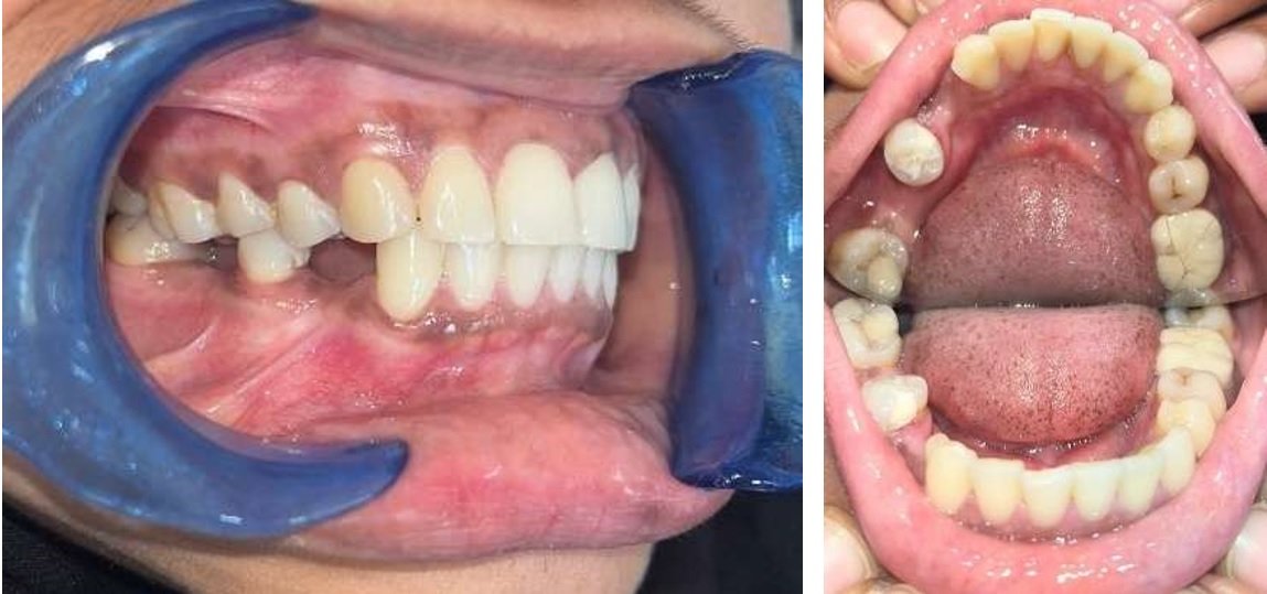
A 27 year old male patient reported to Department of Prosthodontics, Sree Mookambika Institue of Dental Sciences, Kulasekharam, with a complaint of dislodged prosthesis in relation to the upper left anterior region. History of presenting illness revealed that the prosthesis was cemented 8 years back and the prosthesis dislodged one week before reporting to the hospital. His prime concern was esthetic replacement as well as maintenance of midline diastema. On examination the available edentulous span was greater than the approximate size of the adjacent central incisor.
Peri-apical radiograph of this region showed endodontically treated left central incisor with a root fracture at the middle third. The patient was not willing for the extraction of the tooth. Endodontic re-treatment was performed in the same tooth and intentional root canal treatment was done on the left canine. The fractured tooth was reinforced with a pre-fabricated post.
The treatment options for such a situation where conventional removable partial denture with replacement of multiple teeth and also implant prosthesis. The current situation precluded all these options as the space between the teeth was excessively more. So a fixed partial denture was designed which will maintain the existing diastema between the teeth.
After assessing the radiographs, maxillary and mandibular irreversible hydrocolloid impressions were made for a mock –up of the final restorations. Diagnostic wax ups was made. The mock-up a fixed partial denture was designed with a loop connector to make the replacement esthetic and in accordance with the overall appearance and alignment of his other teeth. This was shown to the patient and the patient approved the design.
Tooth preparation was modified for a Fixed partial denture involving the left central incisor and left canine for the replacement of congenitally missing lateral incisor. Impression was made with addition silicone impression material and final cast was poured with die stone. Temporary restoration was fabricated on temporary cast and cemented on central incisor and canine.
Wax pattern of the future restoration was fabricated with loop connector design and a metal framework was fabricated. This metal framework, after finishing was tried in the patient’s mouth. Now the final restoration was fabricated with ceramic facing and the finished restoration was cemented with luting Glass Ionomer cement.

FIG 1: Pretreatment Image illustrating congenitally missing lateral incisor and midline diastema
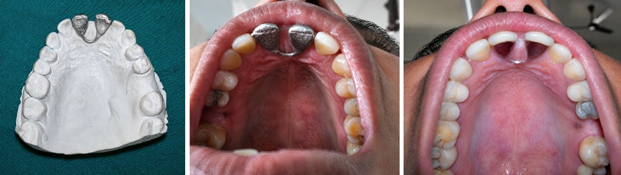
FIG 2:- Metal try-in and occlusal images of the Loop connector intra-orally
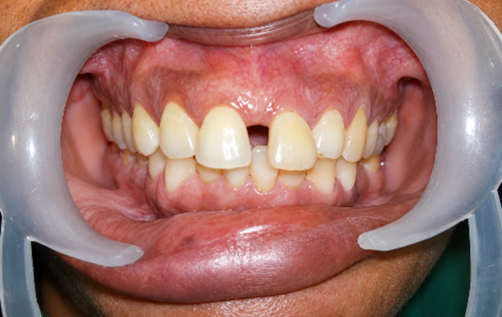
FIG 3:-Post operative images after cementation
CASE REPORT NO. 2: REPLACEMENT OF FRACTURED FPD WITH OPTIMIZED LOOP DESIGN
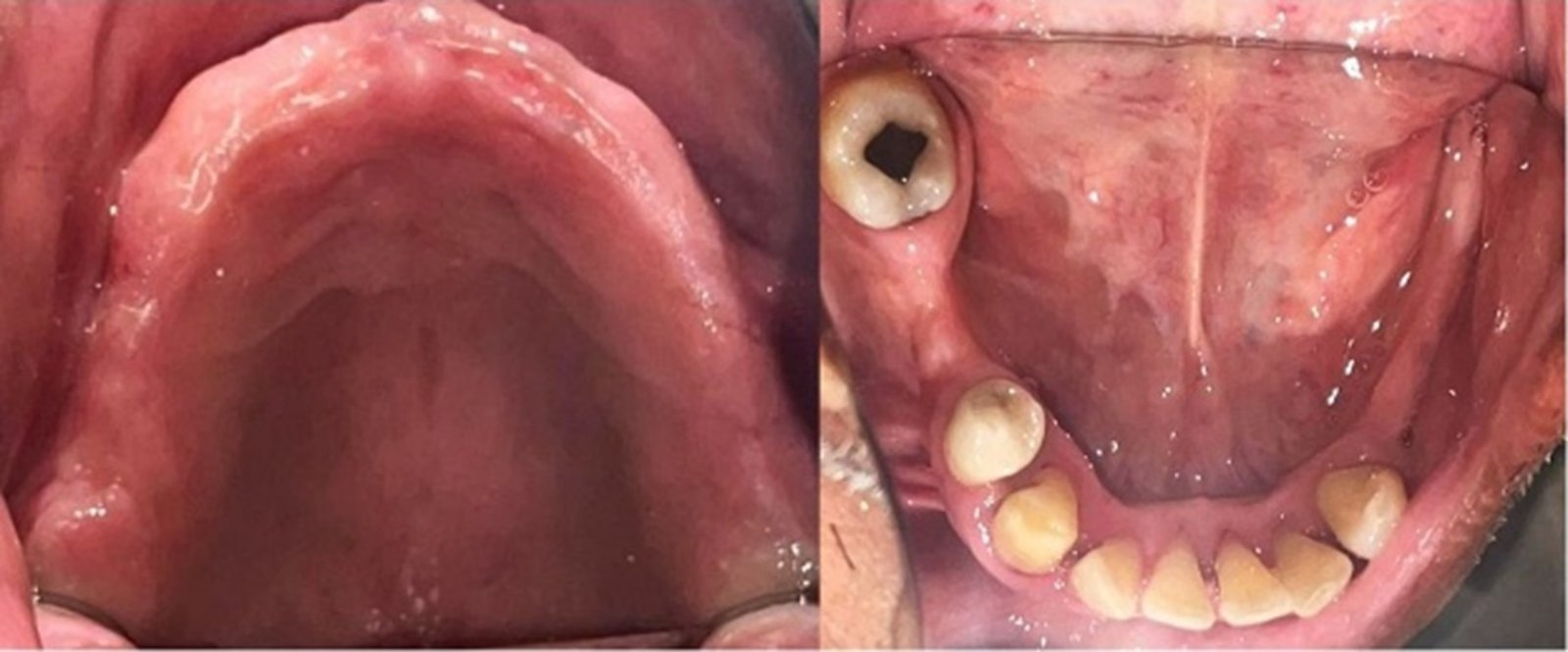
A 37 year old female patient reported with a complaint of fractured fixed partial denture in relation to the upper left anterior region. History of presenting illness revealed that the prosthesis was cemented 3 years back and the prosthesis fractured two days before reporting to the hospital. Her prime concern was esthetic replacement as well as maintenance of midline diastema as the patient was not satisfied with the prosthesis due to bulkiness of the restoration. On examination the available edentulous span was greater than the approximate size of the adjacent central incisor.
Peri-apical radiograph of this region showed endodontically treated right central incisor and left lateral incisor.
The treatment options for such a situation where conventional removable partial denture with replacement of multiple teeth and also implant prosthesis. The current situation precluded all these options as the space between the teeth was excessively more. So a fixed partial denture was designed which will maintain the existing diastema between the teeth.
After assessing the radiographs, maxillary and mandibular irreversible hydrocolloid impressions were made for a mock –up of the final restorations. Diagnostic wax ups was made. The mock-up a fixed partial denture was designed with a loop connector to make the replacement esthetic and in accordance with the overall appearance and alignment of her other teeth. This was shown to the patient and the patient approved the design.
Tooth preparation was modified for a fixed partial denture involving the right central incisor and left lateral incisor for the replacement of missing central incisor. Impression was made with addition silicone impression material and final cast was poured with die stone. Temporary restoration was fabricated on temporary cast and cemented on.
Wax pattern of the future restoration was fabricated with loop connector design and a metal framework was fabricated. This metal framework, after finishing was tried in the patient’s mouth. Now the final restoration was fabricated with ceramic facing and the finished restoration was cemented with luting Glass Ionomer cement.

FIG 4:- Preoperative image illustrating missing left central incisor and excessive spacing due to midline diastema.
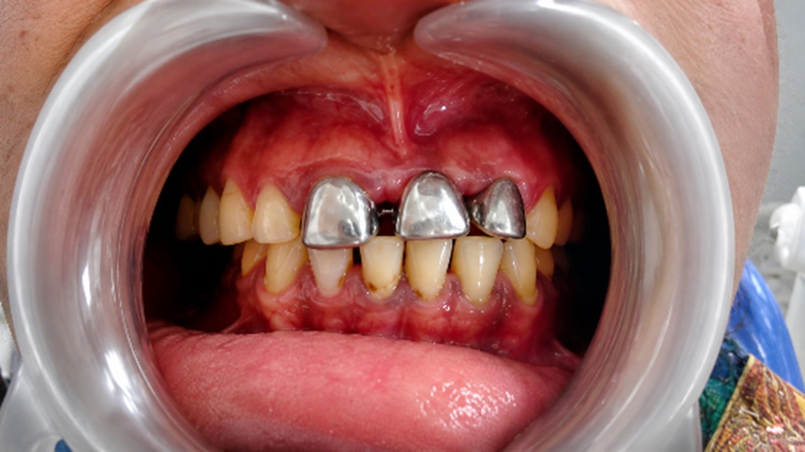
FIG 5:- Metal Tryin of the loop connector intra-orally
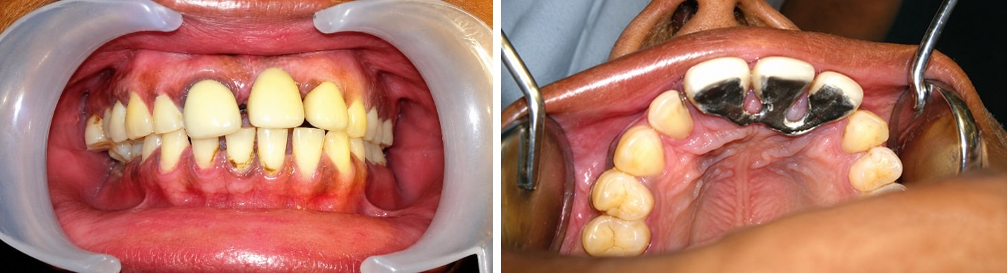
FIG 6:- Post-operative image with cemented Prosthesis.
CASE REPORT NO. 3: MIDLINE DIASTEMA AND PERIODONTALLY COMPROMISED ABUTMENTS
A 42 year old female patient reported with a complaint of midline diastema in the upper arch. History of presenting illness revealed endodontically treated central incisors. Her prime concern was esthetics. On radiographic examination there was horizontal bone loss in relation to 11 and 21.
Various treatment options were analyzed and a fixed partial denture with loop connector was designed which will maintain the existing diastema between the teeth without increasing the mesio-distal dimension of the teeth and also taking the horizontal bone loss into consideration.
Maxillary and mandibular irreversible hydrocolloid impressions were made for a mock –up of the final restorations. Diagnostic wax ups was made. The mock-up a fixed partial denture was designed with a loop connector to make the replacement esthetic and in accordance with the overall appearance and alignment of her other teeth. This was shown to the patient and the patient approved the design.
Tooth preparation was done. Impression was made with addition silicone impression material and final cast was poured with die stone. Temporary restoration was fabricated on temporary cast and cemented on.
Wax pattern of the future restoration was fabricated with loop connector design and a metal framework was fabricated. This metal framework, after finishing was tried in the patient’s mouth. Now the final restoration was fabricated with ceramic facing and the finished restoration was cemented with luting Glass Ionomer cement.

FIG 7:- Preoperative image illustrating midline diastema.

FIG 8:- Metal try-in of loop connector intra-orally

FIG 9:- Prosthesis on the cast and post operative image after cementation.
DISCUSSION: OUTCOMES, VULNERABILITIES, AND MAINTENANCE IMPERATIVES
Esthetic and Functional Outcomes
The case series highlights the loop connector's success in achieving optimal esthetic results where the maintenance of diastema was the paramount concern. By isolating the pontic and creating the connection palatally, the restoration allows for ideal tooth shape and a natural emergence profile, avoiding the bulky appearance of conventional crowns attempting to fill an excessive space.1
Functionally, a key potential disadvantage of this design is the possibility of interference in tongue movement and subsequent discomfort or speech impediment (dysphonia).1 However, by adhering to the critical design principle of keeping the connector round and minimizing its size while maintaining the essential biomechanical cross-sectional area, these phonetic risks can be effectively mitigated, allowing for rapid patient adaptation.1 Patient surveys regarding fixed prostheses generally show high satisfaction with esthetics (around 80%)14, and the loop connector contributes positively to this by solving the unique problem of spacing.
Biomechanical Performance and Longevity
The durability of the loop connector FPD is inextricably linked to the strict adherence to the contraindication against spans longer than one tooth.4 The complexity of the design, which creates a non-rigid connection, means that the restoration is biomechanically stable only when replacing a single unit, even if the space is wide.
The materials science choices are equally critical. In cases utilizing ceramic (e.g., Zirconia), the connector must be designed to the minimum 5 mm2 CSA to prevent framework fracture originating from the tensile stress concentrated at the gingival surface.8 Conversely, the use of advanced materials like Fiber-Reinforced Composite in some scenarios offers superior bond strength and a more favorable stress distribution, as FRC models generally exhibit lower overall stress values than metal equivalents, potentially increasing the functional lifespan of the abutments.13
The Critical Vulnerability: Oral Hygiene Challenges
Excessive spacing in the anterior region makes esthetic replacement a great challenge to the dentist. Use of innovative techniques to achieve esthetic results in fixed partial denture treatment procedures makes loop connectors a suitable and a viable treatment option. Loop connectors have several advantages when it comes to the esthetic appearance, but the patient might object to projecting minor connector in the palatal region, and it might be a potential food trap for the patient.1
If the patient can get adapted to the palatally projecting connector, incorporation of loop connector is an excellent treatment option in cases where excessive space is present in the esthetic zone. The most significant long-term vulnerability of the loop connector FPD is the difficulty inherent in maintaining proper oral hygiene around the complex loop structure.1 Studies indicate that a high percentage of patients (up to 94%) fail to use the necessary interdental aids required for cleaning fixed prostheses.14 The primary reason cited for this lack of compliance is insufficient post-treatment instruction and a lack of knowledge from the dentist regarding the significance of maintenance.14
For this specialized restoration to succeed, the clinician must treat patient education as a mandatory and non-negotiable step.
Mandatory Post-Treatment Oral Hygiene Protocol
The long-term health of the abutments and the longevity of the FPD depend entirely on effective plaque control around the connector. The use of specialized interdental aids is crucial.15
Table 1: Mandatory Post-Treatment Oral Hygiene Protocol for Loop Connector FPDs
|
Cleaning Objective |
Recommended Aid |
Technique Specification |
Clinical Rationale |
|
Cleaning Connector Space |
Interdental Brush (Sized to fit) |
Gently insert the brush horizontally, pushing down slightly until the gum blanches (turns white). Brush all the way through, then pull against the side of each adjacent tooth.16 |
Interdental brushes are highly effective for mechanically disrupting biofilm in wide gaps and around bridges.15 |
|
Palatal Access |
L-Shaped or Bent Interdental Brush |
For back teeth or the palatal side of the loop, gently bend the neck of the brush for better angulation, or approach the area from the lingual side (near the tongue/palate).16 |
Ensures effective access to the less visible and often complex geometry of the palatal loop.16 |
|
Sub-Pontic Cleaning |
Bridge Floss or Floss Threaders |
Thread the specialized floss or threader under the pontic and the loop. Use a saw-like motion to clean the gingival margin and proximal surfaces. |
Essential for removing debris from the gingival aspect of the pontic, which is inaccessible to standard toothbrushes. |
Patients must be trained to select the correct size of interdental brush, which should be large enough to feel slight friction upon insertion.16 Regular professional recall appointments are mandatory to monitor compliance, reinforce technique, and ensure the periodontal health of the abutment teeth.
CONCLUSION
The loop connector fixed partial denture represents a highly specialized and effective tool for the esthetic rehabilitation of broad saddle and major diastema cases in the anterior region. By utilizing a non-rigid connector on the palatal surface, the restoration achieves optimal esthetics, maintains desired spacing, and manages occlusal stress distribution effectively. However, the successful integration of this restoration into the stomatognathic system requires stringent adherence to biomechanical principles—specifically the single-tooth span rule and the use of material-appropriate connector cross-sectional areas. Ultimately, the long-term success of the loop connector FPD is not a reflection of its technical fabrication alone, but a direct consequence of mandatory and rigorous post-treatment patient education in oral hygiene techniques.
REFERENCES
- Bumb S, Sarraf S, Jain S, et al. Orthodontic fixed partial denture in a patient with congenitally missing lateral incisor: a case report. J Indian Prosthodont Soc. 2015;15(2):177-80..
- Müller H, Schulze R, Klenner H, et al. Conservative interdisciplinary management for a congenitally missing maxillary lateral incisor in an adolescent patient. Aust Dent J. 2024;69(1):108-118.
- Anil N, Singh V, Anand R. Epub ahead of print: an approach for managing palatally erupted canines. Int J Health Sci (Qassim). 2014;8(2):227-31.
- Javed F, Al-Kheraif AA, Al-Rasheed A, et al. Orthodontic treatment for an adult patient with missing maxillary lateral incisors: A case report. J Conserv Dent. 2012;15(4):405-8.
- Chopde N, Jain R, Choudhary S. A Novel and Efficient Approach for the Management of Maxillary Palatally Impacted Canines. Contemp Clin Dent. 2022;13(1):86-9.
- Jang J, Wang W, Kim S, Kim M. Comparative Evaluation of Stress Acting on Abutment, Bone, and Connector of Different Designs of Acid-Etched Resin-Bonded Fixed Partial Dentures: Finite Element Analysis. Front Bioeng Biotechnol. 2022;10:798988.
- Kato H, Kawano T, Kamada K, Nishi M, Kanazawa T. Effect of Connector Design on Fracture Resistance in All-ceramic Fixed Partial Dentures for Mandibular Incisor Region. Dent Mater J. 2014;33(5):599-606.
- Chen J, Zhu T, Li R, et al. The effects of restorative material and connector cross-section area on the stress distribution of fixed partial denture: a finite element analysis. Head Face Med. 2025;21(1):15.
- Turkaslan S, Tezvergil-Mutluay A. Intraoral Repair of All Ceramic Fixed Partial Denture Utilizing Preimpregnated Fiber Reinforced Composite. Eur J Dent. 2008;2(Jan):63-68.
- Garoushi S, Vallittu P. Fiber-reinforced composites in fixed partial dentures. Libyan J Med. 2006;1(1):73-82.
- Geiballa GH, Abubakr NH, Ibrahim YE. Patients' satisfaction and maintenance of fixed partial denture. Eur J Dent. 2016;10(2):250–253.
Acknowledgements
I would like to express my sincere gratitude to Dr. T. Sreelal, Former Head of the Department of Prosthodontics, Sree Mookambika Institute of Dental Sciences, Kulasekharam, Tamil Nadu, India, for his invaluable guidance and support throughout these cases and procedures being done
Funding
Self funding
Competing and conflicting Interests
The authors declare that there are no conflicts of interest regarding the publication of this article.
Ethical Approval
Ethical approval was not required for this case report in accordance with the policies of our institution. Written informed consent was obtained from the patient for publication of the case details and any accompanying images.
Informed Consent
Written informed consent for publication was obtained from all patients mentioned in this article.
Disclaimer from Guident
The views and opinions expressed in articles published in Guident are solely those of the authors and do not necessarily reflect the views of the editors, the editorial board or the publisher, Ivory India. Guident does not endorse any product, technique or treatment described. Clinical content is intended for qualified dental and medical professionals and is not a substitute for individual clinical judgement. Guident, its editors and the publisher accept no liability for any consequence arising from the use of information published in the journal.
How to cite this article
Dr. Aparna Mohan, Dr. Anuroopa Shenbaga Kumar, Dr. Junu Henry. Esthetic and Functional Rehabilitation of Broad Anterior Saddles with Diastema Maintenance: A Case Series Utilizing Fixed Partial Dentures with Palatal Loop Connectors. Guident. 2026 Aug;19(9). Available from: https://guident.in/articles/prosthodontics/esthetic-and-functional-rehabilitation-of-broad-anterior-saddles-with-diastema-maintenance-a-case-series.html