Enhancing Iris Positioning Accuracy with Face Shield and Orthodontic Wire Loop: A Novel Technique
Department of Prosthodontics, Crown and Bridge, YCMM and RDF’s Dental College and Hospital, Ahilyanagar Mah. India
ABSTRACT
Accurate iris positioning is critical for the aesthetics and functional success of ocular prostheses. This technique aims to verify iris positioning using a face shield and an orthodontic wire loop as reference guides. A face shield was modified to serve as a stable reference plane, while an orthodontic wire loop was adapted to determine the horizontal and vertical iris position. The technique was applied to a clinical case, and the resulting iris positioning was assessed for symmetry and alignment with the contralateral eye. The combination of the face shield and orthodontic wire loop demonstrated improved accuracy and reproducibility in iris positioning compared to conventional methods. This method provides a cost-effective, easily reproducible, and non-invasive approach to enhance the precision of iris placement in prosthetic rehabilitation. Further clinical validation is recommended to establish its effectiveness across diverse patient profiles.
Keywords: iris positioning, ocular prosthesis, face shield, orthodontic wire loop.
INTRODUCTION
“Eyes” are said to be “mirror of the soul.” They are an organ of vision, the center of facial expressions and an epitome of cosmetic appearance in human beings. Anophthalmia is a consequence of carcinoma, trauma, sympathetic ophthalmic, painful blind eye, or congenital defects.1
Surgical treatment in these cases may involve orbital evisceration, enucleation, or exenteration. This can lead to significant psychological effects, as patients may experience trauma due to the resulting disfigurement. Prosthodontic rehabilitation using an ocular prosthesis can help restore the lost or misshaped eye.2-4
Ocular prostheses are crucial in restoring both the appearance and function of the eye after trauma or illness.5-7 One of the critical challenges in prosthetic fabrication is the precise positioning of the iris, which affects both the cosmetic appearance and the functional integration of the prosthesis.8 Traditionally, iris positioning is achieved through various methods, including manual alignment and digital imaging techniques. However, these methods can be cumbersome and require specialized equipment.9
This case report presents a technique which is a simplified approach to verify the iris positioning using face shield and orthodontic wire loop.
Fabrication of the device:
The fabrication of the iris positioning verification device begins with the careful preparation of a standard, transparent face shield that fits securely on the patient's head. It is essential to ensure that the face shield is clear, free of any distortions, and offers an unobstructed view of the patient’s eyes to facilitate accurate positioning. The face shield acts as the base platform, providing stability and a consistent point of reference during the alignment process.
Next, a transparent or paper ruler marked in millimetres is attached horizontally across the upper edge of the face shield using adhesive or another secure attachment method. The ruler is meticulously aligned to ensure it is parallel to the patient's line of sight, allowing for precise horizontal measurement. This ruler serves as a guide, enabling accurate movement and positioning of the loop during iris alignment.
The critical component-the orthodontic wire loop-is then fabricated using a malleable orthodontic wire of 22 gauge. The wire is carefully shaped into a loop approximately 5 mm in diameter, ensuring it maintains a consistent and symmetrical circular shape. The loop is crafted to slide smoothly across the ruler, allowing for incremental adjustments in the horizontal plane. To facilitate this, the loop is securely attached to the face shield in such a way that it remains stable yet easily movable along the ruler’s length [Figure 1].
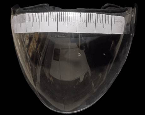
Figure 1: faceshield with orthodontic wire loop
During the final assembly, all components are meticulously aligned to ensure that the wire loop maintains stability while allowing for fine horizontal adjustments. The loop’s ability to glide smoothly along the ruler without deviating from its intended axis ensures that iris positioning remains consistent and reproducible. The entire setup is then double-checked for accuracy, ensuring that the ruler and wire loop remain parallel to the patient’s eyes when the face shield is worn. This ensures that the device provides a precise, reliable, and objective method for verifying iris positioning in ocular prosthesis fabrication, significantly reducing human error and enhancing overall accuracy.
Technique :
A 47 year old lady reported to the Department of Prosthodontics, with a chief complaint of missing left eye in the past 6 months. Medical history revealed a injury to her left eye four years back lead to damage of optic nerve resulted in loss of vision followed by enucleation nine months back. Examination of her eyelids revealed healthy tissue and adequate depth between her upper and lower eyelids to support a custom ocular prosthesis. No other relevant medical history was found. No gross facial asymmetry was noted (figure 2).

Figure 2: Preoperative Photograph
Following a thorough discussion with the patient’s parent and gaining insight into his aesthetic and functional requirements, the decision was made to plan for the fabrication of an ocular prosthesis. An informed consent was taken from the patient’s parent before starting the treatment.
Irreversible hydrocolloid impression material was used to create a preliminary impression, which was then poured with type III gypsum. Clear acrylic was used to create a special tray on primary cast. Light body polyvinyl siloxane material (Zhermack) was used to create the final impression, and type IV gypsum was used to pour the final cast. An iris shell space was carved into a wax design, and a stock iris shell that matched the contralateral eye was selected. An iris shell space was carved into a wax design (Modeling Wax No. 4), and a stock iris shell that matched the contralateral eye was selected. Patient was instructed to sit up straight once the wax pattern was placed and iris positioning was done. To verify the correct positioning, Face shield was placed and the distance between the midline and iris of the normal eye was measured (figure 3).
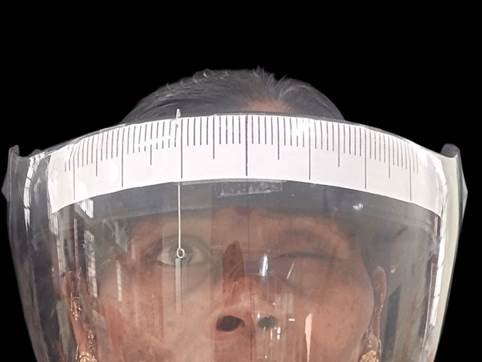
Figure 3: Distance between the midline and iris of the normal eye was measured
The same procedure was done on the contralateral eye and measurement was calculated multiple times for the symmetry (figure4).
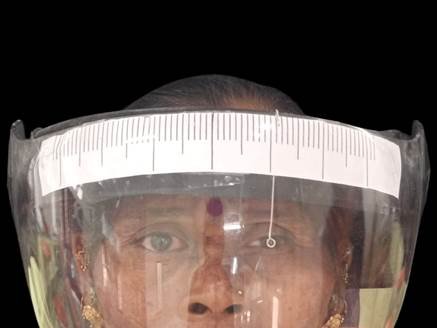
Figure 4: Contralateral eye and measurement was calculated multiple times for the symmetry
The final prothesis was delivery after fabrication (figure 5).
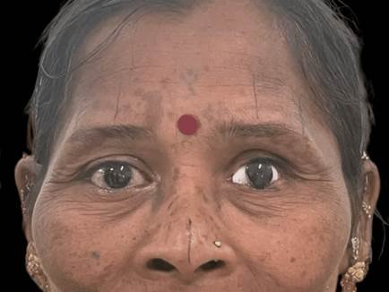
Figure 5: Postoperative Photograph
Post operative instructions were given.
DISCUSSION
The use of a face shield equipped with a movable wire loop and a ruler provides a practical and cost-effective method for verifying iris positioning, particularly in ocular prosthesis fabrication. Precision is one of the key advantages, as the movable wire loop and ruler facilitate fine adjustments, allowing for highly accurate and consistent measurements. This reduces variability and ensures symmetry, which is critical in achieving aesthetically pleasing outcomes. Furthermore, this method enhances objectivity by offering measurable data, minimizing the subjectivity associated with traditional visual estimation techniques. Another notable benefit is its simplicity. The setup involves commonly available materials such as orthodontic wire and a face shield, making it an accessible and affordable alternative to expensive digital devices. Additionally, the method is non-invasive, causing no discomfort to the patient, and can be effortlessly integrated into pre-operative routines or prosthesis fabrication workflows.
Despite these advantages, there are a few limitations that need consideration. This method relies on manual operation, where adjustments to the wire loop are performed manually, introducing a risk of human error, particularly if the operator is inexperienced or lacks a steady hand. Additionally, although the face shield is transparent, it may introduce limited visibility due to minor distortions or reflections, especially under varying lighting conditions, which could compromise the accuracy of measurements.
This method, however, is not suitable in all cases. Contraindications include patients with severe orbital deformities or post-traumatic defects where natural anatomical landmarks are absent, making it difficult to achieve accurate alignment. Similarly, individuals with highly irregular corneal surfaces, such as those with severe keratoconus or corneal scarring, may experience compromised results. Additionally, paediatric or non-cooperative patients who are unable to maintain a steady gaze may hinder the accuracy of manual adjustments, limiting the effectiveness of this approach.10
Amar Bhochhibhoya et.al used a PD ruler.5This technique is highly economical and requires minimal skill, making it more convenient than the graph grid method, which involves tedious preparation and landmark transfer. However, its major limitation is its unsuitability for patients with facial asymmetry or hypertelorism, where accurate iris positioning becomes challenging.
Nimonkar SV et al concluded that objective techniques that make use of simple apparatus without the need for patient participation and cooperation should be opted to achieve best aesthetics.11 Shetty et al. in 2018 used Hanau wide-view spring bow with modifications but cannot be used in patients without ears.7 Laser pointer apparatus by Belkhode et al.,in 2020 was introduced in which a laser pointer helped to measure the distances of the iris mathematically.12 On the contrary, iris sync apparatus is straightforward and less time consuming than other devices in the same category. The horizontal rod provides several readings like that of iris and pupil size on a single ruler while being user-friendly.
The face shield and wire loop method can be effectively applied in several clinical situations and is the only verification technique. It is particularly beneficial during the fabrication of ocular prostheses, where it ensures symmetry and enhances the aesthetic outcome. Furthermore, this method can be integrated into pre-operative surgical planning, allowing for precise assessment and planning of complex ophthalmic procedures. Additionally, it can be used for post-operative evaluations to verify the stability of iris positioning and make necessary adjustments over time.
CONCLUSION
The incorporation of a face shield with an attached ruler and a mobile orthodontic wire loop into the integration is a pioneering method of iris positioning in ocular procedures. Iris positioning has been traditionally dependent on visual estimation, artistic impression, or manual marking, which are all sources of subjectivity and inconsistency between operators. By incorporating a physical reference system consisting of a calibrated ruler and a flexible wire loop, this technique improves iris alignment reproducibility and accuracy. The orthodontic wire loop, having the capability of being adjusted, permits delicate adjustments in horizontal and vertical planes, while the ruler offers a stable scale of measurement for ensuring objective evaluation. Additionally, since the clinician wears the face shield, the configuration remains fixed and enables direct three-dimensional visualization of the patient's face, enhancing accuracy over two-dimensional photographic or hand tracing techniques.
In addition to its benefits, the technique is not faultless. Manual adjustment of the orthodontic wire loop is necessary in the system, creating the potential for human error, particularly in situations requiring extremely precise delicacy. It also depends on the dexterity and attention of the clinician, making it operator-dependent to some extent. In contrast to digital techniques like photogrammetry or augmented reality, this is not an automatic-recording or -analysis method, so its effectiveness might be limited when long-term records or automated integration into digital workflows are necessary. However, the ease, affordability, and flexibility of this face shield-based system render it a highly promising adjunctive tool in clinical and research environments, especially in areas where advanced digital technology access is scarce. Its equilibrium of innovation and pragmatism makes it a valuable means of enhancing iris positioning accuracy without being overly considerate of resources.
REFERENCES
- Sathe S, Pisulkar S, Nimonkar SV, Belkhode V, Borle A. Positioning of iris in an ocular prosthesis: A systematic review. J Indian Prosthodont Soc. 2020;20:345-52.
- Guttal SS, Patil NP, Vernekar N, Porwal A. A Simple Method of Positioning the Iris Disk on a Custom?Made Ocular Prosthesis. A Clinical Report. J Prosthet Dent. 2008;17:223-7.
- Bartlett OS, Moore JD: Ocular prosthesis: a physiologic system.J Prosthet Dent 1973;29:450-459
- Chamaria A, Aras MA, Chitre V, Da Costa GC. Iris positioning using a grid attached to a spring bow for a custom ocular prosthesis. J Clin Diagn Res. 2017;11:214-12.
- Bhochhibhoya A, Mishra S, Mathema S, Acharya B, Maskey B. Alternative technique of iris orientation in a custom?made ocular prosthesis. Journal of Prosthodontics. 2019 Jun;28(5):601-4.
- Gupta L, Aparna IN, Dhanasekar B, Prabhu N, Malla N, Agarwal P. Three?dimensional orientation of iris in an ocular prosthesis using a customized scale. Journal of Prosthodontics. 2014 Apr;23(3):252-5.
- Shetty PP, Chowdhary R, Yadav RK, Gangaiah M. An iris positioning device and centering approach: A technique. The Journal of prosthetic dentistry. 2018 Jan 1;119(1):175-7.
- John AV, Anilkumar S, Rajesh C, Raghavan SM. A novel technique of custom ocular prosthesis fabrication. Journal of Oral Research and Review. 2016 Jul 1;8(2):82-5.
- Chihargo S. CRC1: iris positioning technique by using optical vernier interpupillary distance ruler on the custom ocular prosthesis. J Indian Prosthodont Soc. 2018 Oct;18(Suppl 1):S35.
- Williams, R., & Johnson, P. (2018). Techniques for Accurate Iris Positioning in Ocular Prostheses. Clinical Ocular Prosthetics, 29(2), 145-150
- Nimonkar SV, Belkhode VM, Dahihandekar C, Nimonkar PV, Pisulkar S. A narrative review on techniques of iris replication in an ocular prosthesis. Vol. 23, Journal of Indian Prosthodontic Society. 2023.
- Belkhode VM, Nimonkar S V., Chaudhary SC, Hakkepatil A, Nimonkar P, Prajapat J. An innovative method for iris positioning in a prosthetic eye. Journal of Contemporary Dental Practice. 2020;21(7).
Disclaimer from Guident
The views and opinions expressed in articles published in Guident are solely those of the authors and do not necessarily reflect the views of the editors, the editorial board or the publisher, Ivory India. Guident does not endorse any product, technique or treatment described. Clinical content is intended for qualified dental and medical professionals and is not a substitute for individual clinical judgement. Guident, its editors and the publisher accept no liability for any consequence arising from the use of information published in the journal.
How to cite this article
Dr. Pranjal Patil, Dr. Amrita Pandita Bhatia, Dr. Yukta Gadhe. Enhancing Iris Positioning Accuracy with Face Shield and Orthodontic Wire Loop: A Novel Technique. Guident. 2025 Dec;19(1). Available from: https://guident.in/articles/prosthodontics/enhancing-iris-positioning-accuracy-with-face-shield-and-orthodontic-wire-loop-a-novel-technique.html