Graftless Approach with Short Dental Implant in Posterior Maxilla: A Case Report
Wisdom Dental Clinics ®, Jaipur, India
ABSTRACT
Aggressive treatment options for reduced alveolar ridge height call for bone grafting the area followed by the placement of dental implant. An alternative approach in cases where a limited amount of bone height is available is to use short implant of less than 10 mm of length, instead of the standard range 10 to 16 mm. This strategy avoids the need for bone augmentation procedures and simplifies treatment. This article presents a case report where short implant were placed instead of bone augmentation procedures in a patient with reduced alveolar ridge height.
KEYWORDS: bone grafting, short implant, reduced alveolar ridge height
INTRODUCTION
The sequelae of tooth loss are often associated with compromised masticatory function and unpredictable alveolar ridge resorption, which may in turn complicate prosthodontic treatment outcomes.1 This has been an ultimate challenge to the prosthodontist in accordance with De Van’s principle of preservation. Patients often prefer fixed prosthesis as a treatment. Over the years, traditional methods of tooth placement are slowly and steadily being replaced by newer modalities. The placement and restoration with endosseous dental implants have become routine dental procedures that offer high success rates when suitable planning and protocols are followed.2
A very common challenge encountered in the use of oral implants is the presence of reduced alveolar ridge height.3 This is of particular concern when observed in the posterior areas of the mandible and the maxilla, where the mandibular nerve and the maxillary sinus, respectively, are to be avoided.4 Aggressive treatment options for reduced alveolar ridge height call for bone grafting the area followed by the placement of dental implants.5,6
While these methods have obtained a level of success, some patients reject multiple surgeries and are discouraged by additional treatment duration date, the evidence relating to the predictability of surgically increasing vertical ridge height (other than sinus augmentation) is inadequate. At the same time, a prosthetic solution sometimes is not applicable because of inadequate inter arch space.7 An alternative approach in cases where a limited amount of bone height is available is to use short implants of less than 10 mm of length, instead of the standard range 10 to 16 mm.8,9 This strategy avoids the need for bone augmentation procedures and simplifies treatment.
Recent clinical studies have demonstrated that short implants may be a viable long term solution for sites with limited bone height.10 In the last two decades, it became clear that clinical implantology had advanced to the point that this treatment represented a predictable approach to the replacement of lost teeth. These conflicting results suggest the need for additional research efforts aimed at elucidating successful applications and recommendations for the use of short, wide-diameter implants.
This article presents a case report where short implant were placed instead of bone augmentation procedures in a patient with reduced alveolar ridge height.
CASE REPORT
A 60-year-old male reported to the clinic with the complaint of missing tooth 17 (right maxillary second molar). The tooth was extracted 4 months earlier due to gross decaying; since then he did not wear any prosthesis and was having difficulty in chewing, so he wanted to have a fixed solution. After intraoral examination, treatment options available were: fixed partial denture and an implant-retained restoration/crown; patient agreed with the implant-retained restoration/crown as this was more predictable and conservative to teeth and long term solution.
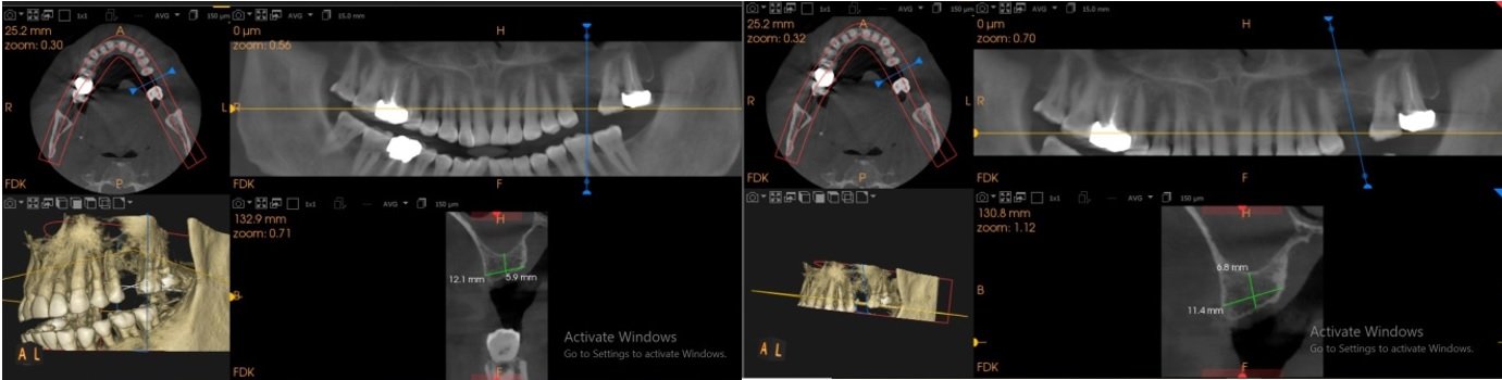
To check for the bone level for implant selection CBCT (cone beam computed tomography) and OPG was done, which showed deficient 3D bone i.r.t 17. (Figures 1)
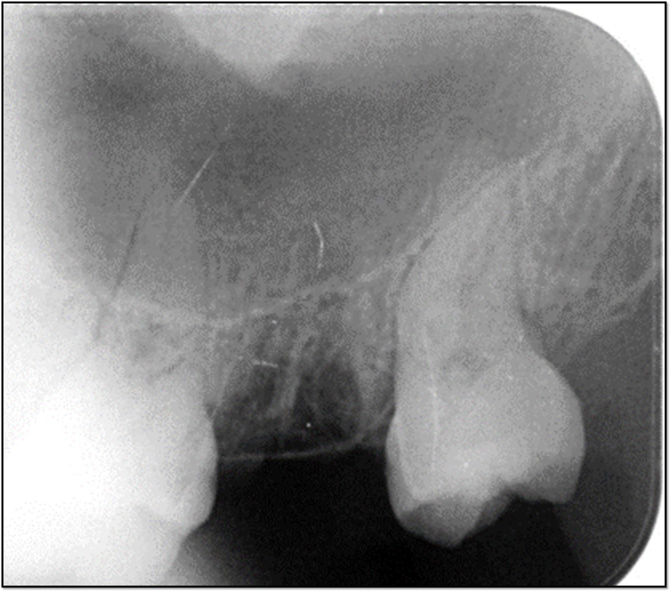
Figure 1: Missing maxillary right second molar
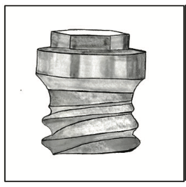
Looking at the compromised bone level, he was offered treatment options with dental implants and bone augmentation. The patient refused bone augmentation and chose the option of short implant placement in the posterior maxilla. After measuring the bone level clinically and radio graphically, it was decided to place a bone level short implant (4.8 × 5 mm) by flapless surgical procedure.
Implant placement was performed under local anesthesia; a tissue punch was used to perforate the gingival tissue so as to gain access to bone without elevating the flap. The osteotomy procedure was initiated with a pilot drill at the punch site under copious irrigation; the final osteotomy was prepared by sequential drilling. The proper angulations and depth of osteotomy were evaluated with a depth gauge and radiographs. After the final osteotomy preparation, implant (4.8 X 5) was placed with an insertion torque of 30Ncm by a torque measuring wrench posing good primary stability, parallel to the roots of the adjacent teeth. (Figure 2)

Figure 2: Implant placement
ISQ measurements were in the range of 75-78, cover screw was then placed on the implant to facilitate the development of a gingival emergence profile for the anticipated restoration. Post-operative OPG was taken to check for the placement of the implant. (Figure 3)
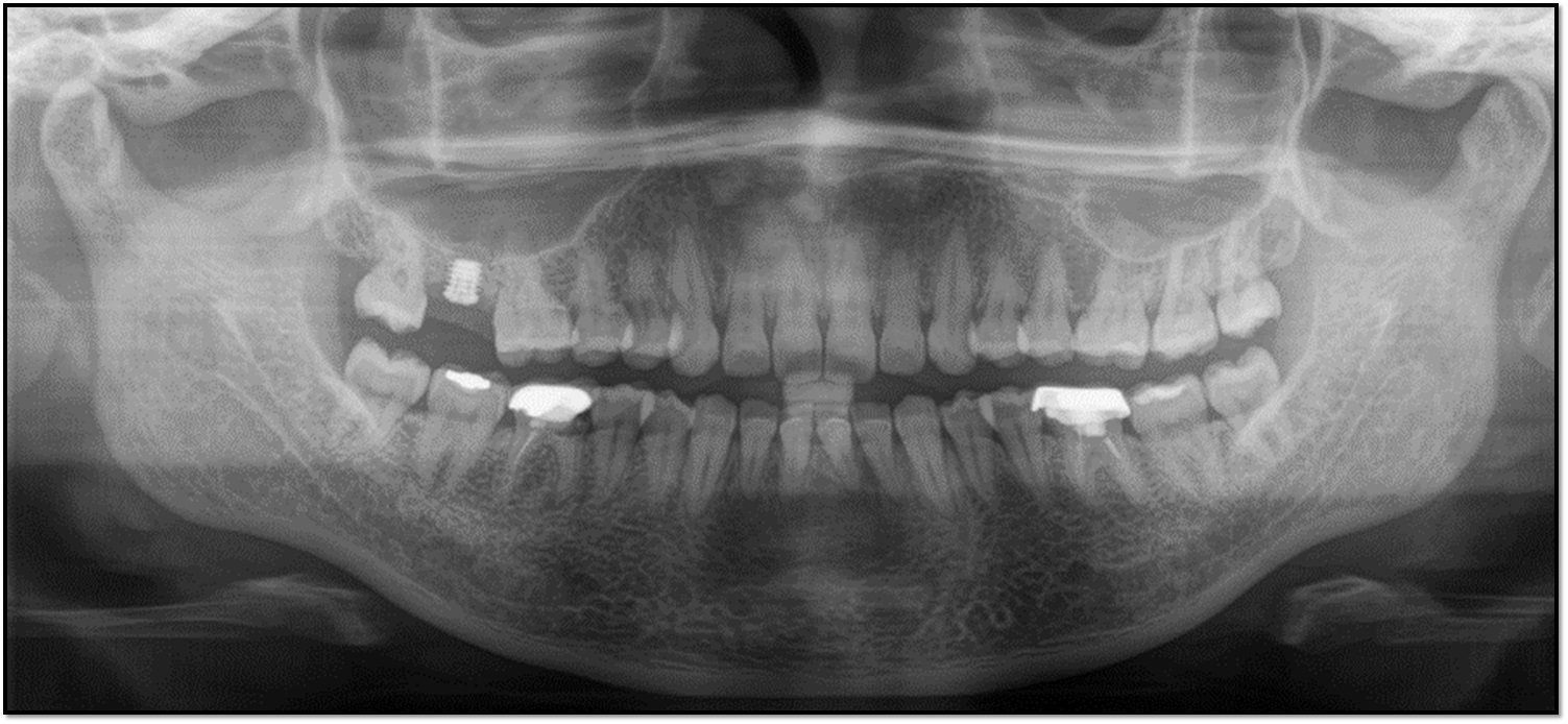
Figure 3: Post-operative OPG
Postsurgical instructions were explained to control postoperative pain. Antibiotics and analgesics were prescribed to the patient for 5 days. The patient was recalled after 1 week for a routine check-up; there was no extra oral swelling or discomfort.
The second stage surgery was done after healing period of 4 months. The implant was exposed without damaging the surrounding bone and gingival healing cap was placed. Indirect impression technique was used for taking the impression of the abutment.
The patient was recalled after 4 months for carrying out the prosthetic procedures .OPG was suggested, ISQ test was done again, which showed excellent biological stability with reading measuring 84, showing excellent secondary stability. (Figure 4)
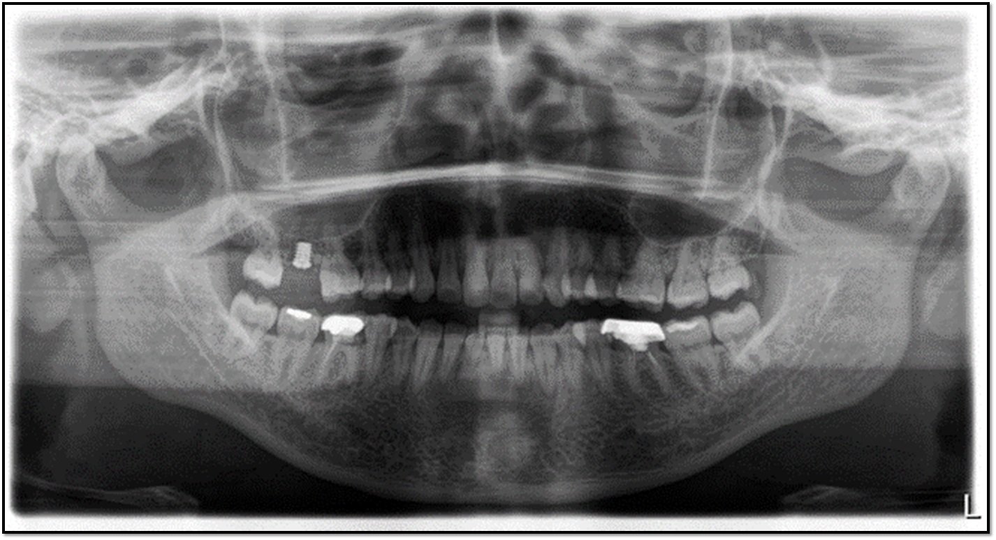
Figure 4: OPG
Healing abutment/gingiva former was replaced with the final abutment and DMLS PFM crown on stock abutment was given. The crown was fabricated and checked for its passive fitting to abutment and occlusal interference checked. Crown was then cemented. After final placement of prosthesis on its desired site, intraorally post-operative radiograph was taken. Patient was asked to come for timely follow-ups. (Figures 5 A, B, C, D)
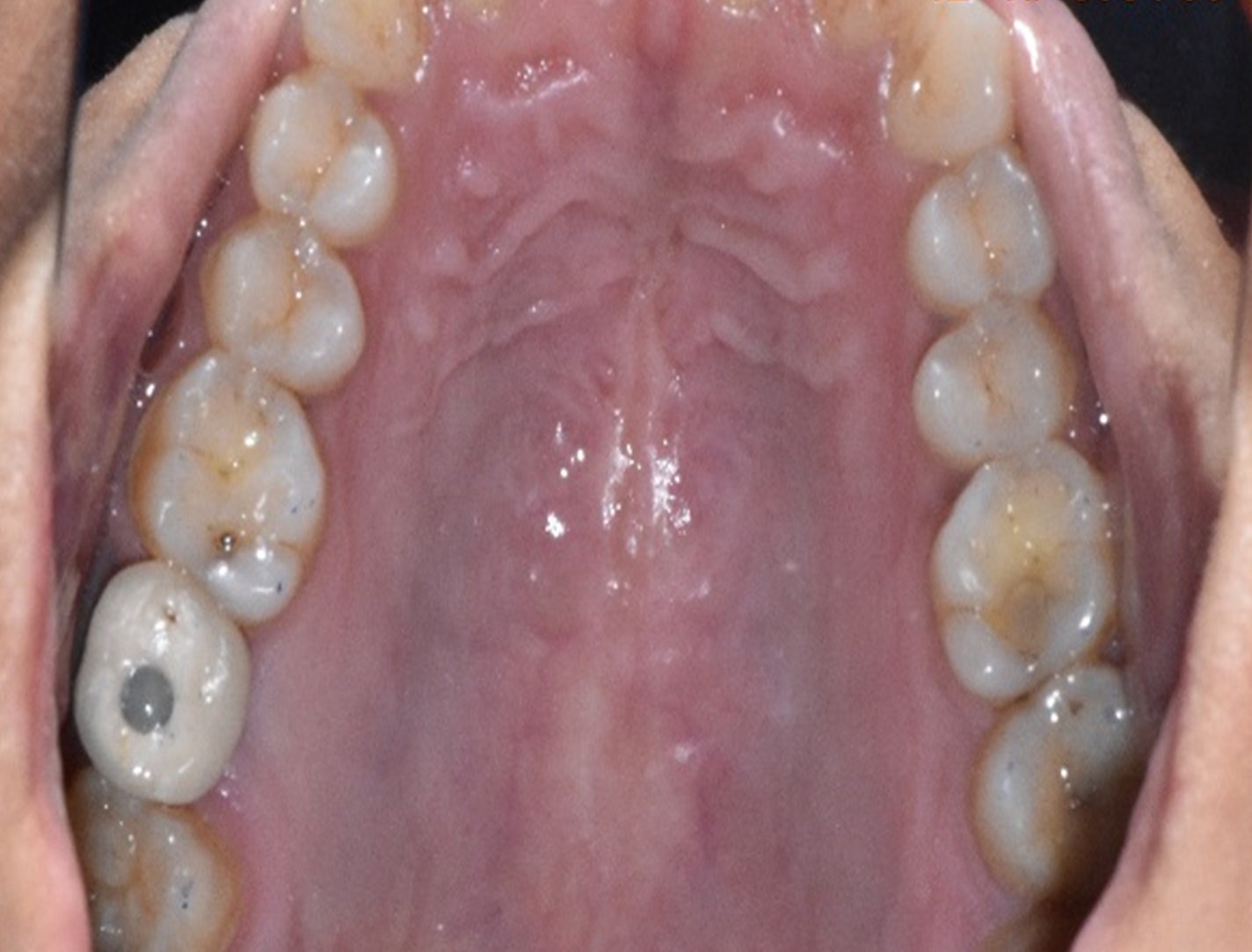
Figure 5A
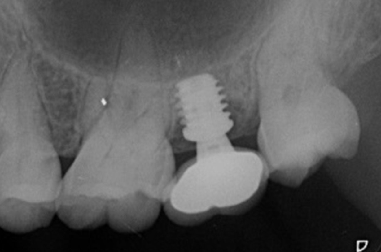
Figure 5B
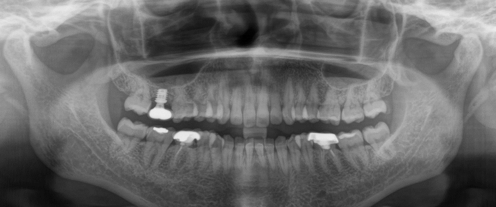
Figure 5C

Figure 5D
Figures 5 A, B, C, D: Post-operative images and x-rays
DISCUSSION
Short dental implants have been proposed as an alternative treatment to simplify implant placement procedures in severely defective alveolar ridge, to avoid vital structures, minimize surgical trauma and the risk of complications associated with invasive surgical procedures. With this in mind, short dental implants would be more suitable in present case.11
Many factors such as implant surface, implant primary stability, bone density and quantity, surgical preparation techniques used, management of the soft tissue and prosthodontic design have been related to implant survival rates.12
Short implant considerations can be categorized as follows:
a. Implant diameter: It is more efficient than length of the implant for dissipation of stress.
b. Crown/implant ratio: Act as vertical cantilever so good surface and implant system with proper force orientation and load distribution might improve the success.
c. Bone quality: Act as the primary factor for the success of short implant.
d. Implant surface: As compared to smooth surface, rough micro topography of surface increase the bone-implant contact thereby, accelerating Osseo-integration.
Here in this case we opted for short implant as there was deficient bone level so long implant could not be placed without bone augmentation so in order to avoid this surgical procedure and looking at the narrow occlusal table with zero cuspal height we chose to go with placement of short implant. Placement of implant without raising a flap requires a certain level of experience and fine motor skills. The possibility of bone perforation and late complications might outweigh the benefit of reduced bone loss with flapless implants, so this procedure must be performed with extreme care and proper preoperative planning.
CONCLUSION
Dental implant treatment with short implant is suitable for patients as it is considered minimally invasive surgery which can reduce the risks of surgical complication sand compromised post-operative healing. The sinus augmentation procedure could be avoided by placement of small dental implants in posterior maxilla. To achieve long term short implant survival, many factors such as good surgical technique, primary implant stability and prosthetic modi?cations to allow for stress reduction during mastication, should be considered.
Short dental implant placement is a successful alternative treatment modality to bone grafting procedures. Short implants are a much less complex and less invasive treatment than placement of longer implants. They also result in the removal of less bone than with longer implants and are less invasive compared to these and therefore probably less traumatic.
REFERENCES
- Gomez-polo M, Bartens F, Sala L, Tamini F, Celemin A, Rio JD. The correlation between crown-3 ratios and marginal bone resorption: a preliminary clinical study. Int J Prosthodont 2010; 23,33-37.
- Pullen G, Debenham C. Short implants: reality and predictability.
- Monje A, Fu JH, Chan HL, Suarez F, Moreno PG,Catena A, Wang HL. Do implant length and width matter for short dental implants (6-9mm)a metanalysis of prospective studies. J Periodont2013.120745.
- Ten Bruggenkate CM, Asikainen P, Foitzic C, Krekeler G, Sutter F. Short (6mm) nonsubmerged dental implants: results of a multicenter clinical trial of 1 to 7 years. Int J Oral Maxillofac Implants 1998; 13:791-798.
- Schliephake H, Neukam FW, Wichmann M. Survival analysis of endosseous implants in bone grafts used for the treatment of severe alveolar ridge atrophy. J Oral Maxillofac Surg 1997; 55:1227-1233.
- Bell RB, Blakey GH, White RP, Hillebrand DG, Molina A. Staged reconstruction of the severely atrophic mandible with autogenous bone graft and endosteal implants. J Oral Maxillofac Surg 2002; 60:1135-1141.
- Griffin TJ, Cheung WS. The use of short, wide implants in posterior areas with reduced bone height: A retrospective investigation. J Prosthet Dent 2004; 92:139-44.
- Gentile MA, Chuang SK, Dodson TB. Survival estimates and risk factors for failure with 6 x 5.7 mm implants. Int J Oral Maxillofac Implants 2005; 20:930-937.
- Morand M, Irinakis T. The challenge of implant therapy in the posterior maxilla: Providing a rationale for the use of short implants. J Oral Implantol 2007; 33(5): 257-266.
- Tawil G, Aboujaoude N, Younan R. Influence of prosthetic parameters on the survival and complication rates of short implants. Int J Oral Maxillofac Implants 2006; 21:275-282.
- Mokcheh A, Jegham H, Turki S. Short implants as an alternative to sinus lift for the rehabilitation of posterior maxillary atrophies:Systematic review and meta-analysis. J Stomatol Oral Maxillofac Surg. 2019;120(1):28–37.
- Srinivasan M, Vazquez L, Rieder P, Moraguez O, Bernard JP, Belser UC, et al. Survival rates of short (6 mm) micro-rough surface implants: a review of literature and meta-analysis. Clin Oral Implants Res.2014;25(5):539–45
Disclaimer from Guident
The views and opinions expressed in articles published in Guident are solely those of the authors and do not necessarily reflect the views of the editors, the editorial board or the publisher, Ivory India. Guident does not endorse any product, technique or treatment described. Clinical content is intended for qualified dental and medical professionals and is not a substitute for individual clinical judgement. Guident, its editors and the publisher accept no liability for any consequence arising from the use of information published in the journal.
How to cite this article
Dr. Gaurav Gupta, Dr. D.K. Gupta, Dr. Neelja Gupta. Graftless Approach with Short Dental Implant in Posterior Maxilla: A Case Report. Guident. 2025 Dec;19(1). Available from: https://guident.in/articles/implantology/graftless-approach-with-short-dental-implant-in-posterior-maxilla-a-case-report.html