Chairside Emax Crown with Cerec Workflow: A Case Report
Jaipur, India
ABSTRACT
This case report describes an innovative approach in creating a smile makeover chairside using a fully digital workflow. An intraoral scan was integrated into the patient’s frontal smile using digital software to create a three-dimensional (3D) digital smile design. Aesthetic function and occlusion remained stable at the 1-year follow-up visit. The use of digital workflow in managing aesthetic cases enhanced the treatment predictability and increased the survival and success of the restorations due to the conservation of tooth structure which reduces number of visits to dentist, thus saves time.
KEYWORDS: intraoral scan, digital smile design, digital workflow
INTRODUCTION
Digital work flow in dentistry has increased in recent years due to headway made in technologies such as intraoral scanners and software programs, which have contributed to improve communication between clinician and dental technician. Digital Smile Design (DSD) is digital tool which provides, from facial perspective, rehabilitative aesthetic planning, better communication between specialists and an improvement in expected outcome of treatments.1
A dynamic documentation of smile is an important step in 2D/3D digital smile design process that can be performed in entire digital flow and will help in re-habilitative procedures. Advantages of using video documentation are that it facilitates and simplifies the documentation process, improves smile design, facial analysis, treatment planning, team communication and patient education.2 The DSD could be converted in to conventional or virtual diagnostic model to facilitate subsequent clinical treatments, i.e. CAD-CAM restorations.3-7
The preparations for minimally invasive treatments have become easily achievable in restorative dentistry because of combination of adhesive technique with restorative materials featuring translucent properties. Materials like lithium disilicate ceramic8-11 have properties like those existing in natural teeth so they have presented positive outcomes.12,13
Another important tool that integrates the digital workflow are intraoral scanners. These are powerful devices that allow an immediate determination of quality of the impression and have the capacity to easily send models to laboratory using e-mail, thus reducing expense and time.14
This report is a presentation of a clinical case that follows a full digital workflow. After a minimal invasive preparation approach, the digital smile protocol, CAD-CAM monolithic lithium disilicate crowns was used to solve a loss of vertical dimension and subsequent aesthetics.
CASE REPORT
A 20-year-old male patient reported to the clinic with aesthetic complains of pain in upper anterior and smile make-over demand. Patient has lost his incisal edge due to trauma 2 days back. Oral hygiene was good and soft tissues were healthy. The patient did not have any other symptoms. [Figure 1]


Figure 1:Pre-Operative photographs
Patient has to travel abroad for further studies so we have to perform all treatment in single day. Considering all the examinations and the data collected, Single sitting root canal treatment with chairside smile make-over with direct aesthetic composite crown was planned. Single sitting root canal treatment was planned on maxillary incisors w.r.t 11,21. Here we opted for CEREC workflow using eMax CAD block to make it convenient and effective for the patient and save time. [Figure 2]

Figure 2: eMax CAD block
Chairside single visit root canal treatment was done on 11 and 21 followed by digital impression using Dentsply Sirona. Single-visit of less than one hour was the time consumed to get the output through the CEREC CAD/CAM chairside system. The system allows scanning, digitally designing, making occlusal adjustments, and calibrating the proximal contacts. [Figure 3]
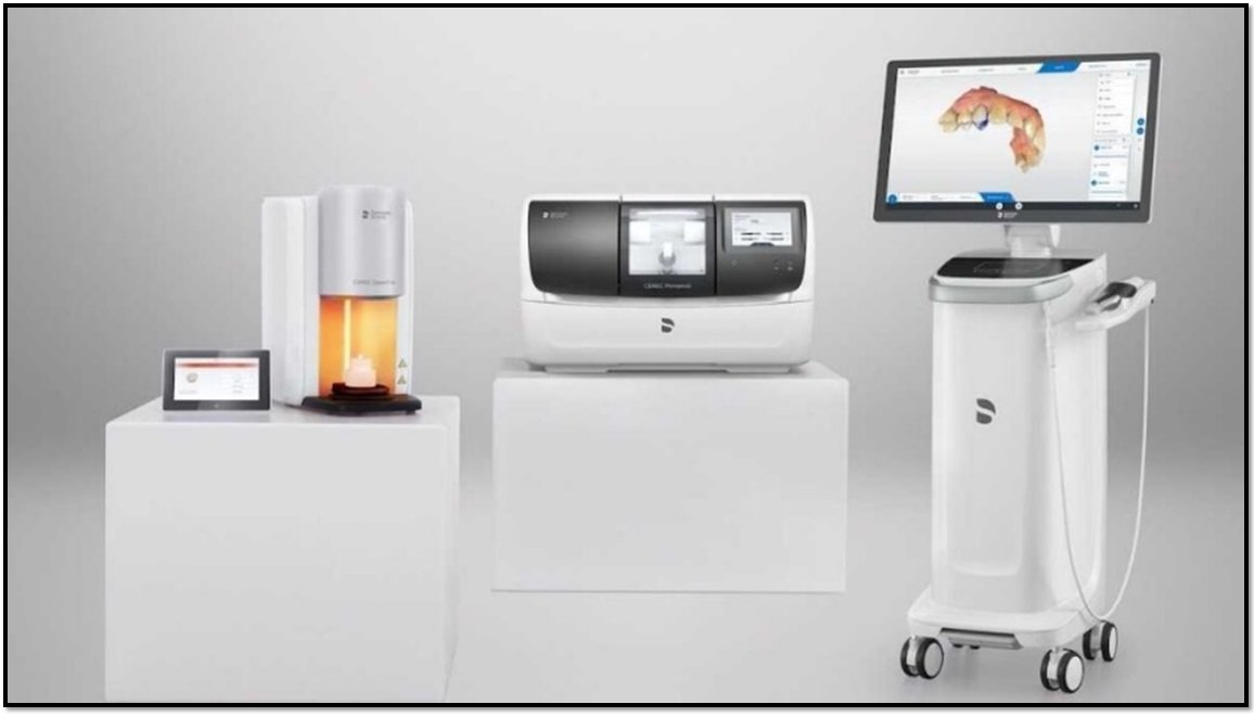
Figure 3: CAD-CAM system
Upper central incisors tooth preparation was done based on the thickness of the prosthetic material. Minimum thickness helps in achieving structural durability and aesthetics. Round angles were made to improve load distribution through the material. Tooth preparation was avoided from any irregular surfaces. We then read the Morphology of dentition on software, and the data is observed by optical impression once the virtual model was developed to select insertion axis. Designing was done in 5 minutes followed. The obtained data had to be sent to the milling machine. Wet milling was done in the primemill which took around 11minutes. An intraoral digital impression was achieved which was converted to a stereolithography (STL) file using special software. The STL files were introduced into additional software systems for design and fabrication of a digital wax-up [Figure 4 a,b,c,d,e].

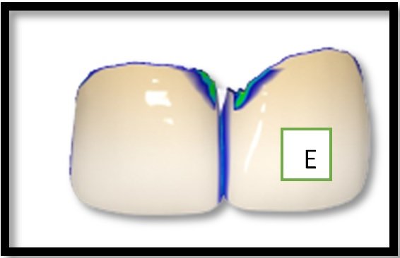
Figure 4a,b,c,d,e: Digital model
After milling the sprue detachment was done from the block followed by sintering and glazing. Sintering was done for strength which took around 12-15 minutes. Both sintering and glazing were simultaneously done in speed fire. Glazing was done to provide aesthetic and gloss in speed fire which took around 8-10 minutes.
Both crowns were made from a single block and treatment was done with eMax CAD block. Later final adhesive luting of veneer was done. The patient was very pleased with the final result. [Figure 5]

Figure 5: Final results
DISCUSSION
Due to the introduction of a whole range of devices, machines and software, the digital revolution is completely changing the dental profession. Thanks to the virtual world, we can plan in detail from surgical to restorative procedures, with the help of 3D modelling and software like CAD-CAM. Intraoral scanners are digital devices used not only to obtain study models but also for the detection of impressions necessary for the modelling of a whole series of restorations. Digital impressions are also a procedure that contribute to a more precise register of the bite, and they can eliminate several analogue procedures that can generate distortions.15
In recent years, the variety of applications, together with the advantages of these machines, have made intraoral scanners highly interesting devices. Nowadays, there are several intraoral scanners in the market, but the most important element to be considered should be the accuracy, i.e. the quality of the data derived from scanning, a combination of trueness and precision. There are already reports in specialized literature that have studied the accuracy of the different intraoral scanners available.16
In modern digital dentistry, the fundamental steps of the therapeutic flow can be summarized into 3 fundamental phases: acquisition of the images and patient data, processing of the information and production of parts, and clinical application onto patients. Digital instruments and the CAD-CAM enable development of more predictable and reliable prosthetic workflows. All these principle work on AI [Artificial intelligence]. Furthermore, the immediate control of the operational procedures guarantees the transmission of accurate information to the dental laboratory and to all the team involved in the therapy.17
The aesthetic demands and expectations of the patients also increase: The digital aesthetic preview thus becomes an expression of everything that the clinician in theory is able to do, legitimizing the patient's demands.In the described clinical case, the accuracy of the Digital Smile System software was assessed by making customized composite veneers from a totally digital workflow and checking the consistency with the initial project, as described experimentally by other authors.18
CONCULSION
Smile is person’s most important ability to express wide range of emotions and determines how well person can function in society. Dentist can provide better aesthetic rehabilitation with minimal tooth preparation by preserving enamel layer for bonding of ceramic with tooth structure. In this case series, anterior aesthetic rehabilitation of patients by zirconia crown placement.
REFERENCES
- Coachman C, Van Dooren E, Gürel G, Landsberg CJ, Calamita MA, Bichacho N. Smile design: From digital treatment planning to clinical reality. In: Cohen M, editor. Interdisciplinary Treatment Planning. Vol 2: Comprehensive Case Studies. Chicago: Quin- tessence; 2012. p. 119–74.
- Coachman C, Calamita A, Sesma N. Dynamic documentation of the smile and the 2D/3D digital smile design process. Int J Periodontics Restorative Dent. 2017; 37:183–93.
- Pimentel W, Teixeira ML, Costa PP, Jorge MZ, Tiossi R. Predictable outcomes with porcelain laminate veneers: a clinical report. J Prosthodont. 2016;25 335–40.
- Lin WS, Zandinejad A, Metz MJ, Harris BT, Morton D. Predictable restorative work flow for computer-aided design/computer-aided manufacture- fabricated ceramic veneers utilizing a virtual smile design principle. Oper Dent. 2015;40: 357–63.
- Arias DM, Trushkowsky RD, Brea LM, David SB. Treatment of the patient with gummy smile in conjunction with digital smile approach. Dent Clin NAm. 2015;59:703–16.
- Coachman C, Calamita MA, Coachman FG, Coachman RG, Sesma N. Facially generated and cephalometric guided 3D digital design for complete mouth implant rehabilitation: a clinical report. J Prosthet Dent. 2017;117:577–86.
- Zimmermann M, Mehl A. Virtual smile design systems: a current review. Int J Comput Dent. 2015;18:303–17.
- Edelhoff D, Liebermann A, Beuer F, Stimmelmayr M, Güth JF. Minimally invasive treatment options in fixed prosthodontics. Quintessence Int. 2016; 47:207–16. https://doi.org/10.3290/j.qi.a35115.
- Culp L, McLaren EA. Lithium disilicate: the restorative material of multiple options. Compend Contin Educ Dent. 2010;31:716–25.
- Zarone F, Ferrari M, Mangano FG, Leone R, Sorrentino R. Digitally oriented materials: focus on lithium disilicate ceramics. Int J Dent. 2016;2016:9840594.
- Soares PV, Spini PH, Carvalho VF, Souza PG, Gonzaga RC, Tolentino AB, et al. Esthetic rehabilitation with laminated ceramic veneers reinforced by lithium disilicate. Quintessence Int. 2014;45:129–33. https://doi.org/10.3290/j.qi.a31009.
- Morimoto S, Albanesi RB, Sesma N, Agra CM, Braga MM. Main clinical outcomes of feldspathic porcelain and glass-ceramic laminate veneers: a systematic review and meta-analysis of survival and complication rates. Int J Prosthodont. 2016;29:38–49.
- Sulaiman TA, Delgado AJ, Donovan TE. Survival rate of lithium disilicate restorations at 4 years: a retrospective study. J Prosthet Dent. 2015;114:364–6.
- Tarantili VV, Halazonetis DJ, Spyropou-Los MN. The spontaneous smile in dynamic motion. Am J Orthod Dentofac Orthop. 2005;128:8–15.
- Ender A, Mehl A. Accuracy of complete-arch dental impressions: a new method of measuring trueness and precision. J Prosthet Dent. 2013;109:121–128. doi: 10.1016/S0022-3913(13)60028-1. [PubMed] [CrossRef] [Google Scholar] [Ref list]
- Goracci C, Franchi L, Vichi A, Ferrari M. Accuracy, reliability, and efficiency of intraoral scanners for full-arch impressions: a systematic review of the clinical evidence. Eur J Orthod. 2016;38:422–428. doi: 10.1093/ejo/cjv077. [PubMed] [CrossRef] [Google Scholar] [Ref list]
- Coachman C, Calamita MA, Sesma N. Dynamic documentation of the smile and the 2D/3D digital smile design process. Int J Periodontics Restorative Dent. 2017;37(2):183?193. [PubMed] [Google Scholar]
- Omar D, Duarte C. The application of parameters for comprehensive smile esthetics by digital smile design programs: A review of literature. Saudi Dent J. 2018;30(1):7?12. [PMC free article] [PubMed] [Google Scholar]
Disclaimer from Guident
The views and opinions expressed in articles published in Guident are solely those of the authors and do not necessarily reflect the views of the editors, the editorial board or the publisher, Ivory India. Guident does not endorse any product, technique or treatment described. Clinical content is intended for qualified dental and medical professionals and is not a substitute for individual clinical judgement. Guident, its editors and the publisher accept no liability for any consequence arising from the use of information published in the journal.
How to cite this article
Dr. Gaurav Gupta, Dr. D.K. Gupta, Dr. Neelja GuptaWisdom Dental Clinics ®. Chairside Emax Crown with Cerec Workflow: A Case Report. Guident. 2024 Mar;17(4). Available from: https://guident.in/articles/prosthodontics/chairside-emax-crown-with-cerec-workflow-a-case-report.html