CBCT- a Boon in Prosthodontics
Department of Prosthodontics & Implantology, K.D. Dental College & Hospital, Mathura (U.P) India
ABSTRACT
CBCT is a medical imaging technique of x-ray CT where the x-rays are divergent forming a cone. CBCT systems have been designed for imaging hard tissues of the maxillofacial region. The increasing availability of this technology provides the dental clinician with an imaging modality capable of providing a three‑dimensional representation of the maxillofacial skeleton. CBCT systems have been designed for imaging hard tissues of the maxillofacial region. CBCT is capable of providing sub-millimeter resolution in images of high diagnostic quality, with short scanning times (10–70 s) and radiation dosages reportedly up to 15–100 times lower than those of conventional CT scans. Increasing availability of this technology provides the dental clinician with an imaging modality capable of providing a three-dimensional representation of the maxillofacial skeleton with minimal distortion with minimal distortion.
INTRODUCTION
With the advancement and discoveries in technology diagnosis has become easy. Diagnostic imaging and techniques help develop and implement a cohesive and extensive treatment plan for the dentist and the patient.Radiology is important in the diagnostic assessment of the patient and information acquired from these help develop a comprehensive treatment plan and objectives. The incorporation of the third dimension into dental and craniofacial imaging is now a practical reality. The future of maxillofacial/dental imaging appears exciting as the paradigm shifts from landmarks, lines, and distances to surfaces, area, and volumes. One such development in the diagnostic field is introduction of CBCT also referred to as C-arm computed tomography [CT], cone beam volume CT, or flat panel CT. Cone beam radiographic technology was first introduced in the European market in 1996. In 2013, Tacconi, Mozzo, Godi and Ronca received an award for this revolutionary invention in maxillofacial radiology.
WHAT IS CBCT?
CBCT is a medical imaging technique of x-ray computed tomography where the x-rays are divergent forming a cone. Most prosthodontists are familiar with the thin‑slice images produced in the axial plane by conventional helical fan beam CTs. CBCT allows the creation of “real‑time” images not only in the axial plane but also two‑dimensional (2D) images in the coronal, sagittal and even oblique or curved image planes – a process referred to as multiplanar reformatting (MPR). In addition, CBCT data are capable of reformation in volume, rather than a slice, providing three‑dimensional (3D) information.Although the CBCT principle has been in use for almost two decades, only recently with the development of inexpensive X‑ray tubes, high‑quality detector systems like flat panel detectors (FPDs) and powerful personal computers, have affordable systems become commercially available.
Most CBCT units for maxillofacial applications use an image intensifier tube (IIT). A system employing a flat panel imager (FPI) was released by i‑CAT The FPI–charge coupled device, currently known as FPD, consists of a cesium iodide scintillator applied to a thin film transistor made of amorphous silicon. Images produced with an IIT generally result in more noise than images from an FPD and also need to be preprocessed to reduce geometric distortions inherent in the detector configuration. Hence, most of the currently available CBCT machinery is made with FPD receptors.

Figure 1. A) conventional computed tomography(CT) scan uses a very narrow “fan beam” that rotates around the patient, acquiring one thin slice (image) with each revolution. Because of the numerous revolutions needed, radiation dose is increased.
B) CBCT captures all data in one rotation, thus reducing radiation and avoiding distortion and errors in reformatting
TYPES OF COMPUTED TOMOGRAPHY
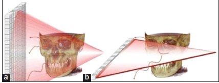
Figure 2 a) Fan Beam Computed Tomography, b) Cone Beam Computed Tomography
FAN BEAM COMPUTED TOMOGRAPHY
- In this the x -ray source and solid state detector are mounted on a rotating gantry.
- Data are acquired using a narrow fan shaped x-ray beam transmitted through the patient.
- The patient is imaged slice by slice, usually in the axial plane and interpretation of the images is achieved by stacking the slices to obtain multiple 2D representations.
- The linear array of detector elements used in conventional helical fan beam CT scanners is actually a multi‑detector array.
- This configuration allows multi‑detector CT scanners to acquire up to 64 slices simultaneously, considerably reducing the scanning time compared with single‑slice systems and allowing generation of 3D images at substantially lower doses of radiation than single detector fan beam CT arrays.
CONE BEAM COMPUTED TOMOGRAPHY
- Cone beam computed tomography scanners are based on volumetric tomography, using a 2D extended digital array providing an area detector combined with a 3D x-ray beam.
- The cone beam technique involves a single 360° scan in which the X‑ray source and a reciprocating area detector synchronously move around the patient’s head,stabilized with a head holder.
ADVANTAGES OF CBCT
1. X-RAY BEAM LIMITATIONReducing the size of the irradiated area by collimation of the primary x-ray beam to the area of interest minimizes the radiation dose
2. IMAGE ACCURACY
The volumetric data comprises a 3D block of smaller cuboid structures know as voxels. All CBCT units provide voxel resolutions that are isotropic, that is, equal in all 3D while in conventional CT its anisotropic
3. RAPID SCAN TIME
As CBCT acquires all basis images in a single rotation, scan time is rapid (10 to 70s) and comparable with that of medical spiral CT systems.
4. DOSE REDUCTION
Report suggests that the effective dose of radiation average range of 36.9- 50.3 milliseivert is significantly reduced by upto 98% compared with conventional fanbeam CT systems.
5. REDUCED IMAGE ARTIFACT
With the manufacturer’s artifact suppression algorithms and increasing number of projections, clinical experience has shown that CBCT images can result in a low level of metal artifacts.
INDICATIONS IN PROSTHODONTICS
Implant ProsthodonticsAnatomic structures suich as inferior alveolar nerve, maxillary sinus, mental foramenand adjacent roots are easily viewed using CBCT. Measurements from the CBCT are routinely accurate throughout the maxilla and mandible and makes the treatment plan and procedures like sinus lift, ridge augmentation, intricate extraction procedures and implant placement with or without a surgical guide. The surgical guide can be fabricated with a CBCT image in the complete absence of patient thus reducing number of appointments precise placement, prefabrication of the abutments and prostheses, and someday delivery of the prosthesis. It also finds application in presurgical imaging, as well as surgical intraoperative and post surgical evaluation(for assessment of osseointegration).
CBCT data combined with data from intraoral scanners is used to interface with interactive machinery like CAD/CAM or 3Dbprinters for precision milling/additive manufacturing resulting in immediate delivery of chairside fixed prostheses and surgical guides.
It is extremely useful in identifying adequate bone support for implant placement, helpful in determining the volume of graft and the type of graft needed. It also provides valuable information about thickening and perforations involving sinus membrane, patency of the osteomeatal complex and also aids in more informed planning with respect to surgical access to the sinus. The anatomical detail gained through a CBCT provides the implantologist ample amount of information to improve the success rate of grafting of the maxillary sinus and sinus implants.
TMJ Imaging
CBCT has the ability to define the true position of the condyle in fossa which often reveals the dislocation of the disk and extent of the translation of the condyle. It facilitates easy measurement of the roof of glenoid fossa and provides the ability to visualize the 3D relation that the condylar head has with the glenoid fossa. It also visualizes soft tissue calcification around TMJ.
It is the choice of imaging in case of trauma, pain and dysfunction and fibro-osseous ankylosis as well as in the detection of condyler cortical subcortical erosion and cysts. The use of 3D features facilitates safe application of the image guided technique which is a treatment modality for TMJ disc adhesion.
Maxillofacial Prosthodontics
CBCT has now replaced standard CT in imaging and planning craniofacial defect reconstruction. Three dimensional augmented virtual models of the patient’s face, bony structure and dentition can be created out of CBCT DICOM data by software volume rendering for treatment planning. DICOM or digital compatibility is the universally accepted data transfer protocol with minimal or nil distortion and nonalterable primary image that helps prevent malpractice.
The shape of the graft can be virtually planned and can also be positioned in the defect creating a virtual reconstruction prior to the actual surgery and also implant placement over the graph can be planned.
Obturators can be accurately milled in larger CAD/CAM UNITS, eliminating the the cumbersome clinical process of construction.
Craniofacial and Airway analysis
Identifying airway obstruction is a great challenge. Various methods like nasopharyngoscopy, cephalometry, nasal airway resistance and polysomnography have been used. Lateral and frontal radiographs are used to assess pharyngeal obstruction.
CBCT shows minimal error and a three dimensional presentation of the airway and surrounding structures which makes volumetric analysis and exact visualization of the airway. It is also very efficient in identifying focal and diffuse narrowing of the airway.
Comprehensive treatment planning in overdenture patients
The placement of denture over retained root and tooth is not new. After extraction the rate of resorption increases.
The process of initial assessment to a follow up during a 4 years review would be precise with the use of a CBCT thereby improving the prognosis of such dentures.
Tellagren found that the average bone loss in the mandible was 9 to 10 mm of vertical height compared to 2.5 to 3 mm on the maxilla.
CONE BEAM COMPUTED TOMOGRAPHY VERSUS COMPUTED TOMOGRAPHY
- Cost of equipment is 3 to 5 times less than traditional medical CT
- CBCT equipment is substantially lighter and smaller
- CBCTs have better spatial resolution and no special electrical requirements are needed.
- No floor strengthening is required as most CBCT are wall mounted.
- Very little technical training is required and easy to operate
- In most CBCT patients are seated while in traditional CT in which the scanning is done by lying down. This eliminates claustrophobia and enhances patient comfort and acceptance.
- Radiation dose is less as compared to traditional CT and also both jaws can be imaged at the same time in CBCT.
- CBCT beats CT in facial region as its primary use in facial region is implant placementand CBCT scores higher as compared to CT.
- Only disadvantage of CBCT over CT is hounsefield units(HU) density measurements. CT gives precise HU.
CONCLUSION
Because of the versatility of CBCT and reduced cost its only a matter of time that CBCT will find its way in all clinical practices and thus helps in transforming the pressing work schedule of a prosthodontist into a relaxed, easier and reliably a more precise one.REFERENCES
- Scarfe WC, Farman AG, Sukovic P. Clinical applications of cone‑beam computed tomography in dental practice. J Can Dent Assoc 2006;72:75‑80.
- White SC, Heslop EW, Hollender LG, Mosier KM, Ruprecht A, Shrout MK, American Academy of Oral and Maxillofacial Radiology, et al. Parameters of radiologic care: An official report of the American Academy of Oral and Maxillofacial Radiology. Oral Surg Oral Med Oral Pathol Oral Radiol Endod 2001;91:498‑511.
- George Puthenpurayil John, Tatu Elenjickal Joy1, Justin Mathew2, Vinod R. B. Kumar3. Fundamentals of cone beam computed tomography for a prosthodontist. 2015: 15(1).
- Hu H, He HD, Foley WD, Fox SH. Four multidetector‑row helical CT: Imagequality and volume coverage speed. Radiology 2000;215:55‑62
- Mozzo P, Procacci C, Tacconi A, Martini PT, Andreis IA. A new volumetric CT machine for dental imaging based on the cone‑beam technique: Preliminary results. Eur Radiol 1998;8:1558‑64.
- Baba R, Konno Y, Ueda K, Ikeda S. Comparison of flat‑panel detector and image‑intensifier detector for cone‑beam CT. Comput Med ImagingGraph 2002;26:153‑8.
- Sukovic P. Cone beam computed tomography in craniofacial imaging.Orthod Craniofac Res 2003;6 Suppl 1:31‑6.
- Ziegler CM, Woertche R, Brief J, Hassfeld S. Clinical indications for digital volume tomography in oral and maxillofacial surgery. Dentomaxillofac Radiol 2002;31:126‑30.
- Cohnen M, Kemper J, Möbes O, Pawelzik J, Mödder U. Radiation dose in dental radiology. Eur Radiol 2002;12:634‑7.
- George Puthenpurayil John, Tatu Elenjickal Joy1, Justin Mathew2, Vinod R. B. Kumar. Applications of cone beam computed tomography for a prosthodontist 2015;16(1):3-7.
Disclaimer from Guident
The views and opinions expressed in articles published in Guident are solely those of the authors and do not necessarily reflect the views of the editors, the editorial board or the publisher, Ivory India. Guident does not endorse any product, technique or treatment described. Clinical content is intended for qualified dental and medical professionals and is not a substitute for individual clinical judgement. Guident, its editors and the publisher accept no liability for any consequence arising from the use of information published in the journal.
How to cite this article
Dr. Shaivy Ambuj, Dr. Manesh Lahori. CBCT- a Boon in Prosthodontics. Guident. 2020 Sep;13(10). Available from: https://guident.in/articles/prosthodontics/cbct-a-boon-in-prosthodontics.html