Peripheral Ossifying Fibroma
Navi Mumbai, India
Abstract
Gngival growths are one of the most frequently encountered lesions in the oral cavity. Most of these lesions are innocuous, but some do have malignant potential. Different lesions with similar clinical presentations make it difficult to arrive at a correct diagnosis. One of the infrequently occurring gingival lesions is peripheral ossifying fibroma. Lesions with histological features similar to POF have been given various names, adding to the confusion. We report the varied clinicopathological features of peripheral ossifying fibroma and investigate the possible etiopathogenesis of this disease.
Key words
Peripheral ossifying fibroma, surgical excision, periodontal ligament.
INTRODUCTION
Peripheral ossifying fibroma is defined as a non- neoplastic enlargement of gingiva that is classified as a reactive hyperplastic inflammatory lesion the accepted classification of localized reactive hyperplastic lesions (LRHL) of the gingiva is into four types: Focal fibrous hyperplasia (FFH), pyogenic granuloma (PG), peripheral ossifying fibroma (POF) and peripheral giant cell granuloma (PGCG).1 Localized hyperplastic lesions of the oral mucosa are common and are usually a consequence of chronic inflammation. Most connective tissue hyperplasias of the oral mucosa rep-resent an exuberant production of granulation tissue in chronic inflammatory reactions. This case report shows us a case of peripheral ossifying fibroma showing its clinical and histopathological picture.
CASE REPORT
A 35 year male patient came to the department of periodontics of Dr.D.Y.Patil school of dentistry, Navi Mumbai with a swelling in maxillary right posterior region palatal aspect since 3 months which was increasing in size. Extent of the lesion was such that it was almost extending to the occlusal 3rd of the teeth (17 and18) (Figure 1). The patient had non-contributing medical history. Surgical excision of the lesion was undertaken under local anesthesia with 2% Lignocaine hydrochloride and 1:80,000 adrenaline using local infiltration (Figure. 2, 3). After complete excision of the tissue (Figure.4) scaling and root planing was done followed by placement of Coe pak (Figure 5). Post-operative antibiotics Cap. Amoxicillin (500mg) and NSAID Tab.Ketorolac (10mg) were given t.i.d. for 3 days. After a week patient was recalled during which Coe pak was removed and wound was inspected (Figure 6). Healing was uneventful. Histopathology showed abundance of connective tissue with some foci of calcification suggestive of peripheral ossifying fibroma (Figure 7).
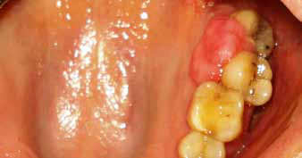
Figure 1: Pre-operative view
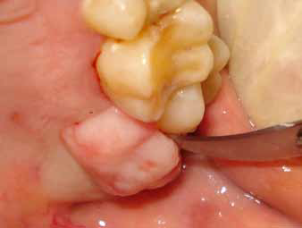
Figure 2: Excision
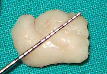
Figure 3: Excised tissue
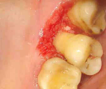
Figure 4: After excision
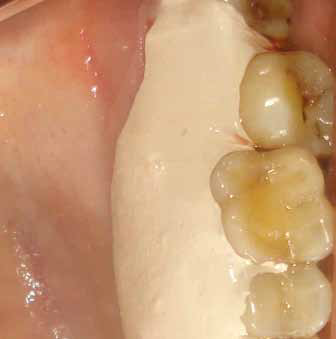
Figure 5: Coe pak
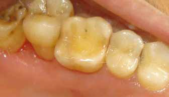
Figure 6: Post-operative view
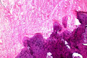
Figure 7: Histopathology
DISCUSSION
In 1982, Gardner coined the term peripheral ossifying fibroma for a lesion that is reactive in nature and is not the extra osseous counterpart of a central ossifying fibroma (COF) of the maxilla and mandible.2 There are two types of ossifying fibromas: the central type and the peripheral type. The central type arises from the endosteum or the periodontal ligament adjacent to the root apex and causes the expansion of the medullary cavity. The peripheral type occurs solely on the soft tissues covering the tooth-bearing areas of the jaws.3 Central ossifying fibroma was found to exhibit increased proliferative activity compared to peripheral ossifying fibroma.4 A polarizing microscopy study revealed that 73% of the 22 peripheral ossifying fibroma cases examined contained a fibrocellular connective tissue stroma surrounding the mineralized mass. The mineralized mass was comprised of woven bone in 50% of the cases, while 18% of the cases showed a combination of lamellar bone and cellular cementum, 18% of the cases comprised only cementum (cellular and acellular), and the remaining 13.6% exhibited a mixture of woven and lamellar bone. This evidence supports the theory that peripheral ossifying fibroma develops from the periodontal ligament/periosteum as undifferentiated mesenchymal cells with an inherent proliferative potential to form bone or cementum.5 There is much uncertainty about the pathogenesis of this lesion. An origin in the periodontal ligament has been suggested. The reasons for considering the periodontal ligament as the origin of peripheral ossifying fibroma include the exclusive occurrence of peripheral ossifying fibroma in the gingiva (interdental papilla), the proximity of the gingiva to the periodontal ligament, and the presence of oxytalan fibers within the mineralized matrix of some lesions.6 The mature fibrous connective tissue proliferates excessively in response to gingival injury, gingival irritation, subgingival calculus or a foreign body in the gingival sulcus. Chronic irritation of the periosteal and periodontal membranes causes metaplasia of the connective tissue and initiates the formation of bone or dystrophic calcification. Thus, local irritants such as dental plaque, calculus, microorganisms, masticatory forces, ill-fitting dentures and poor quality restorations have been implicated in the etiology of peripheral ossifying fibroma.7 Clinically peripheral ossifying fibroma appears as a solitary nodular mass that is either pedunculated or sessile. The surface mucosal color ranges from red to pink, and the surface is frequently ulcerated. The mass usually arises from the interdental papilla. Lesions occur slightly more frequently in the maxillary arch (60%) and the incisor cuspid region (50%).8 One of present cases showed a deviation from these preferred sites and occurred in the maxillary posterior region. Multicentric peripheral ossifying fibroma. has been reported very rarely.6 peripheral ossifying fibroma is definitively diagnosed through a histopathological examination. The histopathological examination usually shows the following features: 1) benign fibrous connective tissue with varying fibroblast, myofibroblast and collagen content, 2) sparse to profuse endothelial proliferation, and 3) mineralized material that may represent mature, lamellar or woven osteoid, cementum-like material, or dystrophic calcifications. Acute or chronic inflammatory cell infiltration can also be observed in these lesions.6 The treatment of choice is complete surgical excision with the removal of the irritating factors.
CONCLUSION
This report highlights the clinical and histologic features of peripheral ossifying fibroma.. Peripheral ossifying fibroma has a high rate of recurrence, making postoperative follow-up mandatory. It is also necessary to use consistent and specific nomenclature in the literature to avoid confusion and the loss of important data.
REFERENCES
- Neville BW, Damm DD, Allen CM, Bouquot JE. Oral and maxillofacial pathology. China: Saunders 2009; 507-9; 517-23.
- Gardner DG. The peripheral odontogenic fibroma: an attempt at classification. Oral Surg Oral Med Oral Pathol 1982; 54:40.
- Keluskar V, Byakodi R, Shah N. Peripheral ossifying fibroma. J Indian Acad Oral Med Radiol 2008; 20:54-6.
- Sousa SC, Mesquita RA, Araujo NS. Proliferative Activity in peripheral ossifying fibroma and ossifying fibroma. J Oral Pathol Med 1998; 27:64-7.
- Shetty DC, Urs AB, Ahuja P, Sahu A, Manchanda A, Sirohi Y. Mineralized components and their interpretation in the histogenesis of peripheral ossifying fibroma. Indian J Dent Res 2011; 22:56-61.
- Kumar SK, Ram S, Jorgensen MG, Shuler CF, Sedghizadeh PP. Multicentric peripheral ossifying fibroma. J Oral Sci 2006; 48:239-43.
- Miller CS, Henry RG, Damm DD. Proliferative mass found in the gingiva. J Am Dent Assoc 1990; 121:559-60.
- Neville BW, Damm DD, Allen CM, Bouquot JE. Oral and Maxillofacial Pathology. 2nd ed. Philadelphia:W.B. Saunders; 2005.p.451-2.
Disclaimer from Guident
The views and opinions expressed in articles published in Guident are solely those of the authors and do not necessarily reflect the views of the editors, the editorial board or the publisher, Ivory India. Guident does not endorse any product, technique or treatment described. Clinical content is intended for qualified dental and medical professionals and is not a substitute for individual clinical judgement. Guident, its editors and the publisher accept no liability for any consequence arising from the use of information published in the journal.
How to cite this article
Dr. Tanay V. Chaubal, Dr. Arvind Shetty, Dr. Devanand Shetty Department of Periodontics, Dr.D.Y Patil School of Dentistry. Peripheral Ossifying Fibroma. Guident. 2015 Jan;8(2). Available from: https://guident.in/articles/periodontics/peripheral-ossifying-fibroma-454.html