Pyogenic Granuloma
Karnataka, India
Abstract
Pyogenic granuloma is a common, usually solitary, benign sessile or pedunculated vascular proliferation of the skin and mucous membranes, presenting as hemorrhagic growth. This term is a misnomer because the lesion is unrelated to infection and it arises in response to stimuli such as low grade local irritation, traumatic injury or hormonal factors. Females are far more susceptible than males because of the hormonal changes that occur in women during puberty, pregnancy. Clinically pyogenic granuloma is smooth or red erythematous papules on a pedunculated or sometimes sessile base, which is usually hemorrhagic. The surface ranges from pink to red to purple, depending on the age of the lesion. Here is a case report of pyogenic granuloma in 35 years old female patient where the lesion was excised using electro cautery.
Keywords: Pyogenic Granuloma, irritational fibroma, peripheral ossifying granuloma
INTRODUCTION
Pyogenic granuloma (PG) is a kind of inflammatory hyperplasia. The term “inflammatory hyperplasia” is used to describe a large range of nodular growths of the oral mucosa that histologically represent inflamed fibrous and granulation tissues.1,2 The term “Pyogenic granuloma” or “granuloma pyogenicum” was introduced by Hartzell in 1904.3 It usually arises in response to various stimuli such as low grade local irritation, traumatic injury, hormonal factors, or certain kinds of drugs4. Pyogenic granuloma may occur in all age groups, though it is predominantly seen in young females in the second decade of life because of the hormonal changes in this period.4 Clinically these lesions usually present as single nodule or sessile papule with smooth or lobulated surface.5 These may be seen in any size from a few millimeters to several centimeters.6 As lesions mature, the vascularity decreases and the clinical appearance is more collagenous and pink.5. Authors report a case of pyogenic granuloma managed by electro cautery.
CASE REPORT
A female patient aged 35 years reported with a chief complaint of enlargement with the upper right back tooth region since three months. The enlargement started as a small painless lesion which gradually increased in size. The patient also complained the spacing being created between the teeth as the enlargement increased. No relevant Dental or Medical history and patient was not on any medications. The patient brushed her teeth once daily using a toothpaste and brush using a horizontal stroke.
CLINICAL EXAMINATION
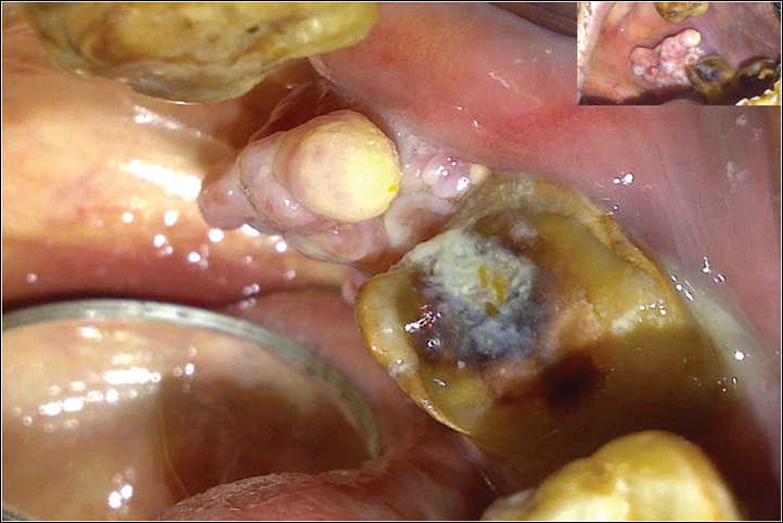
On examination of the enlargement, it was present in the interdental area between 13 & 14, measuring about 1x1cms was pale pink in colour, smooth shiny in surface, well defined margins (Fig 1,2 ), on palpation it was non tender, fibrous in consistency, no signs of bleeding and exudation with the same. Blood investigations showed normal levels and radiographic examination showed a horizontal bone loss in the lesion area. Based on the findings a provisional diagnosis of Pyogenic granuloma was made. Differential diagnosis included irritational fibroma, giant cell granuloma, peripheral ossifying granuloma was made.
TREATMENT
The irritational factors (Plaque and Calculus) were eliminated by scaling and root planning. Patient was educated about the oral hygiene, treatment plan was explained to the patient and informed consent was obtained. After 15 days the enlargement was excised using electrocautery under local anesthesia (fig 3) and the specimen was sent for histopathological Examination (Fig 6). There was no intraoperative complication, periodontal dressing was placed (Fig 5), patient was prescribed with analgesics for three days and 10ml of 0.2% Chlorhexidine mouth wash for 15 days twice daily. Patient was recalled after 1 week for removal of pack and checkup.
The case was followed for 6 months and no signs of recurrences or any discomfort was seen.
HISTOPATHOLOGICAL EXAMINATION
H&E stained section reveals ulcerated parakeratinized stratified squamous epithelium with pseudo membrane on the surface. The connective tissue stroma shows loose bundle of collagen fibers, numerous dilated & engorged blood vessels, chronic inflammatory cells and areas of extravasated RBCs (Fig 9).


Fig 3: Excision by using Electrocautery

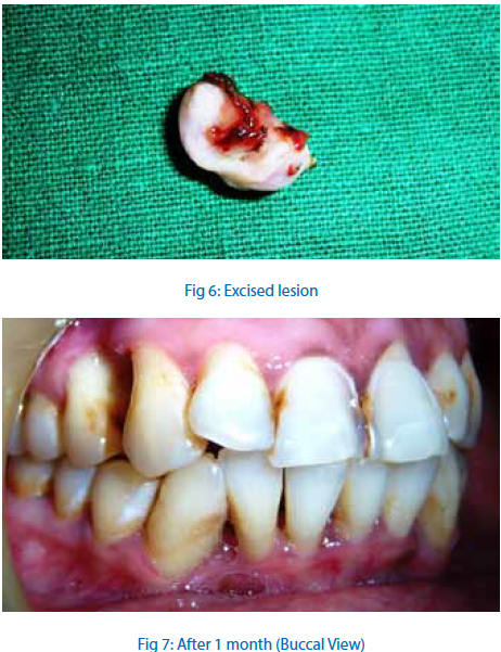

Fig 9: Photomicrograph of the lesion stained with H&E
DISCUSSION
The pyogenic granuloma is a relatively common, tumor like, exuberant tissue response to localized irritation or trauma. The name pyogenic granuloma is a misnomer since the condition is not associated with pus and does not represent a granuloma histologically.7 It occurs in all age groups, children to older adult, but are more frequently encountered in females in their second decade due to the increased levels of circulating hormones estrogen and progesterone8 Lesions are slightly more common on the maxillary than the mandibular region, anterior areas are more frequently affected than posterior areas. Also, Oral pyogenic granuloma is the most common gingival tumor accounting for 75% of all cases. The lips, tongue, and buccal mucosa are the next most common site9 .In the oral cavity pyogenic granulomas show a striking predilection for the gingiva, with interdental papillae being the most sites in 70% of the cases. Gingival irritation and inflammation that result from poor oral hygiene, dental plaque and calculus or over hanging margins restorations may be precipitating factors in many cases10 Pyogenic granulomas of head and neck are uncommonly seen extra gingivally in areas of frequent trauma such as the lower lip, tongue and palate11 majority of PGs are found on the marginal gingiva with only 15% of the tumors on the alveolar part12 Clinically, Pyogenic Granuloma is a smooth or lobulated exophytic lesion manifesting as small, red erythmatous papules on a pedunculated or sometimes sessile base, which is usually hemorrhagic and Compressible12.The size varies in diameter from few millimeters to several centimeters, rarely exceeding 2.5 cm12. Clinically development of the lesion is slow, asymptomatic and painless but it may grow rapidly. The surface is characteristically ulcerated and friable which may be covered by a yellow, fibrinous membrane and its color ranges from pink to red to purple, depending on age of the lesion. Young pyogenic granulomas are highly vascular in appearance, because they are composed of predominantly hyperplastic granulation tissue in which capillaries are prominent. Thus, minor trauma to the lesion may cause considerable bleeding, whereas older lesions tend to become more collagenised and pink12. Radiographic findings are usually absent13 However, Angelopoulos14 concluded that in some cases long standing gingival pyogenic granulomas caused localized alveolar bone resorption. Differential diagnosis included peripheral giant cell granuloma, peripheral ossifying fibroma, metastatic cancer, hemangioma, pregnancy tumor, conventional granulation tissue hyperplasia, Kaposi’s sarcoma, bacillary angiomatosis and non‑Hodgkins lymphoma. Peripheral giant cell granuloma can be histologically identified due to the presence of multinucleated giant cells and lack of an infectious source. Ossifying fibroma or peripheral odontogenic fibroma occurs exclusively on the gingiva, however it has a minimal vascular component unlike a pyogenic granuloma. Due to the proliferating blood vessels differential diagnosis of pyogenic granuloma from a hemangioma is made histologically in which hemangioma shows endothelial cell proliferation without acute inflammatory cell infiltrate, which is a common finding in pyogenic granuloma. Metastatic tumors of the oral cavity are rare and attached gingiva is commonly affected, clinically they resemble reactive or hyperplastic lesions such as pyogenic granuloma, but microscopically they usually resemble the tumor of origin, which usually is distant from the metastatic lesion seen in the oral cavity.15 Management of pyogenic granuloma depends on the severity of symptoms. If the lesion is small, painless and free of bleeding, clinical observation and follow up are advised. Other treatment modalities include laser surgery, electrodessication . Injection of absolute ethanol, sodium tetradecyl sulfate (sclerotherapy) and corticosteroids have also been tried with successful results in cases with recurrent lesions. Recurrence occurs in upto 16% of the lesions, which might be due to incomplete excision, failure to remove etiologic factors, or due to reinjury to the area, making follow up necessary.16,17
CONCLUSION
A pyogenic granuloma is an exuberant growth of granulation tissue secondary to irritation. Individuals with poor oral hygiene and chronic oral irritants most frequently are affected. Intraorally, it can present with a wide array of clinical appearances, ranging from a sessile lesion to an elevated mass. They are usually more common in females, in the present case excision of the granuloma was done and the case was followed up for six months and growth did not recur.
REFERENCES
- Eversole L R. Clinical outline of oral pathology: Diagnosis and treatment (3rd ed). Hamilton:BC Decker 2002; 113-14.
- Greenberg MS, Glick M. Burkit’s oral medicine : Diagnosis and treatment (3rd ed). Hamilton: BC Decker 2003;141-42.
- Hartzell MB. Granuloma pyogenicum. J Cutan Dis Syph 1904;22:520-525.
- Sumanth Shivaswamy, Nazia Siddiqui, A. Sanjay Jain, Ajit Koshy, Sonal Tambwekar, Akhil Shankar. A rare case of generalized pyogenic granuloma: A case report. Quintessence Int 2011;42:493–499.
- Ramirez. K, Bruce G: carpenter. Wpyogenic granuloma: casereport in a 9-year-old girl. General Dentistry 2002, 50(3):280-1.
- Nevile BW, Damm DD, Allen CM, Bouquot JE: oral and maxillofacial pathology Second edition. W.B. saunders co; 2004:444-449.
- Regezi AJ, Sciubba J. Clinical pathologic correlations. 2nd ed.Philadelphia: WB Saunders Co; 1985. p. 194–6.
- Ojanotko Harri AO, Harri MP, Hurttia HM, Sewon LA. Altered tissue metabolism of progesterone in pregnancy gingivitis and granuloma. J Clin Periodontol 1991;18:262‑66.
- Ragezi JA, Sciubba, James J, Jordan Richors CK. Oral Pathology, Clinical pathologic correlation. Fourth. Sanders Company; 2003. 115-176.
- Greenberg MS, Glick M. Burket’s oral medicine:diagnosis and treatment. 10th ed, BC Decker, Hamilton, 2003: 141-42.
- Patil K, Mahima VG, Lahari K. Extragingival pyogenic granuloma. Indian J Dent Res 2006; 17: 199-202.
- Jafarzadeh H., Sanatkhani M. and Mohtasham N. Oral pyogenic granuloma: a review. J Oral Sci. 2006; 48:167-75.
- Kamal R, Dahiya P, Puri A. Oral pyogenic granuloma:Various concepts of etiopathogenesis. J Oral Maxillofac Pathol 2012;16:79-82.
- Angelopoulos AP. Pyogenic granuloma of the oral cavity: Statistical analysis of its clinical features. J Oral Surg 1971;29: 840-47.
- Sheiba R. Gomes, Quaid Johar Shakir, Prarthana V. Thaker, Jamshed K. Tavadia. Pyogenic granuloma of the gingiva: A misnomer? – A case report and review of literature. J Indian Soc Periodontol - Vol 17, Issue 4, Jul-Aug 2013 ,514-19.
- Taira JW,Hill TL, Everett MA. Lobular capillary hemangioma with satellitosis. J Am Acad Dermatol. 1992; 27: 297-300.
- Selvamuthukumar SC,Nalini Ashwath, Anand V. Unusual presentation of pyogenic granuloma of buccal mucosa. JIAOMR. 2010; 22 (4): S 45-47.
Disclaimer from Guident
The views and opinions expressed in articles published in Guident are solely those of the authors and do not necessarily reflect the views of the editors, the editorial board or the publisher, Ivory India. Guident does not endorse any product, technique or treatment described. Clinical content is intended for qualified dental and medical professionals and is not a substitute for individual clinical judgement. Guident, its editors and the publisher accept no liability for any consequence arising from the use of information published in the journal.
How to cite this article
Dr. Sachin Shivanaikar, Dr. Sanketa Patil Belgaum. Pyogenic Granuloma. Guident. 2015 Mar;8(4). Available from: https://guident.in/articles/periodontics/pyogenic-granuloma.html