
Surgical Management of an Incomplete Oblique Intra Alveolar Root Fracture with PRF Placement
AMC Dental College Ahmedabad, Gujarat, India, Subharti Dental College, Meerut, Utter Pradesh, India, Department of Conservative Dentistry and Endodontics
Abstract
Horizontal intra alveolar root fractures are rare in comparison to other types of injuries and the reported prevalence in the literature is between 0.5% and 7%. Fracture occurs often in the middle-third of the root and rarely at the apical-third. The present paper reports a clinical case of a oblique radicular fracture located between the middle- and apical-third of a upper left-central incisors which was managed by surgical removal of separated fragment followed by sealing with mineral trioxide aggregate (MTA) and platelet rich fibrin (PRF) placement.
KEY WORDS:intra alveolar root fractures, Mineral trioxide, Platelet rich fibrin.
INTRODUCTION
Root fractures are defined as fractures involving the dentine, cementum and pulp1. They comprise 0.5 to 7% of the injuries affecting the permanent dentition and commonly occur between the age group of 11 to 20 years2. Root fractures can be broadly classified as horizontal/ oblique or vertical. Horizontal/ oblique root fractures are the most common type and occur mainly in the anterior region of the maxilla in fully erupted teeth with complete root formation, owing to a frontal impact2. They occur most commonly in the middle-third and rarely in the apical and coronal third of the root3.Horizontal/oblique root fractures can be further subclassified on the basis of: Location of fracture line (cervical, middle and apical); Extent of fracture (complete or incomplete); Number of fracture lines (simple, multiple and comminuted) and Position of coronal fragment (displaced and not displaced)4.
Majority of root fractures have been shown to undergo healing. It has been reported that 77% of root fractured teeth healed, with pulp necrosis occurring in 20% cases5. Healing with granulation tissue is a sign of pulp necrosis and an indication that endodontic treatment of coronal portion of the tooth is necessary. This paper present a case of incomplete oblique root fracture healed with connective tissue formation, which was preserved in functional state by surgical removal of fractured segment followed by sealing of fractured root end with MTA (mineral trioxide aggregate).
CASE REPORT
A 25-year-old female patient came to the department with the chief complain of discolored upper anterior teeth. She reported a history of trauma on maxillary anterior teeth region 15 years back. On intra-oral examination sinus was present in relation to maxillary left central incisor.
Radiographic examination revealed discontinuity in the middle third of the root on mesial aspect and associated radiolucency in relation to maxillary left central incisor and open apex was present in relation to maxillary right central incisor (fig 1).

Fig 1. Preoperative radiograph revealing incomplete root fracture of maxillary left cental incisor.
No increased mobility of the coronal segments was found. Coronal segments were found necrosed on vitality testing for both the central incisors. Endodontic treatment of coronal segment till the fracture line and surgical removal of fractured segment followed by sealing with MTA was planned for left cental incisor. Apexification with MTA followed by obturation was planned for right central incisor.patient consent was obtained.
Access opening were made for both the incisors. Working length was measured till fractured level of root for 21. After confirming working length, CaOH2 dressing was placed for a week. Surgical removal of fractured root segment was planned for 21. Apexification with MTA followed by obturation was done for 11 through orthograde approach.
Under local anesthesia, full thickness periosteal flap was reflected to exposed the root area. Buccal cortical bone was found resorbed at fractured level and fractured segment was clearly seen (fig 2). Fractured segment was removed (fig 3) and curettage of bony cavity was done. Root end was prepared and MTA was placed to seal the root end by retrograde approach (fig 5). Bony cavity was filled with plasma rich fibrin made up of patient own blood, to facilitate bony healing. Flap was gently placed back to its original place and suture were placed. Patient was reviewed after one week for suture removal and the healing at the surgical site was uneventful. After one year patient was asymptomatic with signs of healing.
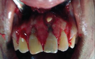
Fig 2: Fractured segment insitu.

Fig 4: Radiograph after fractured root fragment removal.

Fig 5: Sealing with MTA.

Fig 6 : Post operative radiograph.
DISCUSSION
Root fracture is one of the consequences of dental trauma.The prognosis for tooth survival following a horizontal/oblique root fracture can be summarized as quite good5.Consequently, the prognosis of the tooth is influenced by the factors such as the patient’s age, the stage of root growth, mobility of the coronal fragment, dislocation of the coronal fragment, and fragment diastasis7.
The choice of treatment depend on radiographic features, such as periradicular changes, the width of the root canal space and the amount of separation of two fragments8, 9. Dental treatment may not be necessary in cases of root fracture where there is no mobility or displacement of the coronal segment, and if the patient has no apparent complaints8,9.
Root fractures heal differently depending on the degree of separation of the fragments, the severity of injury, and the ability of the pulp to heal6. The histological reactions at the fracture line are categorized into four types: (I) interposition of calcified tissue (callus formation) (II) interposition of connective tissue, which is characterized by peripheral rounding of the fracture’s ends (III) interposition of bone and connective tissue, radiologically characterized by the clear separation of the two fragments and (IV) interposition of granulation tissue, caused by an infected or necrotic pulp. The last mode of healing with granulation tissue is a sign of pulp necrosis and an indication that endodontic treatment of the coronal portion of the tooth is required. However, if the pulp injury is severe, healing does not occur and granulation tissue separates the fractured segments, obviously without the hard-tissue healing6, 9. The extent of fracture and development of pathology at fracture site necessitate a surgical approach. Various materials can be used to close the root canal at the fracture site like MTA and biodentin. In first case MTA was used to create an apical barrier at the fracture site. MTA has been reported to have superior biocompatibility, sealing ability and is less cytotoxic when used for root end filling10. Bone regeneration after surgical intervention takes place in a very slow manner. Hence, to enhance these processes a number of bone substitutes are being tried out. Enhancement of the regenerative process of human body by utilizing the patient’s own blood is a unique concept in dentistry. Blood and blood products have been used for over a century for various purposes. PRF is a matrix of autologous fibrin, in which are embedded a large quantity of platelet and leukocyte cytokines during centrifugation. PRF acts as a bioactive scaffold for periodontal tissue regeneration. Application of PRF has two major benefits: (a) The promotion of soft tissue healing, (b) the induction of new alveolar bone formation11,12.PRF is a by-product of the patient’s own blood; therefore, chances of infectious disease transmission is rare. Results of PRF have been found promising in various case reports13.In the present case PRF from patient’s own blood was used to fill bony cavity for rapid and good healing.
CONCLUSION
Treatment of root fracture depends on various factors such as position of fracture line, mobility of tooth and pulpal status. Horizontal root fracture with partial fragment detachment can be managed by surgical removal of detached fragment followed by sealing of the exposed root area.
REFERENCES
- Andreasen JO, Andreasen FM, Andersson L. Textbook and Color Atlas of Traumatic Injuries to the Teeth (4thed). Oxford, Blackwell 2007. 337-37.
- Majorana A, Pasini S, Bardellini E, Keller E. Clinical and epidemiological study of traumatic root fractures. Dent Traumatol 2002; 18: 77–80.
- Yates JA. Root fractures in permanent teeth: a clinical review. Int Endod J 1992; 25: 150–157.
- Malhotra N. A Review of root fractures: Diagnosis, treatment and prognosis. Dent Update 2011;38:615-28.
- Caliskan MK, Pehlivan Y. Prognosis of root fractured permanent incisors. Endod Dent Traumatol 1996: 12: 129-136.
- Trop M, Chivian N, Sigurdsson A. Traumatic injuries.In: Cohen S, Burns RC, editors. Pathways of the pulp. St. Louis: Mosby; 1998.p. 567-72.
- Andreasen JO, Andreasen FM, Mejàre I, Cvek M. Healing of 400 intra‑alveolar root‑ 1. Effect of pre‑injury and injury factors such as sex, age, stage of root development, fracture type, location of fracture and severity of dislocation. Dent Traumatol 2004;20:192‑202.
- Aras MH, Ozcan E, Zorba YO, Aslan M. Treatment of traumatized maxillary permanent lateral and central incisors horizontal root fractures.Indian J Det Res 2008;19(4):354-6.
- Andreason FM, Andreason JO, Bayer T. Prognosis of root fractured permanent incisors- predictation of healing modalities. Endod Dent Tramatol 1989;5:11-22.
- Agrawal Vineet S ,Kapoor Sonali. Clinical management of severe external root resorption and immature open apex with MTA and calcium hydroxide- A case report. Endodontology 2013; 83-88.
More references are available on request.
Disclaimer from Guident
The views and opinions expressed in articles published in Guident are solely those of the authors and do not necessarily reflect the views of the editors, the editorial board or the publisher, Ivory India. Guident does not endorse any product, technique or treatment described. Clinical content is intended for qualified dental and medical professionals and is not a substitute for individual clinical judgement. Guident, its editors and the publisher accept no liability for any consequence arising from the use of information published in the journal.
How to cite this article
Dr. Akansha Malik, Dr. Parul Bansal, Dr. Kalpana Kanyal. Surgical Management of an Incomplete Oblique Intra Alveolar Root Fracture with PRF Placement. Guident. 2015 Mar;8(4). Available from: https://guident.in/articles/endodontics/surgical-management-of-an-incomplete-oblique-intra-alveolar-root-fracture-with-prf-placement.html


