
Non-Surgical Management of a Lateral Open Bite Using Micro Implants
Department of Orthodontics Dr. Bharath Rao (Assistent Professor) Department of Oral Biology. Manipal College of Dental Sciences, Manipal University, Manipal Karnatka, India
Abstract
Lateral or posterioropenbite is commonly associated with a tongue thrust habit involving premolars and molars. Treatment of adults is limited to orthodontic extrusion of the posterior teeth or surgical impaction of maxilla. Non-surgical management using microimplants has become popular in the last decade. A 22 year old female reported to the Orthodontic clinic with a complaint of crooked front teeth. She had a convex profile, protrusive lips with super class I molar and a lateral anterior openbite of 4mm. Anterior crowding of 11 mm required extraction of 1st premolars.
Fixed orthodontic appliance (0.022”slot, MBT prescription) along with micro-implantswere inserted in August 2012. 016” Niti wires were used for leveling and alignment and retraction with 019x025”SS. After 18 months of treatment, appliance was debonded and lingual bonded retainers were placed. Maxillary and Mandibular superimpositions show maxillary molar intrusion of 3mm and anterior retraction of 3mm and a 2mm counter-clockwise rotation of mandible.
INTRODUCTION
Openbite is a lack of contact in a vertical direction between the incisal/occlusal surfaces of the maxillary and mandibular anterior/posterior teeth.1 Lateral openbite in children is commonly associated with a tongue thrust habit and affects erupting premolars and molars. Unless intercepted, it may develop into a skeletal malocclusion requiring surgery in adulthood. The prevalence of openbite is 3.5% of white American children and 16.5% of African-Americans,2. Less often a class III malocclusion may be associated with lateral open bite,crowding and posterior crossbite.
Treatment planning in such cases is further complicated by over-eruption of posterior dento-alveolar heights, resulting in a longer vertical facial dimension and a steeper mandibular plane.3,4Passive bite blocks,active bite blocks with magnets or springs, high-pull headgear and vertical elasticsare some of the treatment options available for treating youngerindividuals.
However in an adult patient theseoptions are limited to orthodontic extrusion of the anterior teethor orthognathic - surgery.As a general rule, surgical impaction of the maxillary posterior segments is needed to obtain counter-clockwise rotation of mandible and a reduction in anterior facial height.
Non-surgical management of openbiteusing microimplantshas become popular for intrusion of posterior teeth. Kuroda et al5found it simpler to intrude the posterior teeth with skeletal anchorage than to correct it surgically. Sugawara et al6 also used miniplates to intrude mandibular molars.
Orthodontic micro-implants allow easy placement with minimal anatomic limitations. Virtually all intraoral bony structures (mandibular symphysis, midpalatal, infrazygomatic, alveolar process, sub-ANS and retromolar areas) can host microimplants. But the inter-radicular bone in the alveolar processis biomechanically advantageous because of its proximity to the center of resistance of teeth. On the other hand,the risk of root contact or damage exists because of the narrow inter-radicular spaces and varying oral anatomies of individual patients.

CASE SUMMARY
A 22 year old female reported to the Orthodontic clinic with a complaint of crooked front teeth. She had a convex profile, protrusive lips and an increased LAFH. Intra-oral examination revealed a super class I molar and a lateral anterior openbite of 4mm. Anterior crowding of 11 mm and upper midline shiftto left by 2mm were also noted (Figure 1).
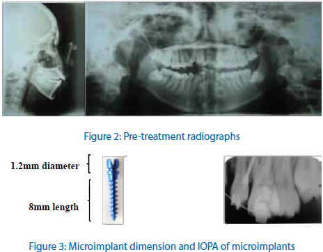
Cephalometric evaluation revealed a class III skeletal base (Wits appraisal=0mm) with retrognathic maxilla (SNA=77°), retrognathic mandible (SNB=72°) and vertical growth pattern (GoGn-SN=57°).Dental analysis revealed lower anteriors were more proclined than upper anteriors (Up1-NA=29° at 12mm, Lw1-NB=33° at 12mm and IMPA=105°).OPG showed impacted 38 and 48.
COGS (dental) analysis showed maxillary posterior dento-alveolar region had extruded by 3mm and maxillary anterior dento-alveolar region had intruded by 2 mm contributing to lateral openbite.
Diagnosis and treatment Objective
A diagnosis of Angle’s class III malocclusion with openbite and anterior crowdingwas given. The objectives of treatment were:
- De-crowd the anteriors and correct the anterior proclination
- Establish positive overjet and a straight profile
- Correct axial inclination of maxillary 1st molar and class III molar relation.
- Establish positive overbite by intrusion of posteriors using micro-implants to obtain counter-clockwise rotation of mandible.
- Protraction of upper left 1st molar by 1.5mm to finish in a class I molar using friction mechanics
- Correction of dental midline shiftusing asymmetric elastics
TREATMENT ALTERNATIVES
The treatment options were either orthognathic surgery or anorthodontic camouflage.Surgical correction would consist of maxillary impaction. Straight profile, edge to edge biteand class I occlusion, could be have been addressed with this approach. The patient, however, refused any surgical procedures due to time constraint and financial reasons.Hence, a camouflage option by premolar extraction and maxillary molar intrusion was chosen.
TREATMENT PLAN
Fixed orthodontic appliance consisting of 4 bands and 16 bondable metal brackets (3M Unitek, Gemini MBT Brackets, Monrovia,CA) with 0.022”slot and a MBT prescription were used. Initial leveling was started with 016” Nitiwires and retraction with 019x025”SS.At the end of leveling and aligning, the lower anteriors de-compensated and produced a negative overjet of 1mm. Her 1st premolars were extracted to correct the proclination and achieve a positive overjet at the end of retraction. Upper anterior were consolidated and retracted using friction mechanics. Class I molar relation was achieved using Class III elastics (3/16M, 3M Unitek, Monrovia, CA). Finishing and detailing was done using artistic bends.
OPG also confirmed root parallelism (Figure 5). Post-Treatment lateral cephalometric analysis revealed a class III skeletal base (Wits Appraisal =0 mm) with upright lower anteriors (IMPA = 95º, U1-NA= 25º, L1-NB= 26º) and a hypo-divergent facial pattern (MP-HP=55º).
Maxillary and Mandibular superimpositions show maxillary molar intrusion of 3mm and anterior retraction of 3mm and a 2mm counter-clockwise rotation of mandible. Soft tissue superimpositions indicated an improvement in the profile with upper lip and lower lip retraction of 1mm. (Figure 6) (Black line indicates pre-treatment tracing and Red line indicates post – treatment tracing).
BIOMECHANICS OF MOLAR INTRUSION USING MICRO-IMPLANTS
Micro-implants (1.2mm diameter and 8mm length) were inserted on buccal side mesial to right upper 1stmolar under LA respectively. They were inserted at height of 5mm from crest of interdental papilla at an angle of 20-30º. Ligature wire was tied between the micro-implant and 1stmolar hook in diagonal fashion to prevent molar extrusion. These ligatures were changed at 3 week interval until occlusal plane was levelled.Occlusal planes were leveled in 6months and the correction was maintained using a TPA (Transpalatal arch) (Figure 3).
DISCUSSION
Lateral Openbite can be caused by a maxillary excess, vertical skeletal pattern, abnormal dental eruption and a tongue thrust habit. After lateral tongue thrust, failure of dental eruption is the most common cause of lateral openbite. The two types of eruption failure are:1) Primary and 2) Secondary.
In 1981, Proffit and Vig coined the term “Primary failure of eruption/retention” to describe the eruption failure of non-ankyosed teeth before emergence without a physical barrier in the eruption path or abnormal position.It is genetic/familial with varying penetrance and expression. It affects the posteriors and the teeth distal to affected teeth are also involved resulting in a posterior openbite. Clinically, the involved teeth erupt two years behind schedule and the response to orthodontic force may be abnormal or absent.
Secondary retention refers to cessation of tooth eruption after emergence without a physical barrier in the eruption path or abnormal position. Ankylosis/retained deciduous molar are the main etiological factors and infra-occlusion is the clinical finding. Our patient had openbite extending to distal aspect of 1st permanent molars, suggesting a secondary failure of eruption. We also ruled out lateral tongue thrust habitand systemic causes.
The lateral openbite caused by lack of posterior dento-alveolar development results in divergent occlusal planes. To level the maxillary occlusal plane, maxillary arch wire (0.018”SS) was segmented distal to lateral incisor and inter-maxillary elastics were worn in a triangular fashion. This extruded the premolars vertically while the mandibular teeth were held in position. The net effect is clockwise rotation of maxillary occlusal plane and increase in overbite (Figure4).
Moderate crowding requires extraction of 1st premolar to achieve ideal overjet, de-crowd and improve the AP relationship7,8. However, Camouflage option in Class III patients may:worsen the profile9, lead to root exposure, resorption of the incisors10,11plaque accumulation and periodontal diseases.12 We attempted to correct anterior proclination, crowding with 1st premolar extraction. The edge to edge bite had to be corrected by retraction of anteriors under maximum anchorage. Class III elastics (3/16) also helped to tip (distally tipped maxillary 1st molar) and protract maxillary molars to class I relation but should be avoided in cases of skeletal openbite and gummy smile. Fixed orthodontic mechanics is predominantly extrusive unless re-inforced. The presence of incompetent lips and a vertical growth pattern required absolute anchorage (micro-implants) to prevent further maxillary molar extrusion during treatment.
Absolute anchorage can be achieved using microimplants (6-12mm in length and 1.5-3 mm in diameter). Every millimeterof posterior intrusion induces counter clockwise rotation of mandible/chin by 3-4 mm.13To achieve it, microimplants were placed distal to center of resistance of dentition. The choice of buccal alveolus between 2nd premolar and 1st permanent molar was based on visibility and 2 mm thick cortical plate. Intrusive forces on the maxillary molar from buccal aspect may produce third order problems like buccal tipping. To counteract this, a trans-palatal arch should be provided. Insertion at an angle of 20-30° helps prevent root damage and increases surface area of implant contact to cortical bone.
One study suggested that openbite correction by a non-extraction approach lead to 38.1% relapse compared to a surgery.14Lefort I superior impaction of maxilla is the gold standard in adult openbitecases but requires hospitalization and holds the risk of mortality. Microimplantsare superior (to miniplate system) in terms of short chair-side time, requires local anesthesia and therefore economical. They are also smaller, easy to place and remove surgically.
Reitan showed that principle fibres of periodontal ligament become rearranged in8-9 weeks but the supra-alveolar structures take longer due to their slower turnover rate15-16.One study reported that intruded teeth were more stable than extruded teeth. But Sugawara et al showed that one third of molar intrusions relapsed during the first year of retention6Various authors have suggested that over-correction, by retaining the micro-implants in the alveolus (until occlusalsettling is complete)and performing muscle exercises could overcome relapseof openbitein adults.
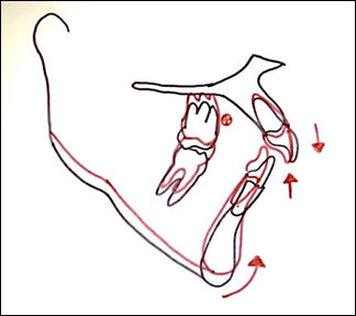
Figure 4: Biomechanics of openbite closure
CONCLUSION
Lateral openbite is a complex malocclusion to treat due to its varied etiology. Treatment planning of such cases will depend on identifying the etiology. Besidesorthognathic surgery, micro-implants can provide anchorage and intrude maxillary posterior segment in adult openbite cases. This produces closure of an anterior open bite with counter-clockwise rotation of mandibulararch which is favorable in vertical growth pattern.
ACKNOWLEDGEMENT
We are thankful to Manipal University for providing the laboratory and journals for our research. We are also grateful to Dr.Nirmala N Rao (Professor and Dean, MCODS,Manipal) and Dr. Arun S Urala (Professor, Dept. of Orthodontics,MCODS,Manipal) for always being source of inspiration and encouragement during our research period. I am deeplyindebted to Mr. Sharath Kumar Hegde (Orthodontic lab technician) for fabrication of Hawley’s retainers.
REFERENCES
- Subtelny JD, Sakuda M. Open-bite: diagnosis and treatment. Am J Orthod. 1964;50:337–358.
- Kelly JE, Sanchez M, Van Kirk LE. An assessment of the occlusion of teeth of children 6-11 Years, Washington, DC: National Center for Health Statistics; 1973. p. 3.
- Buschang PH, Sankey W, English JD. Early treatment ofhyperdivergent open-bite malocclusions. Semin Orthod.2002;8:130–140.
- Lopez-Gavito G, Wallen TR, Little RM, Joondeph DR. Anterior open-bite malocclusion: a longitudinal 10-years postretention evaluation of orthodontically treated patients. AmJ Orthod. 1985;87:175–186.
- Kuroda S, Sakai Y, Tamamura N, Deguchi T, Takano-Yamamoto T. Treatment of severe anterior open bite with skeletal anchorage in adults: comparison with orthognathic surgery outcomes. Am J OrthodDentofacialOrthop 2007;132:599-605.
- Sugawara J, Baik UB, Umemori M, Takahashi I, NagasakaH,Kawamura H, et al. Treatment and posttreatmentdentoalveolar changes following intrusion of mandibular molars with application of a skeletal anchorage system (SAS) for open bite correction. Int J Adult OrthodOrthognathSurg 2002;17:243-53.
- Park HS. Orthodontic Treatment Using Micro-implant: Clinical Applications of Micro-implant Anchorage. Seoul, Korea: DaehanNare Pub Co; 2006:5–33.
- Costa Pinho TM, Ustrell Torrent JM, Correia Pinto JG. Orthodontic camouflage in the case of a skeletal Class III malocclusion.World J Orthod 2004;5:213-23.
- Popp TW, Gooris CG, Schur JA. Nonsurgical treatment for a Class III dental relationship: a case report. Am J OrthodDentofacialOrthop 1993;103:203-11.
- Battagel JM, Orton HS. Class III malocclusion: a comparison of extraction and non-extraction techniques. Eur J Orthod 1991;13:212-22.
- McFadden WM, Engstrom C, Engstrom H, Anholm JM. A study of the relationship between incisor intrusion and root shortening. Am J OrthodDentofacialOrthop 1989;96:390-6.
- Kaley J, Phillips C. Factors related to root resorption in edgewise practice. Angle Orthod 1991;61:125-32.
- Ashley FP, Usiskin LA, Wilson RF, Wagaiyu E. The relationship between irregularity of the incisor teeth, plaque, and gingivitis: a study in a group of schoolchildren aged 11-14 years. Eur J Orthod 1998;20:65-72.
- Janson G, Valarelli FP, Henriques JF, de Freitas MR, Canc¸ado RH. Stability of anterior open-bite nonextraction treatment in the permanent dentition. Am J OrthodDentofacialOrthop 2003;124:265-76.
- Reitan K. Principles of retention and avoidance of posttreatment relapse. Am J Orthod 1969;55:776-90.
- Reitan K. Clinical and histologic observations on tooth movement during and after orthodontic treatment.AmJ Orthod 1967;53:721-45.
Disclaimer from Guident
The views and opinions expressed in articles published in Guident are solely those of the authors and do not necessarily reflect the views of the editors, the editorial board or the publisher, Ivory India. Guident does not endorse any product, technique or treatment described. Clinical content is intended for qualified dental and medical professionals and is not a substitute for individual clinical judgement. Guident, its editors and the publisher accept no liability for any consequence arising from the use of information published in the journal.
How to cite this article
Dr. Madhumitha Natarajan. Non-Surgical Management of a Lateral Open Bite Using Micro Implants. Guident. 2015 Jan;8(2). Available from: https://guident.in/articles/orthodontics/non-surgical-management-of-a-lateral-open-bite-using-micro-implants.html


