
Camouflage Treatment of a Class II Divison 1 Malocclusion
Manipal College of Dental Sciences Manipal -576104, Karnatka, India.
This case report demonstrates treatment of an adult patient with pre-adjusted edgewise mechanotherapy. Clinical and cephalometric evaluation revealed Class II Skeletal pattern, high mandibular plane angle, incompetent upper lip and everted lower lip, increasedoverjet and deep overbite. The goal of treatment was to bring about maximum retraction of upper anterior teeth. The case was successfully managed by extraction of all first Premolars and fixed appliance therapy using preadjusted edgewisemechanics. Stable esthetic results were attained at the end of the treatment.
INTRODUCTION
Awareness and demand for high quality orthodontic treatment for adult patients has increased. A growing patient with class II malocclusion can be treated by orthopedic appliance, myo-functional appliance or with inter-maxillary class II elastics and fixed appliance therapy. Otherwise for a non-growing patient, treatment options remain as camouflage or orthognathic surgery. However, to accept orthognathic surgery as a treatment modality is not easy for every patient.On the other hand, camouflage treatmentoffers patient satisfaction similar to that achieved with surgery1. This article shows camouflage treatment of class II division 1 malocclusion.
CASE-REPORT
A 19-year-old patient presented with a Class II division 1 malocclusion witha high angle between the maxillary and mandibular planes. The patient requested orthodontic treatment to improve her dental appearance. She had no relevant medical history. Extra oral examination revealed convex profile, high mandibular plane angle andincompetent upper lip. Intraoral examination revealed end on molar and canine relation, curve of spee of 1.5 mm, lower midline deviated to the right by 2 mm, poor oral hygiene, inflamed soft tissues, increasedoverjet and deep overbite (figure 1).
TREATMENT OBJECTIVES
- Retraction of Upper and Lower anteriors
- Improvement in facial profile and achieving lip competence
- AttainingClass I molar and canine relation
- Establish normal overjet and overbite
Correction of lower midline
TREATMENT PLAN
- Extraction of all first Premolars
- Fixed appliance therapy using Pre-adjusted edgewise mechanics.
TREATMENT PROGRESS
Treatment involved the extraction of first premolars in the upper and lower arch to correct the proclination and achieve angle’s Class-I molar and canine relation. Treatment started with the placement of 0.022Ë preadjusted edgewise brackets and molar tubes (Victory series, 3M, UNITEK, Monrovia, CA, USA). In the upper arch banding was done on first molar and molar tube with head gear slot was placed on upper first molar to facilitate head gear use. Patient was asked to wear a high pull head gear with a force of 400 gms for 8 hours daily throughout the treatment period.
Treatment began with leveling and alignment with 0.016Ë NiTiarchwire(d-tech, Wagholi, Pune, India)and well-coordinated 0.018Ë stainless steel (S.S.) arch wires (A.J. Wilcock Premium Plus Australian arch wire, Whittlesea, Victoria, Australia). Individual canine retraction was started and finished with an elastic chain (3M, UNITEK, Monrovia, CA, USA) on 0.019x0.025” stainless steel wire. Class II elastics were given simultaneously to burn the anchorage and correct the molar relation (figure 2).

Fig 1: Pretreatment intraoral and extraoral photographs.
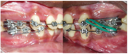
Fig 2: Canine retraction with class II elastics.
After canine retraction, 0.019 × 0.025Ë S.S. T loop archwires (Truforce, Ortho Technology, Inc., Tampa, USA) were placed in both arches (figure 3).Patient was recalled every 6 weeks for reactivation of loops and to check the progress.Settling was done with triangular elastics on upper and lower 0.016Ë S.S. wire, after which the appliance was debonded.
RESULTS
The first molar was successfully brought into a Class-I relationship. Treatment time was 2 years. Endof treatment, a proper overjet and overbite was establishedwith coincident midlines and no opening of mandibular plane angle. Mandibular plane was maintained with the high pull head gearand a profile change was noted at the end of treatment (figure 4, 5).
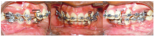
Fig 3: Anterior retraction with T loop stainless steel arch wire

Fig 4: Post treatment intraoral and extraoral photographs.

Figure 5- Profile comparison.
DISCUSSION
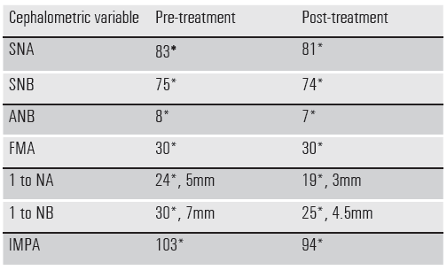
A paradigm shift is noted in the present era with more emphasis on soft tissue appearance rather than the ideal cephalometric values. Mihaliket al2 compared patients’ perceptions of outcomes with orthodontic and the surgical groups and found results with both the options as highly positive. The orthodontics-only (camouflage) patients reported fewer temporomandibular joint problems and had similar satisfaction asthe surgical patients, but patients who had surgery had significantly more positive dentofacial images.In our case, an acceptable soft tissue profile was achieved and patient was satisfied with the treatment results. More improvement in chin, nose and lip relation was observed by Saelenset al3 with extraction protocol than non-extraction method.In this case, all first premolars were extracted to improve the patient’s profile and achieve lip competence. Individual canine retraction was performed on the 0.019x0.025” stainless steel wire with elastic chain. The force was kept in the range of 50-100 grams to avoid canine tipping4. This was assisted with class II elastic wear which corrected the molar relationship by anchorage loss. Also, the placement of elastic chain made oral hygiene maintenance better. Thus, individual canine retraction and head gear in the upper arch reinforced upper anchorage. Anterior retraction was started on 0.019x0.025” stainless steel wire with a T loop. The advantage of using T loop is a better moment to force ratio5.The overall duration of treatment was 2 years with no adverse effects. Upper and lower incisors were retracted to achieve normaloverjet and overbite.
Table: Comparison of cephalometricvariables
CONCLUSION
Camouflage treatment of Class II divison 1 malocclusion yields stable esthetic results with proper diagnosis and sound mechanics.
NOTE: There is no conflict of interest. This patient was treated after her approval to go ahead with the orthodontic treatment. The patient was informed about all the advantages, disadvantages and limitations before the start of the treatment. Patient also consented to taking pre, post and ongoing stage photographs during the course of treatment.
REFERENCES
- Sood S. Treatment of Class II division 1 malocclusion in a non-growing patient. Virtual Journal of Orthodontics [serial online] 2010 October.
- Mihalik CA, Proffit WR, Philips C. Long-term follow-up of Class II adults treated with orthodontic camouflage: A comparison with orthognathic surgery outcomes. Am J OrthodDentofacialOrthop. Mar 2003; 123(3): 266-78.
- Saelens NA, De Smit AA. Therapeutic changes in extraction vs non extraction orthodontic treatment. Eur J Orthod 1998;20:225-36.
- Nattrass C, Ireland AJ, Sheriff M. An investigation into the placement of force delivery systems and the initial forces applied by clinicians during space closure. BJO1997;24:127-31.
- Patel AS, Ravindranath VK, Karandikar GR, Malik AS. Comparative assessment of four different designs of retraction loops made of beta titanium arch wire: a finite element study. Journal of contemporary dentistry; Jan-april 2014;4(1):6-9.
More references are available on request
Disclaimer from Guident
The views and opinions expressed in articles published in Guident are solely those of the authors and do not necessarily reflect the views of the editors, the editorial board or the publisher, Ivory India. Guident does not endorse any product, technique or treatment described. Clinical content is intended for qualified dental and medical professionals and is not a substitute for individual clinical judgement. Guident, its editors and the publisher accept no liability for any consequence arising from the use of information published in the journal.
How to cite this article
Dr. Siddharth Mehta, Dr. Anjali Mehta, Dr. Surendra Lodha Department of Orthodontics and Dentofacial Orthopedics. Camouflage Treatment of a Class II Divison 1 Malocclusion. Guident. 2015 Feb;8(3). Available from: https://guident.in/articles/orthodontics/camouflage-treatment-of-a-class-ii-divison-1-malocclusion.html


