Lateral Bridging Flap: A Minimally Invasive Solution for Root Coverage and Gingival Enhancement: A Case Report
Department of Periodontology, Yenepoya Dental College, Mangalore, India
ABSTRACT
Background: Gingival recession is a prevalent periodontal condition that can lead to root hypersensitivity, aesthetic concerns, and an increased risk of root caries. Surgical interventions for root coverage often require a second surgical site, leading to increased patient discomfort and morbidity. The lateral bridging flap technique offers an alternative by utilizing adjacent keratinized tissue to achieve root coverage without the need for additional grafting procedures. Objective: This case report evaluates the effectiveness of the lateral bridging flap technique in achieving complete root coverage and improving the width of attached gingiva in a patient with multiple recession defects. Case Presentation: A 37-year-old male patient presented with Miller’s Class II gingival recession involving teeth 23 and 24, with a recession depth of 4 mm and a narrow band of attached gingiva measuring 2 mm. A lateral bridging flap procedure was performed, which involved an arch-shaped incision, split-thickness flap elevation, and lateral mobilization of keratinized tissue to cover the exposed root surfaces. Periosteal releasing incisions ensured tension-free adaptation of the flap. Postoperative healing was uneventful, with complete root coverage and an increase in the width of attached gingiva by 2 mm at follow-up. Conclusion: The lateral bridging flap technique provides a predictable and minimally invasive solution for managing gingival recession while simultaneously increasing the width of attached gingiva. This approach eliminates the need for a second surgical site, reduces patient morbidity, and demonstrates stable long-term outcomes, making it a valuable addition to periodontal plastic surgical techniques.
INTRODUCTION
The recession of gingival tissue is defined as the apical migration of marginal gingiva in relation to cementoenamel junction. There can be multiple etiologic factors for this, generally, it can be related to mechanical factors (such as toothbrush trauma, traumatic occlusion), and localized plaque induced inflammatory lesions, and generalized forms of destructive periodontal disease.1 It is frequently associated with esthetic concerns, root hypersensitivity and risk of tooth loss.
Various mucogingival surgeries are available for root coverage, with the coronally advanced flap being a common choice for treating multiple teeth. However, these procedures often require an adequate width of attached gingiva, which may be insufficient in some cases. When this occurs, additional techniques to increase the width of attached gingiva are typically needed before proceeding with root coverage. This often means two separate surgeries, which can be a burdensome for both the patient and periodontist.2
This case report presents a combined approach, merging root coverage and gingival augmentation into a single procedure. Specifically, a coronally repositioned flap was used for root coverage, along with a vestibular deepening technique to increase the width of attached gingiva, addressing multiple issues simultaneously.
CASE REPORT
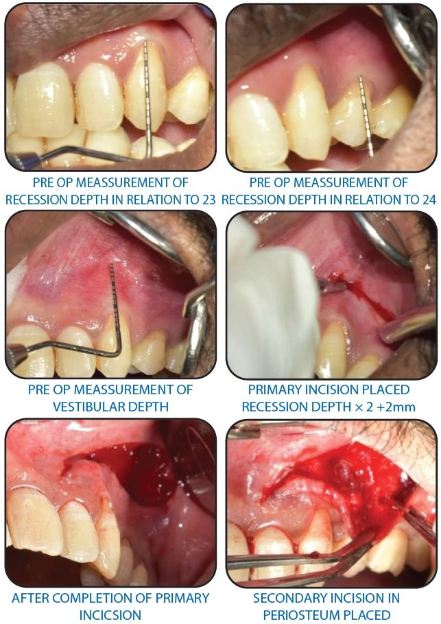
A 37-year-old male patient presented to the Department of Periodontology at Yenepoya Dental College with the chief complaint of gingival recession involving maxillary upper teeth. Clinical examination revealed a deficient zone of attached gingiva, as indicated by a positive tension test, and a shallow vestibule. Clinical parameters such as Probing Pocket Depth, Gingival Recession, width of attached gingiva and Clinical Attachment Level were recorded using a UNC-15 periodontal probe. The recession in relation to teeth numbers 23 and 24, was classified as Miller’s class II. measuring approximately 4 mm in relation to 23 and 24, with the width of the attached gingiva recorded at 2 mm. The probing pocket depth was 3 mm in relation to 23 and 24, with loss of attachment was 7 mm. The surgical procedure was explained in detail to the patient, and informed consent was obtained. Scaling and root planing were performed as part of the phase I therapy.
SURGICAL TECHNIQUE
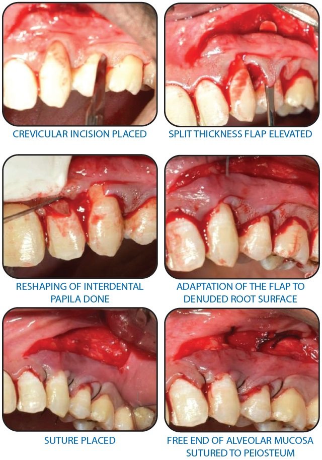
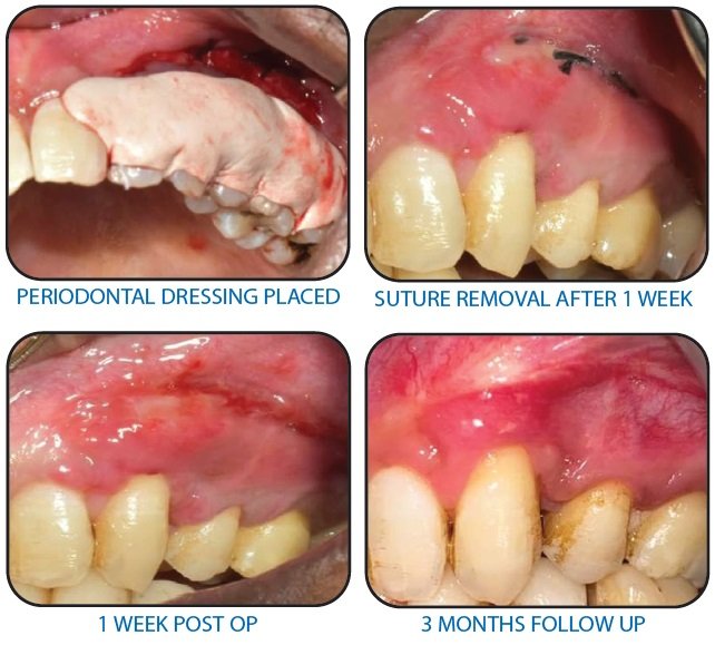
The first incision that was placed was arch-shaped, with a distance to the vestibule of approximately 2x GR + 2 mm. This is necessary in order to produce a sufficiently wide bridging flap ensuring a sufficient blood supply. Following this a crevicular incision was placed and full thickness “bridge” flap was elevated in the coronal direction. The third incision into the periosteum was placed at the base of flap. The whole bridge flap was then coronally repositioned to cover the denuded root surface finally the interdental papillae were reshaped in order to achieve good positioning of the reposed flap in these areas. Simple interrupted stabilization sutures were placed, and in the vestibule, epithelium was sutured with the periosteum. Periodontal dressing was placed after achieving complete haemostasis. Patient was discharged with post-surgical instructions and medications. Patient was recalled after 7 days for suture removal. Healing was found to be satisfactory with no postoperative complications. Complete root coverage was achieved. the gain in width of the attached gingiva was 2 mm in relation to 23 and 24. A PPD of 2 mm was present on same teeth.
ROOT COVERAGE WITH LATERAL BRIDGING FLAP
DISCUSSION
The current case report underscores the effectiveness of the lateral sliding bridge flap technique in managing multiple gingival recession defects through a single surgical procedure. This approach provides a patient-friendly alternative to more invasive methods, such as those involving secondary donor sites or grafting techniques. The technique’s ability to achieve both root coverage and vestibular deepening in a single step offers significant advantages in terms of reducing patient discomfort and recovery time.
The result from this report align with the earlier studies by Marggraf (1985) and Romanos et al. (1993), which consistently demonstrated substantial root coverage and stability over extended follow-up periods. A key advantage of the Lateral sliding flap is the simultaneous extension of the vestibule, which helps to relieve tension from apical frenal attachments, reducing the likelihood of recurrent recession. This dual benefit reinforces the long-term viability of the technique, even in patients with limited attached gingiva.
A critical aspect of the success of the technique lies in the keratinization of the transposed alveolar mucosa. As supported by studies like Bokan (1997), the remnants of attached gingiva, de-epithelialized tissue, and contributions from the periodontal ligament play a crucial role in promoting keratinization, thereby ensuring the durability of the procedure.
However, while this technique offers several advantages, careful patient selection remains vital. Cases with inadequate bone support or thin biotypes may require additional consideration or adjunctive treatments. Proper surgical planning, precise execution, and postoperative care are paramount to ensuring the long-term success and functional stability of this procedure.
CONCLUSION
The lateral sliding bridge flap (LSBF) technique is an effective and minimally invasive method for addressing gingival recession, offering predictable root coverage and increased keratinized tissue without the need for secondary donor sites. Although most effective for Miller’s Class I and II defects, the technique’s versatility makes it a valuable option for periodontal plastic surgery.
REFERENCES
- Wennstro¨m J, Pini Prato G. Mucogingival therapy: Periodontal plastic surgery. In: Lindhe J, Karring T, Lang N, eds. Clinical Periodontology and Implant Dentistry, 4th ed. Copenhagen: Munksgaard; 2003:576-649.
- Cairo F, Pagliaro U, Buti J, Baccini M, Graziani F, Tonelli P, et al. Root coverage procedures improve patient aesthetics. A systematic review and Bayesian network meta-analysis. J Clin Periodontol. 2016;43(11):965-75
- Cheng YF, Chen JW, Lin SJ, Lu HK. Is coronally positioned flap procedure adjunct with enamel matrix derivative or root conditioning a relevant predictor for achieving root coverage? A systemic review. J Periodontal Res 2007;42: 474-85.
- Marggraf E. A direct technique with a double lateral bridging flap for coverage of denuded root surface and gingival extension: Clinical evaluation after 2 years. J Clin Periodontol. 1985;12(1):69-76.
- Romanos GE, Bernimoulin JP, Marggraf E. The double lateral bridging flap for coverage of denuded root surface: Longitudinal study and clinical evaluation after 5 to 8 years. J Periodontol. 1993;64(7):683-8.
- Bokan I. Potential of gingival connective tissue to induce keratinization of an alveolar mucosal flap: A long- term histologic and clinical assessment. Quintessence Int. 1997;28(10):731-6.
- Vijayalakshmi R, Uma S, Saravanakumar R, Ramakrishnan T, Emmadi P, Anitha V. Double lateral sliding bridge flap for the coverage of denuded roots: Two case reports. Perio - Periodontal Practice Today. 2008;5(1):29-33.
- Wennström JL, Pini Prato GP. Mucogingival therapy – periodontal plastic surgery. In: Lindhe J, Karring T, Lang NP, editors. Clinical Periodontology and Implant Dentistry. 4th ed. Oxford: Blackwell Munksgaard; 2003. p. 576-649.
- Greenwell H, Fiorellini J, Giannobile W, Offenbacher S, Salkin L, Townsend C, et al. Oral reconstruction and corrective considerations in periodontal therapy. J Periodontol. 2005;76(9):1588-600.
- Gupta V, Bains VK, Mohan R, Bains R. Bridge flap technique as a single-step solution to mucogingival problems: A case series. Contemp Clin Dent. 2011;2(2):110-4.
- Sato N. Periodontal surgery: A clinical atlas. London: Quintessence Publishing Co. Ltd; 2000. p. 82-125.
- Wennström JL. Lack of association between width of attached gingiva and development of soft tissue recession: A 5-year longitudinal study. J Clin Periodontol. 1987;14(3):181-4.
Disclaimer from Guident
The views and opinions expressed in articles published in Guident are solely those of the authors and do not necessarily reflect the views of the editors, the editorial board or the publisher, Ivory India. Guident does not endorse any product, technique or treatment described. Clinical content is intended for qualified dental and medical professionals and is not a substitute for individual clinical judgement. Guident, its editors and the publisher accept no liability for any consequence arising from the use of information published in the journal.
How to cite this article
Dr. Belim Wasim Siraj, Dr. Rajesh K.S. Lateral Bridging Flap: A Minimally Invasive Solution for Root Coverage and Gingival Enhancement: A Case Report. Guident. 2025 Feb;18(3). Available from: https://guident.in/articles/periodontics/lateral-bridging-flap-a-minimally-invasive-solution-for-root-coverage-and-gingival-enhancement-a-case-report.html