Pyramidal Bilateral Deciduous First Molar: A Case Report of a Rare Morphology
Dept. of Pedodontics and Preventive Dentistry, Yogta, Dental College and Hospital, KHED Maharashtra, India.
Abstract
Morphological dental anomalies with respect to roots may involve a single tooth, or group of teeth or the entire dentition. Single tapering root forms may be found in any molar tooth and are frequent in permanent molars. Their occurrence in the primary dentition is extremely rare. The objective of this paper is to present a case with bilateral primary mandibular molars having a single root.
Key words: Decidous first molar, Dental anomalies, Primary teeth, Tooth Root
INTRODUCTION
Morphological dental anomalies with respect to roots may involve a single tooth, a group of teeth or the entire dentition1. Single tapering root forms may be found in any molar tooth, but are seen most frequently in second and third permanent molars. But the occurrence of single rooted primary mandibular first molars is extremely rare. Ackerman2 et al in 1973 was the first one to report a case of single rooted primary molar in a 10-year-old child. `Pyramidal, is used to denote teeth with complete fusion of the roots with a solitary enlarged root canal3. The other terms used are ‘conical’ or ‘fused’. The term conical would appear to be self-explanatory. The term fused roots refers to teeth with roots connected but still maintaining at least two distinct canals2. Racial variation exists, and females are more frequently affected than males. The cause for this female predilection is unknown3. The objective of this paper is to present a case with bilateral primary mandibular molars having single root.
CASE REPORT
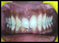
An eight-year old female patient reported to the Department of Pedodontics and Preventive Dentistry, K.D. Dental College, Mathura, with a complaint of pain in relation to the lower back tooth region on both the sides. Medical history and past dental history were not relevant. On clinical examination, the patient was in mixed dentition [Figure 1] and the following teeth were present:

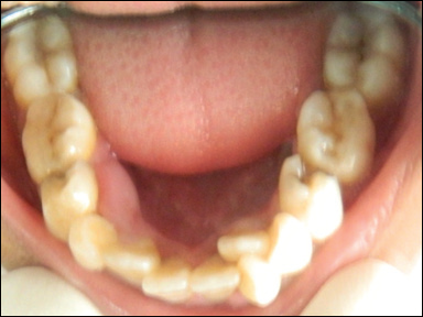
Fig 1: Maxillary and Mandibular occlusal view
16 55 54 53 12 11 21 22 63 64 65 26
46 85 84 83 42 41 31 32 72 73 74 75 36
Deep proximal caries were seen in relation to both primary first mandibular molars, which were indicated for further investigation. Intraoral periapical radiographs were obtained in relation to 74 and 84 using bisecting angle technique. Radiographs revealed caries involving the pulp with both the teeth, but while evaluating the root morphology, both the molars had a single root [Figure 2]. The treatment plan included endodontic therapy for 74 and 84 followed by stainless steel crowns but due to poor socioeconomic status of the patient the treatment was deferred to a later date.
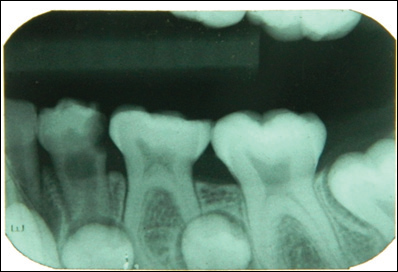
Fig 2(a): IOPA of 74
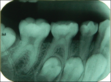
Fig 2(b) IOPA of 84
DISCUSSION
Hertwig,s epithelial root sheath (HERS) cells play a pivotal role during root formation of the tooth and are able to form cementum-like tissue. Hertwig’s epithelial root sheath, a double layer of cells is formed by the epithelial cells of the inner and outer enamel epithelium, which proliferate from the cervical loop of the enamel organ. Prior to the beginning of root formation, the root sheath forms the epithelial diaphragm, which encloses the primary apical foramen. Differential growth of this epithelial diaphragm in multirooted teeth causes the division of the root trunk into two or three roots4.
Disturbances in morphodifferentiation may affect the form and size of the tooth without impairing the function of ameloblasts or odontoblasts. New parts may be differentiated i.e. supernumerary cusps or roots, or suppression of parts may occur i.e loss of cusps or roots5. As seen in this case also, both the decayed mandibular primary molars had single root instead of two roots. Literature regarding etiology of single rooted molars reveals failure in invagination of HERS, other environmental factors such as chemotherapy, radiation therapy and trauma may also affect the morphology of the developing roots4,6.
For the endodontic treatment of such single rooted primary teeth, many factors should be considered. In search of an extra canal, we tend to perforate the tooth by excessive tooth removal. These iatrogenic errors can be minimized if the clinician has the knowledge of the general location and dimension of the pulp chamber, which can be well appreciated with the pre-operative radiograph. It is still unknown whether such anomalies in primary dentition will affect the underlying permanent dentition or not, but the documentation by clinicians will surely help to assess its prevalence.
CONCLUSION
Single rooted permanent molars have often been reported in the literature. However, reports on single rooted primary mandibular first molar are very few. The variation in root or root canal morphology is challenging for diagnosis and successful endodontic therapy. Knowledge of unknown variation like the case discussed is essential. Thorough knowledge of root canal morphology and anatomical variations of primary teeth can help a pediatric dentist in successful management.
REFERENCES
- Robbins IM, Keene HJ. Multiple morphologic dental anomalies: report of a case. Oral Surg Oral Med Oral Path Oral Radiol Endod 1964;17:683-90.
- Ackerman JL, Ackerman AL, Ackerman AB. Taurodont, pyramidal and fused molar roots associated with other anomalies in kindred. Am J Phys Anthrop 1973;38:681-94.
- Nguyen AM, Tiffee JC, Arnold RM. Pyramidal molar roots and canine-like dental morphologic features in multiple family members: a case report. Oral Surg Oral Med Oral Pathol Oral Radiol Endod 1996;82(4):411- 416.
- Nanci A. Ten Cate’s oral histology: Development, structure and function. 7th ed, Mosby publications, New Delhi 2008;79-107.
- Bhaskar SN. Orban’s. Oral Histology and embryology. 11th ed, Elsevier publications, New Delhi 2001;28-48.
- Minicucci EM, Lopes LF, Crocci AJ. Dental abnormalities in children after chemotherapy treatment for acute lymphoid leukemia. Leuk Res 2003;27:45-50.
Disclaimer from Guident
The views and opinions expressed in articles published in Guident are solely those of the authors and do not necessarily reflect the views of the editors, the editorial board or the publisher, Ivory India. Guident does not endorse any product, technique or treatment described. Clinical content is intended for qualified dental and medical professionals and is not a substitute for individual clinical judgement. Guident, its editors and the publisher accept no liability for any consequence arising from the use of information published in the journal.
How to cite this article
Dr.Priyanka Razdan, Dr.Sridhar shetty. Pyramidal Bilateral Deciduous First Molar: A Case Report of a Rare Morphology. Guident. 2016 Aug;9(9). Available from: https://guident.in/articles/pedodontics/pyramidal-bilateral-deciduous-first-molar-a-case-report-of-a-rare-morphology.html