Periapical Cyst Management in a Fourteen Year Old Adolescent
K.D Dental College and Hospital, Mathura, India
ABSTRACT
Periapical cysts are most common odontogenic cyst located at the apex of carious or traumatized tooth. It arises from epithelial remnants stimulated to proliferate by an inflammatory process originating from pulpal necrosis of a non-vital tooth. Radiographically, the classical description of the lesion is a round or oval, well-circumscribed radiolucent image involving the apex of single or multiple teeth. Cyst can be managed surgically and or non-surgically depending on site and size of cyst. This case report deals with a fourteen year old adolescent with periapical cyst associated with maxillary right central and lateral incisors that was successfully managed with single sitting root canal therapy (RCT) along with surgical enucleation of the cyst.
Keywords: Periapical cyst, root canal treatment, cyst enucleation
INTRODUCTION
Cyst is defined as a pathologic cavity lined by epithelium usually containing fluid or semi-solid material and sometimes air. Odontogenic cysts are derived from the epithelium associated with the development of dental apparatus and can arise from 1) Tooth germ 2) Epithelial rests of malassez 3) Reduced enamel epithelium of a tooth crown 4) Remnants of dental lamina or 5) possibly the basal layer of oral epithelium1. A periapical or radicular cyst arises from epithelial cell rests of Malassez in the periodontal ligament as a consequence of inflammation following pulpal necrosis of a non-vital tooth. This condition is usually asymptomatic but can result in a slow-growth tumefaction in the affected region.2 Many radicular cysts are symptomless and are discovered with periapical radiographs of teeth with non-vital pulps. The treatment of the cysts can be either non-surgical management or surgical management being either marsupialization or enucleation depending on the size and localization of the lesion, the bone integrity of the cystic wall and its proximity to vital structures.3 Shear M and Geward GR also reported that periapical cyst has high incidence in the maxillary anterior region, presumably as a result of trauma.4 This case report presents a case of surgical management of a periapical cyst associated with non-vital permanent right maxillary central and lateral incisors in a 14 year old.
CASE REPORT
A 14 year old male reported to the Department of Pedodontics and Preventive Dentistry, K. D. Dental College and Hospital, Mathura, with a complaint of pus discharge from upper front tooth region and bad smell from the mouth for past two year. Past history revealed that he had trauma two year before in maxillary anterior region for which no treatment was sought. Post trauma he had dull intermittent pain in relation to the upper anteriors which subsided without medication. During examination, he was found to be in good general and physical health. On intraoral examination, Ellis class IV fracture was present with maxillary right central incisor and vitality test gave a negative response in relation to maxillary right central and lateral incisor (figure 1). An intraoral periapical radiograph of maxillary central and lateral incisor revealed a well-circumscribed periapical radiolucency of about 3x2 cm in dimension, involving both the roots apices and extending from mesial aspect of root apex of right canine to left central incisor, suggesting a cystic lesion (figure 2). Based on the history, clinical examination and radiographic examination, a provisional diagnosis of Ellis class IV fracture of maxillary right central incisor associated with cyst involving root apex of both right central and lateral incisor was made but the final call for type of cyst was left to histopathologic report. Treatment plan comprised of RCT with right central and lateral incisor and cyst enucleation. With the consent of the parents, the endodontic therapy was carried out followed by cyst enucleation procedure. Under all aseptic conditions local anesthesia was administered and crevicular incision was given on labial aspect extending from right canine to left central incisor to reflect full thickness flap that exposed a wide labial bone defect. Cyst lining was excavated along with its content followed by thorough curettage (figure 3 and 4). Flap closure was done using 3-0 silk suture. Specimen was sent for histopathological examination which confirmed periapical cyst. Considering patient’s young age and good health, bone graft was not used. After a week, patient was recalled for suture removal and a periapical radiograph was taken (figure 5). Esthetic restoration with composite was done with right central and lateral incisor after a week (figure 6). This case is a good example of a large lesion been treated with conventional RCT and cyst enucleation without the use of materials which enhance bone regeneration like Platelet Rich Fibrin (PRF) G-bone (Synthetic Granules and blocks made of Multiphasic Calcium Hydroxyapatite in low crystalline form). Intraoral periapical radiograph for successive two years postoperatively was taken which revealed regression of the cystic lesion and formation of bony trabaculae (figure 7, 8 and 9).
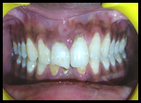
Figure 1: Front view of fractured right maxillary central incisor

Figure 2: IOPA radiograph showing cyst in relation to maxillary central and lateral incisor
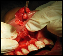
Figure 3: Enaculation of bony defect and cystic lesion
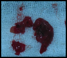
Figure 4: Excavated cystic lining

Figure 5: IOPA radiograph after one week showing progressive healing
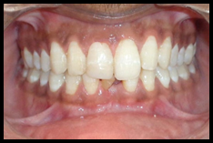
Figure 6: Esthetic restoration done in maxillary right central incisor
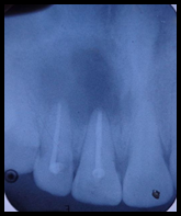
Figure 7: IOPA radiograph after 6 months showing progressive healing

Figure 8: IOPA radiograph after 12 months showing progressive healing
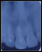
Figure 9: IOPA radiograph after 24 months showing healthy bony trabaculae
DISCUSSION
A cyst is a pathologic closed cavity lined by an epithelium that can occur anywhere in the body and vary in size and contents such as blood, seroma, semisolid or gaseous contents, but is not normally filled with pus, unless it is infected. Cysts constitute about 17% of all the tissue specimens submitted to oral pathology biopsy services. The periapical cyst is the most common odontogenic cyst (52.3–70.7% of all odontogenic cysts) followed by the dentigerous cyst (16.6–21.3% of all odontogenic cysts) and odontogenic keratocyst (5.4–17.4% of all odontogenic cysts).5 Periapical cysts are odontogenic cysts that are derived from the inflammatory activation of epithelial root sheath residues (cell rests of Malassez). They are inflammatory in nature and usually arise within a periapical granuloma relating to stimulation resulting from a necrotic tooth.6-8 They are most commonly associated with permanent teeth and are rare in the primary teeth.9 The choice of treatment may be determined by factors such as the extension of the lesion, relation with noble structures, origin, and clinical characteristics of the lesion, and co-operation and systemic condition of the patient. The treatment of these cysts is still under discussion and many professionals opt for a conservative treatment by means of endodontic therapy alone. However, in large lesions the endodontic treatment alone is not efficient and is associated with decompression, marsupialization or enucleation.10-12 In this regard, it is suggested that the treatment of periapical cyst should be defined according to the clinical and radiographic evaluations of each case.
CONCLUSION
The clinical case reported in this article was managed successfully by endodontic therapy with emphasis on thorough debridment, disinfection and three dimensional obturation of the root canal system which was followed by surgical enucleation of the cyst. Two year follow up and radiographs showed healing of lesion with bony trabaculae formation.
REFERENCES
- Shafer’s textbook of oral pathology, 6th edition, Pub Elsevier; pp: 487-490
- Azad A, Chourasia HR, Singh D, Sharma I, Azad A, Pahlajani V. Management of a Large Periapical Cyst: A Case Report. People’s J Scient Res 2014; 7(1): 47-50.
- Bonder L. Cystic lesions of the jaws in children. Int J Pediat Otorhinolaryngol 2002; 62: 25–29.
- Shear M. Cysts of the oral regions. 3rd ed. Boston: Wright; 1992. Radicular and residual cysts; pp. 136–62.
- Verghese GM, Thomas G, Kuttappa MA, Govind GK Management of a large periapical cyst (apical matrix & surgical complications) - A case report. Endodontology; 84-89.
- Ten Cate AR. The epithelial cell rests of Malassez and the genesis of the dental cyst. Oral Surg Oral Med Oral Pathol 1972; 34: 956–964.
- Main DM. Epithelial jaw cysts: 10 years of the WHO classification. J Oral Pathol 1985;14:1–7.
- Nair PN. New perspectives on radicular cysts: Do they heal? Int Endod J 1998; 31: 155–160.
- Lustmann J, Shear M. Radicular cysts arising from deciduous teeth: Review of the literature and report of 23 cases. Int J Oral Surg 1985;14:153–161.
- Michael MH, Gary LL. Conservative treatment of persistent periradicular lesions using aspiration and irrigation. J Endod 1990;16:182–186.
- Rees JS. Conservative management of a large maxillary cyst. Int Endod J 1997;30:64–7
- Danin J, Linder LE, Lundqvist G, Ohlsson L, Ramskold LO, Stromberg T. Outcomes of periradicular surgery in cases with apical pathosis and untreated canals. Oral Surg Oral Med Oral Pathol 1999; 87: 227–232.
Disclaimer from Guident
The views and opinions expressed in articles published in Guident are solely those of the authors and do not necessarily reflect the views of the editors, the editorial board or the publisher, Ivory India. Guident does not endorse any product, technique or treatment described. Clinical content is intended for qualified dental and medical professionals and is not a substitute for individual clinical judgement. Guident, its editors and the publisher accept no liability for any consequence arising from the use of information published in the journal.
How to cite this article
Dr. Priti Yadava, Dr. Chanchal Singh Department of Pedodontics & Preventive Dentistry. Periapical Cyst Management in a Fourteen Year Old Adolescent. Guident. 2016 Jul;9(8). Available from: https://guident.in/articles/pedodontics/periapical-cyst-management-in-a-fourteen-year-old-adolescent.html