Computer Applications in Orthodontics
Department of Orthodontics & Dentofacial Orthopedics, A.B Shetty Memorial Institute of Dental Science Mangalore Karnatka, India
Abstract
Advances in computer technology continue at a remarkable rate; breakthroughs in computing power, speed, graphics capabilities and storage capacity accelerate, while the ratio of hardware cost to performance declines, often precipitously. Great efforts are being made to make computers easier to use.Continued improvement in the quality of orthodontic services will require implementation of new diagnostic and treatment approaches. New technologies will provide improved precision, accuracy, and perspective. This will provide the clinician with the means to optimize the way treatment is planned through computer modeling, testing, and simulations. In this way, treatment results can be predicted with greater accuracy, thereby reducing unfavorable outcomes and increasing efficiency. The usage of computers as applicable to the orthodontist are briefly discussed here.
INTRODUCTION
Computer is a machine that performs calculations and processes information with astonishing speed and precision. A computer can handle vast amounts of information and solve complicated problems. They enable designers and artists to see things that have never been seen before.
The value of computers lies in their ability to perform certain tasks extremely quickly and accurately. These tasks include
- Solving numerical problems.
- Storing and retrieving information.
- Creating and displaying documents and pictures.
Computers are used to store large quantities of information. Word processing is one of the most important and widespread uses of computers. the other area which is beneficial to laypersons is the ease of accounting, which has been recently included in many of the patient data management software’s.
Computer graphics - the use of computers to make pictures is another fascinating and fast growing area. Computer programs that perform computer-aided design (CAD) are important in many field, particularly engineering and architecture. Recently its importance in medical as well as dental fields has been proved.The usage of computers as applicable to the orthodontist are briefly discussed here.
APPLICATIONS IN RESEARCH
FINITE ELEMENT ANALYSIS (F E A):
Finite Element Analysis is a numerical method of structure analysis based on principle of dividing an infinite structure into a finite number of small elements connects each other at the corner points or nodes.It is a means of discretizing a continuous structure into sub-domains called Finite Elements.
Essentially an attempt at simulating a physical object and analysingit’sbehaviour when subjected to various circumstances.
This is a well-sophisticated engineering tool, which has been used extensively in design optimization and structural analysis. It is one of the most significant developments in the history of computational methods.
This method was first introduced to the dental arena in the 1970’s and growth model was documented by MOSS in 1980.
CAD / CAM
This refers to Computer Aided Designing/ Computer Aided Machining. This field of engineering has been widely used for dental applications also and orthodontic purposes for precision attachments.
- Occlusal splints used for inter maxillary positioning after orthognathic surgery requires accurate positioning, stabilization and control of mobilized segments.
Conventional fabrication is a complicated process. For bi-maxillary surgery it is
more difficult.
Recently (CAD) computer aided designing program has developed to assist in planning maxillo-facial and cranio-facial surgeries.
- Bone implants, dental restoration mere fabricated by entering CAD data into a CAM machine (computer aided manufacturing machine).
THE BENDING ART SYSTEM (BAS)
BAS – It is the first ever developed robotic CAD / CAM system for the fabrication of customized orthodontic arch wires.
- Prof. Helge Fischer-Brandies invented this in 1984 and his co-worker Dr. Wolfgang Orthuber, together with an engineering company developed this hardware and software.
- 1st prototype of BAS was manufactured in 1993.
- It is used for fabrication of both labial and lingual orthodontic wires.
DESIGN AND TESTING OF ORTHODONTIC LOOPS USING A COMPUTER PROGRAM
Demetrios J. Halazonetis developed this computer program to allow quick and easy assessment of the force characteristics of orthodontic loops. It helps us to know how the loops work and to design better loops.
The program was developed with Borland Delphi for Windows. With this program, you can simulate almost any orthodontic loop that connected between two brackets. Tools available with this software are Reposition brackets, spool of wire, cutter and plier.
GRAPHIC APPLICATIONS
IMAGE ARCHIVING APPLICATIONS
Image archiving applications are designed for the purpose of acquisition, viewing, storage, printing and communication of digital representations of orthodontic case records including facial and intra oral image, digital photographs and images of study models. The principle advantage of image archiving over conventional methods is the ease with which records can be stored and retrieved.
DIGITAL IMAGING
The widespread use of digital imaging for medical application started in early 1970’s. It includes computed tomography, ultrasound, nuclear medicine, magnetic resources and digital subtraction angiography. It applies to conventional radiography with the benefits of image storage, transmissions and processing.
Image Enhancement
There is potential for improving the diagnostic quality of digital images by enhancing the images using various algorithms. It can be enhanced using algorithm that mathematically manipulates the gray-level values of pixels. Images of poor quality due to incorrect exposure or blurring could be manipulated and re-formatted, there by avoiding further exposure.
Image enhancement divides into three
- Contrast improvement
- Smoothing
- Edge enhancement
DIGITAL PHOTOGRAPHY
It is the preferred method of acquiring facial and intra-oral images. The major difference from conventional photography is that the camera stores all its images onto a computer chip or a storage medium in a digital format.
Advantages of digital photography
- No film or processing is required.
- Instant viewing of the pictures and mistakes can be rectified
- immediately.
- Editing is possible
- Immediate image review, allows the operator to assess the
- lighting, focus and patient positioning
- Saves money in long run by not buying rolls.
- No chemical storage for developing.
VIDEO CAPTURES
It is used less frequently for facial and intra oral image acquisition. Video capture system connects a video camera to a computer through a frame grabber. Frame grabber is an interface board in a computer that converts the analog video signal from a video camera into a 24- bit RGB digital image. The frame grabber grabs an image frame from the continuous video signal.
VIDEO IMAGING
With continued improvements in orthodontic and surgical techniques, there is a greater demand for the orthodontist to communicate with the patient and other professionals about the projected treatment goals and outcome. Experienced clinician having and good mental image, about the treatment outcome, but the patient’s ability to visualize or interpret and thus accept the plan have been limited, clear communication of treatment goals potential options of treatment is an important aspect of today’s concepts of informed consent and clinical practice.
RADIOGRAPHIC IMAGE ACQUISITION
DENOPTIX DIGITAL RADIOGRAPHIC SYSTEM
With all the digital technology available today, the orthodontists still rely on silver halide film for radiography. This system (Denoptix system) is a digital radiographic technology, which completely eliminates silver halide film.
Digital intra oral radiography is possible since Trophy introduced charge couple device (CCD) sensors. But CCD cannot store data so it is connected to a computer and since CCD’s are expensive which made a limitation for its use.
CEPHALOMETRIC APPLICATIONS:
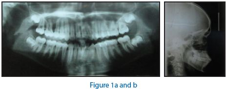
Cephlometric applications offer treatment-planning manipulations such as growth estimation, orthopedic intervention, orthodontic tooth movement and orthognathic surgery.
Currently there are 2 methods of radiographic digitization
- Direct computer digitization of radiograph
- Indirect computer digitization of radiograph
Direct digitization of radiograph on a computer:
Digitization is a process by which analog information is converted into digital form. This is done with the help of a digitizing tablet or a mouse to digitize landmarks from a digitalcephalogram displayed on CRT monitor or flat panel display.
A digitizing tablet or digitizer is an inexpensive, accurate tool for digitizing cephalograms.
Advantages of Cephalometric Applications:
Cephalometric analyses and treatment plans are useful components of an orthodontic case diagnosis and treatment planning. Currently, no evidence supports the notion that an analysis or treatment plan generated by a cephalometric application is more reliable than one arrived at through careful manual methods. The primary advantage of cephalometric applications over manual methods lies in the speed with which these determinations can be made.
Photocephalometric Treatment Visualization:
The preferred approach to treatment visualization involves software that extends the algorithms used in cephalometric treatment planning software to embrace lateral facial images. These photocephalometric applications work by linking digitized lateral cephalograms to lateral facial bitmapped images for automatic facial transformation.
Moustafa et al – cephalometric head films with a video camera and a frame grabber and described soft wax for detection of soft-tissue profile contours and automated hard and soft tissue landmark location and is reported to be reliable.
Indirect computer digitization of radiograph:
For this a video camera or scanner captures an image of the radiograph and stores it in the computer. The use of digital radiography is another method of cephalometric radiograph entry into the computer and analysis.
This stored image displayed on monitor and is indirectly digitized via a mouse or an on screen electronic pen.
DIGICEPH:
It is one of the advanced systems for computerized cephalometric digitization, automatic analysis and plot superimposition. It is capable of producing 13 cephalometric analyses. The system is developed by the center for Bio medical Engineering IIT New Delhi and Dept. of Dental surgery at the AIIMS – New Delhi.
DIGIGRAPH
Cephalometric analysis conventionally requires radiographic exposure, which may not be compatible with growing concern over radiation hazards. Recently Dolphin imaging system introduced a non-radiographic system “DIGIGRAPH” workstation that may be an alternative to conventional Radiography.
RMO’s Jiffy Orthodontic Evaluation:
Rocky Mountain Orthodontics (RMO) was the first to provide the dental profession in the late 1960s with a computer-aided cephalometric diagnosis. RMO Diagnostic Services Department continues to provide various diagnostic services, including computerized cephalometric diagnosis and forecast of growth and treatment.
PorDios
PorDios (Purpose On Request Digitizer Imput Output System) is a cephalometric IBM- compatible system whose development is aimed to provide orthodontists withan user-friendly programme. This programme can be easily changed by the user in order to satisfy individual preferences and needs.
PorDios works with a digitizer in the standard way and also enables the use of a video or scanner as means of digitization of X-rays. It uses well-known cephalometric analyses, including Bjork, Burstone, Coben, Downs, Frontal McNamara, Profile, Ricketts, Steiner, and Tweed and it has the capability to produce occlusograms from photocopies of dental casts. The user of PorDios can alter the existing programme analyses or can develop his own.
Dentofacial Planner
Dentofacial Planner is a computer-aided diagnostic and treatment planning software system for orthodontics and orthognathic surgery.
The programme enables the user to use one of the pre-programmed analyses, including Steiner, McNamara, COGS, Downs, Rick10, Rick32, Grummons, Harvold, Legan, and Jarabak. Furthermore, the orthodontics subsystem allows the user to do superimpositions, estimate facial growth, to simulate the skeletal and soft tissue effects of orthopaedic appliances, and to simulate orthodontic tooth movements.
Both the orthodontics and surgery subsystems allow the operator to manipulate a variety of skeletal regions interactively. The surgery subsystem allows the user to estimate the skeletal and soft tissue effects of orthognathic surgery, creating a so-called Surgical Treatment Objective .
Quick Ceph Image
Quick Ceph Image is a programme designed especially for high-end Macintosh Computers that does computerized cephalometrics and mapping.
Thirteen different analyses can be performed, including Ricketts, Steiner, Jarabak, McNamara, Downs, Soft Tissue, Iowa, Roth, Burstone, Sassouni, Frontal and SMV and model analyses of arch length and Bolton discrepancies. Four of these analyses are reprogrammable in order to provide customized analysis.
The system also provides an effective method for accumulating and storing patient picture records.
COMPUTER AIDED SPACE ANALYSIS:
It was introduced by Chen-Hsing-Yen.
Space analysis is a critical factor in taking the decision of extraction.
Space analysis can include:
- Bolton ratios
- Tooth size relationships of upper and lower arches
- Overjet and overbite
- Posterior intercuspation
These calculations are not difficult but it is time consuming. A simple computer program can simplify and speed up the process.
INVISALIGN TECHNOLOGY
Adult patients are often averse to wearing traditional fixed appliances with wires, bands and brackets. Invisaligin system makes it possible for orthodontists to offer adult patients requiring full mouth orthodontic treatment an esthetically agreeable solution using computer assisted technology that produces a series of plastic overlays.
Clear plastic overlay appliances take variety of form: Retainers, night guards, TMJ splints and bleaching trays. This needs dental technician to manually do and are both costly and vulnerable to human error and inaccuracies.
Recent developments in technology made it possible with invisalign system with the help of single impression.
MAGNETIC RESONANCE IMAGING
- Magnetic Resonance Imaging (MRI) a very recent addition to the medical diagnostic armamentarium, Sir Peter Mansfield and Paul Lauterbur
- It uses nonionizing radiation from the radiofrequency band of the electromagnetic spectrum.
- Nuclear magnetism is caused by spinning motion of specific nuclei present in biological tissues, these nuclei are called as MR active nuclei .
- MR images are generated from the protons of the hydrogen nuclei, since these nuclei are the major atomic constituent of the both the water molecule whichmakes up 75% of the human body, and of tissues which are altered by many disease states.
INDICATIONS
- TMJ- MRI has revolutionized the understanding of the diseases of the TMJ. The objective of MR imaging of the TMJ is to determine the relationship between the TMJ disc and the temporal and mandibular components of the TMJ.
- Congenital disorders
- Cleft lip and palate. A cleft may be associated with other congenital abnormalities or may be a feature of a severe syndrome. These anomalies can be studied with MRI.
- Tonsillitis and Adenoiditis
- Cysts and Infections
- Benign and malignant tumors.
COMPUTED TOMOGRAPHY
Computed Tomography (CT) has been one of the most spectacular advances in medicine in recent years. The technique was invented by Sir Godfrey Hounsfield who was awarded a Nobel prize in 1979.
Computed Tomographic image is a display of the anatomy of a thin slice of the body developed from multiple x-ray absorption measurements made around the body’s periphery.
INDICATIONS
- Localization of impacted canines.
- Evaluation of condylar position in glenoid fossa in skeletal Class III malocclusions.
- Measurement of joint spaces of TMJ.
- Resorption of roots of maxillary canines after ectopic eruption of maxillary canines.
- Assessment of maxillary movements induced by rapid maxillary expansion.
- Assessment of nasopharyngeal airway and cleft palate deformities.
- Evaluation of ossification and closure of various structures in craniofacial complex.
MISCELLANEOUS APPLICATIONS
Apart from the above mentioned applications, computers have come in a big way in being labelled as the Data engines, as they can not only be used to retrieve information stored in the computer itself but also form any part of the world, which has been made possible by means of the World Wide Web ( WWW).
Internet in Dentistry
The Internet is a global information resource, which changed the way human society communicates, interacts and functions. It brought changes to society generally and dentistry specifically. It adds a new, powerful resource for communicating and accessing current and relevant information. The availability of the Internet has opened up a new horizon of scientific co-operation and collaboration at a global level. Various Internet tools allow us to interact with one another unrestrained by geographical boundaries, time and distance.
Various tools in Internet:
- News groups
- FTP sites
- www.
CONCLUSION
Computers have become an indispensable tool for the orthodontist and have wide spread applications ranging from simple functions such as correspondence to specialized applications in research.
Several novel technologies have developed recently. These new technology will provide benefits in the areas of diagnosis; treatment planning, simulation and therapeutics; development of future technologies and research-treatment outcomes.
REFERENCES
- It’s time to Discuss Bytes – Michel N. Jacoby JCO – August 1977 : 546-550
- Computer-aided space analysis – Chen-Hsing Yen JCO – April 1991 : 236-238
- Biomechanics – computer based mechanotherapy – BirteMelson& Giorgio Fiorelli JCO – March 1994 : 136-141
- A computerized tooth-width Analysis – Christopher T. C. Ho, Terrence J. Freer JCO – September 1991 : 498-503
- The Denoptix Digital Radiographic System – Jon J. Menig JCO – July 1999 : 407-40
- Digital imaging of cephalometric radiography Part I: Advantages & limitations of digital imaging – D. B. Forsyth, W. C. Show, S. Richmond Angle Orthod – 1996 ;Vol 66 : 37-42
- Digital imaging of cephalometric radiographs, Part II: Image quality - D. B. Forsyth, W. C. Show, S. Richmond Angle Orthod – 1996 ;Vol 66 : 43-50
- The Digigraph workstation, Part I: Basic concepts – Spiro J. Chacons JCO – June 1990 : 360-367
- The Digigraph workstation, Part II: Clinical management – Richard G. Alexander JCO – July 1990 : 402-407
- The Digigraph workstation, Part III: Accuracy of cephalometric analysis – Spiro J. Chaconas JCO – August 1990 : 467-471
- Automatic computerized radiographic identification of cephalometric landmarks – D. J. Rudolph, P. M. Sinclair, J. M. Coggins AJO – Feb 1998 ; 113 : 173-179
- Design & test orthodontic loops using computer (Ortho Bytes) – Demetrias J. Halazonetis AJO – Mrch1997 : 346-348
- Video imaging for planning & counseling in Orthognathic surgery – Davi M. Sarver, Mark W. Johnston, Victor J. Matukas JOMS – 1998 ; 46 : 939-945
- CAD / CAM in dentistry – Francois Durest et al JADA – Vol. 117 ; Nov. 1998 : 715-720
- CAD / CAM Fabrication of occlusal splints for Orthognathic surgery – Hideki Okumura et al JCO – April 1999 : 231-235
- A comparison of two computer cephalometric programs. JCO – April 1997 : 231-233
- Automated cephjalometric soft tissue Analysis – Yehya A. Mortata et al JCO – September 1990 : 539-543
- Digital photography in orthodontic proctice – GlorgioFiorelli et al JCO – November 1998 : 651-655
- Is the Internet useful for clinical practice – Titus K. L., Schleyer et al JADA – Oct 1999 ; Vol. 130 : 1501-1511
- A profile of current internet users in dentistry – Titus K. L. Schleyer et al JADA – Dec. 1998 ; Vol. 29 : 1748-1753
- Digital imaging and orthodontics (Ortho Bytes) – Oseas Coimbra and Carlos Lomheim AJO – Jan. 1999 : 103-105
- Electronically accessing the scientific literature: Web based MEDLINE searches (Ortho Bytes) – Yossi Bar – Zion AJO – August 1999 : 229-231
- Searching the Internet (Ortho Bytes) – Fredericle J. Regennitter AJO – May 1996 : 565-569
- Orthodontic resources on the World Wide Web (Ortho Bytes) – Fredericle J. Regennitter. AJO – Oct. 1996 : 449-453
- Customized software for case presentation and treatment planning – Ortho-tech news AJO – September 1997 : 353
- Electronic mail on the Internet – Frederick J. Regennitter AJO – August 1995 : 219-222.
Disclaimer from Guident
The views and opinions expressed in articles published in Guident are solely those of the authors and do not necessarily reflect the views of the editors, the editorial board or the publisher, Ivory India. Guident does not endorse any product, technique or treatment described. Clinical content is intended for qualified dental and medical professionals and is not a substitute for individual clinical judgement. Guident, its editors and the publisher accept no liability for any consequence arising from the use of information published in the journal.
How to cite this article
Dr. Priyank Patel, Dr. Aneesh Katyal, Dr. U.S Krishna Nayak. Computer Applications in Orthodontics. Guident. 2014 Aug;7(9). Available from: https://guident.in/articles/orthodontics/computer-applications-in-orthodontics.html