Endodontic Management of Complex Root Canal Anatomy of a Permanent Mandibular First Molar Exhibiting Six Root Canals.
Depaertment of Endodontics, Department of Oral & Maxillofacial Surgery, Nerul, Navi Mumbai
Abstract
The success of an endodontic therapy depends on locating all the canals, their thorough debridement and obtaining a fluid tight seal. Mandibular molars with aberrant root canal anatomies can present a diagnostic as well as clinical challenge for an endodontist. The case reported in this paper presents the successful endodontic management of a permanent mandibular first molar with six root canals. The third canals were located both, mesially in between mesiobuccal and mesiolingual and distally in between distobuccal and distolingual canals.
Keywords: Complex canal anatomy, mandibular first molar, middle mesial canal and middle distal canal.
INTRODUCTION
The main objective of an endodontic treatment is thorough mechanical shaping and chemical cleansing of the complete root canal system followed by attaining an impervious fluid tight seal with an inert obturating material. Knowledge of the internal root canal anatomy and the possible anatomical variations associated with the tooth is prodigiously consequential in planning, executing and resulting in successful endodontic therapy.1 Failure to locate, instrument and successfully obliterate by obturation all the possible root canals is the most common cause leading to endodontic failure.2
The mandibular first molar, being the earliest permanent posterior tooth to erupt and is the tooth often requiring an endodontic treatment. The expected canal distribution in two-rooted permanent mandibular molar is the presence of two canals in the mesially and one or two canals in the distally. Both Vertucci and William and Barker et al have described the presence of an independent middle mesial canal in the mandibular first molar in 1974. The incidence rate of the middle mesial canal in the mandibular first molars was reported to be 1%-15% by Baugh D and Wallace J.3 Stoner et al and Beatty et al have been succeeded in reporting cases with the third middle canal being located in the distal root of a mandibular first molar.4,5 Martinez et al added to the literature, two cases of mandibular molars with six canals, three placed in both mesial and distal roots.6 Reeh has reported a case with seven canals in the mandibular first molar, the mesial root encompassing four and the distal three root canals.7
The secondary dentine apposition during the tooth maturation stages form dentinal vertical partitions inside the root canals leading to development of extra root canals. These canals are usually located centrally between the two main canals, the buccal and the lingual canals. The diameter of these canals is always smaller than the two main canals.6
The case reported, presents the successful endodontic management of permanent mandibular first molar with six root canals, three in the mesial and distal roots.
CASE REPORT
A 20 year-old young male patient was reported to the department of conservative dentistry and endodontics presenting a chief complaint of pain in the mandibular right posterior region for past two weeks. Clinical examination presented a deep occlusalcarious cavity with the right mandibular first molar (46). The pre-operative radiograph showed a deep carious lesion involving the pulp. The clinical and radiographic examinations led to a diagnosis of chronic irreversible pulpitis necessitating endodontic intervention.
After the administration of local anesthesia the tooth was isolated using rubber dam and a standardized endodontic access cavity was made. Clinical examination of the access cavity revealed four distinct root canal orifices, two located mesially and two distally. On inspection with magnification (2.5x loupes, Carl Zeiss, Germany) and an endodontic explorer (DG 16), additional orifices were detected between both the main mesial and distal root canals. (Fig. 1a)
The glide path and patency of the root canals were achieved by #06, #08 and #10 k-files followed by working length determination by and electronic foramen locator and confirming it with a radiograph. (Fig. 1b and c) The shaping was done using waveone files, selected according to the manufacturer’s protocol using Xsmart Plus (Dentsply Tulsa Dental Specialties and DentsplyMaillefer) reciprocating endomotor. The irrigation was done using 5.25% sodium hypochlorite and EDTA gel (RC-Help, Prime Dental Products Pvt. Ltd, India) was used as a lubricant for the files. Following the completion of the shaping procedure, the canals were dried using sterile paper points, mastercones were selected and obturation was done with cold lateral condensation using AH-Plus (De Trey-Dentsply, Konstanz, Germany) as sealer. A post obturation radiograph was taken to
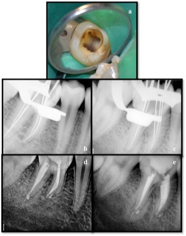
Figure 1 a-e. a. Access opening with 46. b and c. working length RVG. d and e. Obturation RVG.
confirm the quality of obturation. (Fig. 1d and e) The endodontic access cavity was temporarily restored with MD-Temp.
DISCUSSION
Clinicians often misinterpret that the tooth being endodontically intervened will contain a predetermined numbers of roots and root canals, but the literature presents the occurrence of variations in the number of roots and root canals. A thorough knowledge of the root canal anatomy and root canal configuration of the teeth to be treated plays an important role in predicting the success of the endodontic intervention.8 Studies and case reports have been reported in literature describing the presence of aberrant middle canals in the mandibular first molar located in mesial or distal roots or both.3,9,10,11,12
The third middle mesial or distal canals may be may be independent throughout their course in the root with separate apical foramen or may join either of the two main canals.7 The presence of middle mesial canal has been frequently reported in literature; however very few have reported the presence of middle canals in distal root or both mesial and distal roots of the same tooth, which presents itself as a rare anatomic variation.
Endodontic success in the teeth with the aforementioned number of canals requires a careful clinical and radiographic inspection. An accurate diagnosis of extra roots and root canals can avoid procedural errors or the chances of missing root canals during endodontic treatments. Use of multiple pre-operative radiographs, examination of the pulp chamber floor under magnification and sharp endodontic explorer, performing sodium hypochlorite bubble champagne test and visualizing the canal bleeding points is important and aids in locating the root canal orifices successfully. Additional methods such as troughing of the grooves with ultrasonic tips and illumination can assist to locate extra root canal orifices.
A significant constraint in conventional radiography is that it produces a 2D image of a 3D object resulting in superimposition of the structures. Recent and sophisticated diagnostic aids like CBCT and Spiral CT can produce undistorted three dimensional non invasive information of the root canal anatomy, can be used as a pre-operative tool that may increase the success of endodontic treatment. The presented case shows a successful endodontic management of a right mandibular first molar showing the presence of six canals three both in the mesial and distal roots.
CONCLUSION
Treating extra canals may be challenging, however inability to find and properly treat the canal may cause failure. It is mandatory that the clinician should possess a thorough knowledge of not only the normal anatomy of the root canal system, but also the possible aberrations. High incidence of variation in canal configuration is reported and seen in mandibular first molars hence a close clinical inspection of the chamber floor under magnification and the use of sophisticated diagnostic aids like CBCT should be the whilst while treating these teeth.
REFERENCES
- Favieri A, Barros FG, Campos LC. Root canal therapy of a maxillary first molar with five root canals: Case report. Braz D J 2006;17:75-8.
- Barbizam JV, Ribeiro RG, Tanomaru FM. Unusual anatomy of permanent maxillary molars. J Endod 2004;30:668-71.
- Baugh D, Wallace J. Middle mesial canal of the mandibular first molar: a case report and literature review. J Endod 2004;30:185– 6.
- Stoner W, Remeikis N, Carr G. Mandibular first molar with three distal canals. Oral surgery 1984;57:554-7.
- Beatty RG, Interian CM. Amandibular molar with five canals: report of case. J Am Dent Assoc 1985;111:769-71.
- Martinez-Berna A, Badanelli P. Mandibular first molar with six root canals. J Endod 1981;8:348-52.
- Reeh E. Seven canals in lower first molar. J Endodon 1985;11:348-52.
- Adanir N. An unusual maxillary first molar with for roots and six canals: A case report. Aust D J 2007;52:333-5.
- Holtzmann L. Root canal treatment of a mandibular first molar with three mesial root canals. IntEndod J 1997;30:422-3.
- Navarro LF, Luzi A, Garcia AA, Garcia AH. Third canal in the mesial root of permanent mandibular first molars: Review of literature and Presentation of 3 clinical reports and 2 in vitro studies. Med Oral Patol Oral Cir Bucal 2007;12:605-9.
- Weine FS. Three canals in the mesial root of mandibular first molar: Case report. J Endod 1981;8:517-20
- Gupta S, Jaiswal S, Arora R. Endodontic management of permanent mandibular left first molar with six root canals. ContempClin Dent 2012;3:S130-3.
Disclaimer from Guident
The views and opinions expressed in articles published in Guident are solely those of the authors and do not necessarily reflect the views of the editors, the editorial board or the publisher, Ivory India. Guident does not endorse any product, technique or treatment described. Clinical content is intended for qualified dental and medical professionals and is not a substitute for individual clinical judgement. Guident, its editors and the publisher accept no liability for any consequence arising from the use of information published in the journal.
How to cite this article
Dr. Ajinkya M Pawar, Dr. Suraj M Pawar, Dr.D.Y Patil Dental College and Hospital. Endodontic Management of Complex Root Canal Anatomy of a Permanent Mandibular First Molar Exhibiting Six Root Canals. Guident. 2014 Aug;7(9). Available from: https://guident.in/articles/endodontics/endodontic-management-of-complex-root-canal-anatomy-of-a-permanent-mandibular-first-molar-exhibiting-six-root.html