Canine Exposure and Frenectomy in an Orthodontic Case
Dept. of Orthodontics, Dept. of Conservative Dentistry & Endodontics, Dept. of Orthodontics, Chandra Dental College & Hospital, Lucknow
Abstract
Canines play a pivotal role in not only dental and facial esthetics, but the overall arch development and functional occlusion. The use of laser in dentistry has been developed over the last three decades. Initially it was introduced as an alternative to traditional halogen curing light but now it has become the instrument of choice in many dental applications.
The advantages of its utilization consist on the possibility of reducing or avoiding the use of anesthetics, the bloodless surgical field, no need for sutures and bonding can be done immediately after the canine exposure. The following case report presents laser-assisted approach of tooth exposure using 980nm diode laser.
Keywords: Diode lasers, 980 nm wavelength, Impacted canines, orthodontic treatment
INTRODUCTION
Impacted canines are one of the common dental malocclusions seen in orthodontics. It is reported to occur twice more frequently in the maxilla than in the mandible.
Clinical signs suggestive of canine impaction are over retention of primary canine, absence of a labial bulge, presence of palatal bulge or a generalized anterior spacing. Conventional exposure with scalpel often lead to extensive bleeding thereby leading to poor operatory field, difficulty in maintaining isolation for bonding and also the use of special hydrophilic moisture insensitive primers to bond orthodontic attachments.
The use of laser in dentistry has been developed over the last three decades. Initially it was introduced as an alternative to traditional halogen curing light but now it has become the instrument of choice in many dental applications.
The application of lasers is wavelength specific and depends on its absorption and interaction with different oral tissues. The diode is a versatile laser and has been successfully used in three surgical wavelengths ( 810, 940, 980 nm ) and recently the fourth wavelength 1064nm is introduced in dentistry.
Use of diode laser ensures easy exposure with minimal bleeding and less patient discomfort. The clear bloodless field ensures fast predictable bonding , thus enabling fast correction of malocclusion.
The following is a case report presenting laser-assisted approach of tooth exposure using 980nm diode laser.
VISIT 1- A 17 years old female patient presented to the clinic with the chief complaint of spacing in the upper front tooth region.
CLINICAL EXAMINATION- There was generalized spacing in the anterior region associated to a pathological insertion of upper vestibular frenum, positive to the traction test. There was also a labially erupting upper right canine covered with a gingival flap (Figure 1).
On clinical observation patient was advised for orthodontic treatment followed by frenectomy and exposure of the labially erupting upper right canine by 810 nm diode laser.
MEDICAL HISTORY- patient was well nourished and with excellent medical condition.
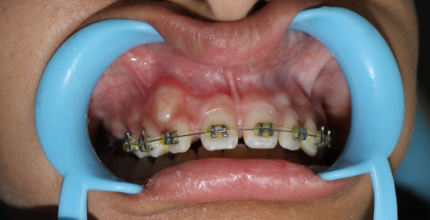
FIGURE 1. PRE TRETAMNT
No known drug allergy at the time of visit.
No history of bleeding and clotting disorders.
DENTAL HISTORY- No relevant dental history.
OCCLUSION- Angle’s class I molar relation.
TMJ- On palpation there was no soreness in muscle group.
- Normal range of motion.
- No clicking and popping sound during opening of mouth was observed.
RADIOGRAPHIC EXAMINATION- no need in this case.
SOFT TISSUE-
- Tongue- no abnormality detected.
- Mucosa- no abnormality detected.
- Gingiva- no abnormality detected.
HARD TISUE- no abnormality detected.
OTHER TESTS- Blanch test was done to confirm for the pathological insertion of upper vestibular frenum,
DIAGNOSIS- Angles Class I malocclusion with ectopic eruption of upper right canine and abnormal upper vestibular frenum.
INDICATION- 810 nm diode laser is suitable choice for this type of surgery as the chosen wavelength ensures easy exposure with minimal bleeding and least patient discomfort .
The clear bloodless field ensures fast predictable bonding thus enabling fast correction of
malocclusion .
CONTRA INDICATION- None.
TREATMENT PLAN OUTLINE- Orthodontic bonding of upper arch followed by frenectomy and canine exposure using 810 nm diode laser wavelength during initial alignment stages and follow up.
TREATMENT ALTERNATIVES-
- A conventional surgical exposure with scalapel.
- Electrosurgery
TREATMENT PROCEDURE-
VISIT 1
The orthodontic bonding was done in the upper arch and an 016 niti wire was placed in the upper arch. Patient was recalled after 1 month.
VISIT 2
After alignment, the patient was shifted from 016 niti archwire to 018 niti archwire . A Local anesthetic gel (20% Benzocaine by the name Ultracare UltradentTM ) was applied on the labial surface of the right canine region. 810 nm diode laser was initiated at 0.8 to 1 watts and the overlying gingival tissue covering the vestibular portion of the crown was removed in a contact and continuous mode. The canine exposure was done till we achieved an adequate clinical crown for bonding an attachment (Figure 2).
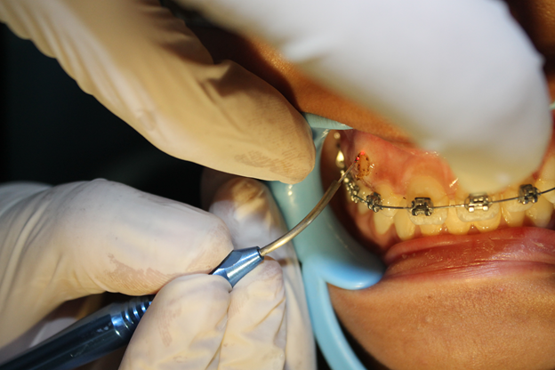
FIGURE 2. CANINE EXPOSURE
Simultaneously the same topical anesthetic gel was applied in the labial frenal area and frenectomy was performed by releasing the ï¬bers by making a small horizontal incision down to the periosteum on the palatal aspect . The release of the frenum was done at 0.8-1.4 watts Continuous Wave (CW) with an initiated disposable tip (Figure 3,4).
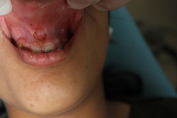
FIGURE 3. AFTER FRENECTOMY
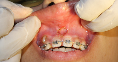
FIGURE 4. IMMEDIATE POSTOPERATIVE
No periodontal pack was applied to support the healing process.
The exposed canine region was immediately bonded by an orthodontic bracket and the ligature wire was used for ligating it to the main archwire (figure 5).
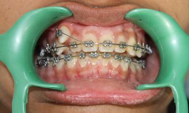
FIGURE 5. AFTER 6 MONTHS
Recall after a week .
VISIT 3
After 1 week, the canine and the frenal part was examined for healing. The frenal part for seen partly covered with a thin layer of fibrin
- Healing was good.
- Recall after 30 days.
VISIT 4
After 1 month healing of the epithelium was completed with the treated frenal attachment appearing pink, healthy and completely free any blanching when the lip was pulled outward and downward.
Healing was excellent.
Recall after 1 year for any recurrence.
VISIT 5
Patient came with no sign of recurrence of any blanching and the canine also came in Class I occlusion ( Figure 6).
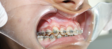
FIGURE 6. AFTER 1 YEAR
LASER OPERATING PARAMETERS-
Wavelength- 810 nm
POWER- 0.8 to 1 watt
Pulse duration- Continuous
POST OPERATIVE INSTRUCTION-
Patient was instructed to avoid alcohol, acidic beverages, hot and spicy food.
Patient was advised to clean the wound area by using 0.12% chlorhexidine mouth wash for two weeks.
Advised to use orthodontic tooth brush for the whole duration of the treatment.
Advised to do gum massage by topical gum ointment.
COMPLICATION- none
PROGNOSIS- was excellent
TREATMENT RECORDS- ATTACHED PHOTOGRAPHS
LONG TERM RESULT- Healing was excellent with no recurrene of any blanching and canine also came in normal Class I occlusion after 1 year.
CONCLUSION
Diode laser is a revolutionary technology providing alternatives for orthodontists in solving different problems in their everyday practice. It is effective and comfortable tool to reduce treatment time and to promote excellent clinical results. To patients is offered method of choice with comfort guarantee, less pain sensations and accelerated healing process.
The choice of surgical and orthodontic treatment modality determines the orthodontic and periodontal prognosis of the impacted teeth and the adjacent teeth as well.
REFERENCES
- Yavuz MS, Aras MH, Buyukkurt MC, Tozoglu S. Impacted mandibular canines. J Contemp Dent Pract 2007; 8(7) : 78-85
- Lalabonova Hr, Firkova E. Lasers in dentistry. Physical. Medicine. Rehabilitation. Health. 2005; 4:4-9.
- Prachis Ph, Prashanth K, Renu P, Vishwanath. Lasers – a contemporary tool in orthodontics. J Int Oral Health 2011; 3(2): 15-21.
Disclaimer from Guident
The views and opinions expressed in articles published in Guident are solely those of the authors and do not necessarily reflect the views of the editors, the editorial board or the publisher, Ivory India. Guident does not endorse any product, technique or treatment described. Clinical content is intended for qualified dental and medical professionals and is not a substitute for individual clinical judgement. Guident, its editors and the publisher accept no liability for any consequence arising from the use of information published in the journal.
How to cite this article
Dr. Shally Mahajan, Dr. Vipul Kumar Srivastava, Dr. Priyank Seth. Canine Exposure and Frenectomy in an Orthodontic Case. Guident. 2014 Aug;7(9). Available from: https://guident.in/articles/orthodontics/canine-exposure-and-frenectomy-in-an-orthodontic-case.html