Facial Swelling Due to Leukemia: A Diagnostic Dilemma for Dentists
Buddha Institute of Dental Sciences Gandhi Nagar, Patrakar Nagar, Kankarbagh, Patna, India, Sarjug Dental College & Hospital, Hospital Road, Laheriasari, Darbhanga, India, Umrao Hospitals and Impressions Dental and Maxillofacial Center, India, Department of Oral and Maxillofacial Pathology
Abstract
Leukemia is a malignant neoplasm resulting fromproliferation of abnormal haemopoietic stem cells with uncoordinated differentiation, regulation and apoptosis. Acute lymphoblastic leukemia (ALL) is the most common malignancy in childhood and more frequently in males. Oral lesions may be the presenting feature of acute leukemias and are therefore important diagnostic indicators of the disease. It frequently involves gingiva and alveolar bones. Gingival overgrowth has several causes, including poor oral hygiene, drugs, systemic illnesses and neoplastic conditions. Here we are presenting a case of acute lymphoblastic leukemiawith facial swelling and gingival enlargement mimicking dental infection along a brief discussion regarding ALL.
INTRODUCTION
Acute leukemias are the most common malignancy in childhood and approximately 30% of malignant diseases in patients under the age of 15 years. Acute lymphoblastic leukemia (ALL) is the most frequent typeof leukemia in children. Leukemia etiology remains unknown and seems to involve both genetic mechanisms and environmental factors.1
Three thousand to 5000 new cases of ALL are diagnosed every year in the United States. Median age at diagnosis is 12 years, and nearly two thirds of cases are diagnosed in children, in whom it represents the most common malignancy, constituting approximately 25% of all childhood cancers. In adults, ALL represents 20% of all leukemias and 1-2% of all cancers.2
The oral cavity in leukemia is prone to complications ranging from exacerbation of pre-existing pathological conditions to new mucosal lesions and infections. Lesions resulting from infiltration of oral tissuesby leukemia cells may occur in the gingival tissues or less frequently in the alveolar bones.1
CASE
A ten years old boy reported in our department with complaint of left side facial swelling and pain as well as difficulty in opening of mouth since last one month. Clinical examination revealed there was extensive swelling over left side of face with restricted mouth opening (7mm inter incisor distance)(Fig 1 A, B & C). On palpation swelling appears tender and hard in consistency and overlying skin is tense.Left submandibular lymph node was tender and palpable size approximately 0.8cm. Intraoral examination shows vestibular obliteration with gingival growth in relation to 75. OPG shows cavitation and pulpal involvement as well as large well demarcated radioleucent lesion involving the root of 75 (Fig 1 D). On the basis of clinical and radiographic interpretation a provisional diagnosis is made as cellulitis secondary to radicular cyst with 75. Then we put patient on medication and advised; fine needle aspiration cytology (FNAC) and hematological investigation. Hematological report shows ESR- 140mm/hour, hemoglobin was 4.5gm%, RBC count-1.34 million/cu. mm, platelet count – 14,000 million/cu. mm, WBC count- 7100 /cu. mm. Differential count shows Neutorphils-06% , Lymphocytes -74%,Eosionphils-02% , Monocytes -01%,Basophils-01%, Lymphoblast-16%, Both the FNAC and peripheral smears confirmed the diagnosis as acute lymphoblastic Leukemia (Fig 2 A&B). Finally after diagnosis as ALL we referred the patient
respective department for further treatment.
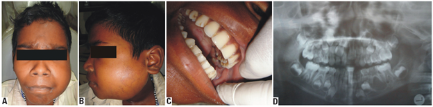
Fig 1: A & B: Left side facial swelling., C: Restricted mouth opening as well as gingival growth in relation to75., D: OPG shows periapical pathology in relation to 75.
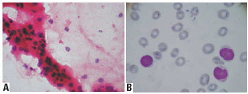
Fig 2: A: FNAC shows a homogenous population of cells with round nuclei mainly of intermediate type showing variable in size. Cells are with granular nuclear chromatin and basophilic cytoplasm., B: Smear showing multiple lymphoblast cells.
DISCUSSION
Acute lymphoblastic leukemia is the most common malignancy in childhood.3
There are many etiologic factors of leukemia, including genetic factors, certain carcinogens like benzene, tobacco smoke, ionizing radiation, advancing age, immune deficiency, viruses like Epstein Barr Virus, and oncogenes.According to the clinical behavior leukemias are classified into acute or chronic formsand characterized as lymphocytic and myelocytic.4
Further there are four main types of leukemia: acute lymphoblastic, acute myeloid, chronic lymphocytic and chronic myeloid. ALL is the most common leukemia seen in children, accounting for 85% of pediatric cases.5This disorder resulting from the clonal proliferation of lymphoid precursors with arrested maturation. The disease can originate in lymphoid cells of different lineages, thus giving rise to B-cell or T-cell leukemias or sometimes to mixed lineage leukemia.142Approximately, 10-20% of all lymphoblastic lymphoma and 80-90% of all ALL have a precursor of pre-B or B cell immunophenotype in contrast to 80-90% oflymphoblastic lymphoma and 10-20% of ALL who have precursor of T cell lineage.6
Clinical manifestations are due to the inhibition of hematopoiesis by leukemic cells and to leukemic infiltration in several organs and systems. Anemia, secondary to decreased red blood cell production, bleeding due to thrombocytopenia and infections due to neutropenia are the cardinal manifestations. Infiltration of organs results in hepatomegaly, splenomegaly and lymphadenopathy.1Other organs may also be involved, such as the kidney cortex, heart, eyes, and gastrointestinal tract. Central nervous system involvement is observedin 5%of children and in less than 10% of adults with ALL at diagnosis but is frequently asymptomatic. Fever may be due to ALL itself. 2
Oral manifestations of the diseaselead majority of the acuteleukemic patients to consult the dentist and in most of thecases the underlying disease is diagnosed from clinical findings during periodontalexamination. According to reports, the most commonfindings in the oral cavity include gingival enlargement, local abnormal color orgingival hemorrhage,petechiae, ecchymoses, mucosal ulceration and oralinfections. In some rare cases atypical features like, chinnumbness and tooth pain has been reported.4
Clinical and radiological characteristics are manifold. Differential diagnosis includes infections and other malignancies.101The radiological signs of ALL include disappearance of the inferior dental canal, an increased periodontal ligament space, loss of lamina dura and destruction of alveolar crestal bone.5
Growth and survival of malignant cells in the bone microenvironment is characterized by the presence of osteolysis and abnormal new bone formation. Bone formation is generally decreased in ALL patients due to high tumor burden.7
The diagnosis and classification of acute leukemia relies on a multidisciplinary approach including morphology, immunophenotyping, karyotype analysis and more specific molecular genetic analysis.8 But the role of the blood smear in the diagnosis of leukemia and lymphoma is to suggest a likely diagnosis or range of diagnoses (we also diagnosed the case initially on the basis of smear and FNAC), to indicate which additional tests should be performed, and to provide a morphologic context without which immunophenotyping and other sophisticated investigations cannot be interpreted.9
ALL treatment involves long-term chemotherapy, which has in the last decades undergone striking advances, leading to the current mark of around 80% of children and adolescents with ALL achieving cure. Despite these high cure rates, approximately 20% still have disease relapse. Dental monitoring of patients with leukemia during and after treatment is extremely relevant forearly detection and appropriate treatment of disease related lesions and side effects of chemotherapy.1
Finally, we conclude on the basis of present case that all the lesions or cases are not common as they appear during routine practice; so one should always fulfill the basic protocol for diagnosis. Which can prevent some uneventful occurrence in the clinic.
Disclaimer from Guident
The views and opinions expressed in articles published in Guident are solely those of the authors and do not necessarily reflect the views of the editors, the editorial board or the publisher, Ivory India. Guident does not endorse any product, technique or treatment described. Clinical content is intended for qualified dental and medical professionals and is not a substitute for individual clinical judgement. Guident, its editors and the publisher accept no liability for any consequence arising from the use of information published in the journal.
How to cite this article
Dr. Swapan Kumar Purkait, Dr. Sumanta Kumar Kolay, Dr. Abdul Hamid. Facial Swelling Due to Leukemia: A Diagnostic Dilemma for Dentists. Guident. 2013 May;6(7). Available from: https://guident.in/articles/oral-surgery/facial-swelling-due-to-leukemia-a-diagnostic-dilemma-for-dentists.html