A Giant Pleomorphic Adenoma of Minor Buccal Salivary Gland
Department of Maxillofacial Surgery, Faculty of Dentistry, Department of Periodontology, Faculty of Dentistry, Department of Maxillofacial Surgery, Faculty of Dentistry, The Saints Cyril and Methodius University of Skopje, Skopje, Republic of Macedonia
ACKNOWLEDGEMENT
Hereby by authors declare that source of case is private and no conflict of interest is there. We would like to thank Dr. Iliev A., and Dr. Popovic D. for immense help and encouragement in making of this article.
AIM
- The purpose of the study is presentation of diagnosis and operative treatment of buccal giant pleomorphic adenoma in 16 years old boy.
INTRODUCTION
- Salivary gland neoplasms are relatively uncommon with an incidence between 0,4 to 13,5 cases per 100.0001 and represent between 1 and 5 percent of all head and neck tumors2
- Tumors of the salivary glands comprise 3% of all neoplasms.3
- The majority of salivary gland Neoplasms are benign with Pleomorphic adenomas the most common, but the majority of minor salivary gland tumors are malignant.4
- The incidence of salivary gland neoplasms in minor salivary glands varies from 9 to 22%3,5,6
- Approximately 8% of pleomorphic adenomas involve minor salivary glands.4
- Tumors arising in minor salivary glands account for 22% of all salivary gland neoplasms7 and majority of them are malignant with only 18% benign, most frequent being pleomorphic adenoma.7
- Joshihara and Suzuki found that the majority of pleomorphic adenoma of minor salivary gland involve the palate followed by lip. The next common sites are the buccal mucose, tongue, tonsil, pharynx, retromolar area and nasal cavity.8
- Waldron et all. report incidence of benign tumor in 426 cases of intraoral minor salivary gland tumor with most common pleomorphic adenoma. The palate was the most common site followed by the upper lip and buccal mucosa. Pleomorphicadenomas, or benign mixed tumors, make up around 65% of all salivary gland tumors and are the most common type of tumors of the minor salivary glands.9
- Pleomorphic adenomas, in general, comprise 40% of minor salivary gland tumors.10
- Patients with pleomorphic adenomas of the minor salivary glands present mostly in their fourth to sixth decades, with a slight predominance in female patients.11
- Waldron-slight female to male preponderence at 1.59 to 1 and the mean age of patients is 49.8 years versus 55.2 years for patients with malignant tumors.9
- Lesions at these three sites: palate, upper lip, and buccal mucosa - account for 76.1% of oral minor salivary gland tumors.9
DIAGNOSIS
SYMPTOMS
- Minor salivary gland pleomorphic adenoma are usually
- slow-growing,
- firm single
- mobile,
- solitary,
- painless
- and nodular mass covered with normal appearing mucosa.
Radiographic
- Ultrasound
- Computerized tomography (CT)
- Magnetic resonance imaging (MRI).
- Aspiration (fine needle aspiration (FNA))
- Use large bore needle
- Biopsy
- May consider frozen section at the time of surgery
Treatment
- Operative treatment with wide and complete surgical excision is method of choice for minor salivary gland pleomorphic adenoma.
CASE
- A 16 year-old boy sought medical attention for a large growth on the left side of the face in November 2007. The painless swelling had gradually increased in size over a period in excess of 3 years.
- The last six months he felt that tumor had extremly growth in size. Examination showed the swelling to be related to the left buccal side and was oval in shape. The growth measured 6.0 × 5.5 x3.5cm in dimensions and was firm in consistency.
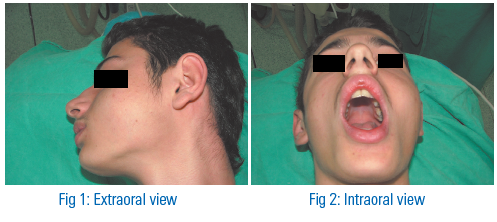
- It was attached to the deeper structures in maxillary and nasal bone, but movable. The overlying mucosa and skin were normal in appearance.On intraoral examination there was firm painless solitary mobile mass nontender to palpation well-encapsulated and clearly demarcated from normal tissue.
- The lesion was clinically diagnosed as a benign tumor arising in the parotid gland and fine needle aspiration cytology diagnosed the lesion as a pleomorphic adenoma (PA).
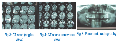
- Computerised tomography shows benign tumor with contact of mandibular body involving sphenoidal bone and sinus maxillaris, mandibular body, pterygoid lateral and temporal muscle with subbuccal and submasseterical position. X-ray pictures with laboratory tests have been also performed.
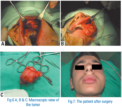
- The tumor was subsequently excised under general anaesthesia along with total extirpation with intraoral approach and reservation of nearby bone structures. One week post-peratively patient recovered fully. Very good aesthetic and functional results were achieved.
- Macroscopically, the excised specimen was ovoid in shape and 6 × 5,5 × 3,5 cm in dimensions and weighed 42 g.
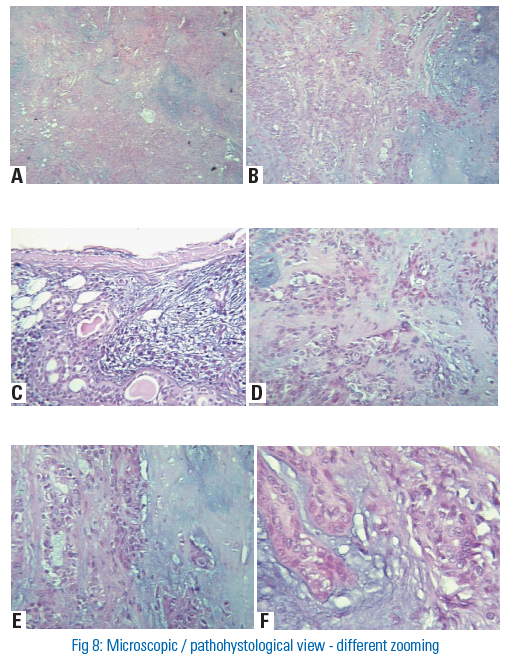
- Microscopically, the tumor was composed of strands and islands of epithelial and myoepithelial cells with many chondrocites sometimes in a myxomatous components. Some areas showed solid sheets of epithelial cells with squamous metaplasia. There was no evidence of malignancy and the tumour was diagnosed as a PA.
Clinical features
- Debate about pathogenesis, aggressive behavior and treatment.
- PA - most common benign tumor of the minor salivary glands.
- Peak incidence in the third to fifth decade
- Most common site palate followed by the upper lip, and buccal mucosa.
Symptoms
- Slowly growing,asymptomatic intraoral swelling in early period.
- Pain,trismus,sensory deficits,infection, numbness,toothache,illfittingdentures,malocclusion,ulcerations,draining sinuses,nasal obstruction,or even epistaxis may bring the patient.
- Pathological fracture is very unusual finding.
Diagnosis
Radiographic
- Ultrasound
- Computer tomography (CT)
- Magnetic resonance imaging (MRI).
- Aspiration
- (use large lumen needle)
- Biopsy
- (may consider frozen section at the time of surgery)
Histology
- Strands and islands of epithelial and myoepithelial cells
- Many chondrocites sometimes in a myxomatous components-corrugated surface layer of parakeratin
- Areas with solid sheets of epithelial cells with squamousmethaplasia.
Treatment
- Treatment must be guided of the
- behavior and growth potential of the PA
- Anatomic site
- Size
- And pathohistological analisys
- The treatment of choice for PA is total extirpation with intraoral
- approach(if it is possible) and preservation of nearby structures
- Treatment modalites are ranged from
- conservative excision to wide block resection and bone grafting.
CONCLUSIONS
- Patient was followed on an outpatient basis
- Very successful esthetic result with intraoral approach was achieved.
- Neither reccurens or functional disorder have been observed.
- Future follow up is necessary.
LITERATURE
- Rousseau A, Badoual C . Head and neck: Salivary gland tumors: an overview. Atlas Genet Cytogenet Oncol Haematol. Septem ber 2010 .
- Rubin R, Strayer DS, (eds): Rubin’s Pathology: Clinicopathologic Foundations of Medicine. 4th edition. Lippincott Williams & Wilkins, Maryland; 2004:1283-85.
- Grewal DS, Pusalkar AG, Phatuk AM Pedunculated pleomorphic adenoma of the tongue base manifesting with dysponea: A case report. J Laryngol Otol 1984;98:425-7.
- Spiro RH, Huvos AG, Berk R, et al.: Mucoepidermoid carcinoma of salivary gland origin. A clinicopathologic study of 367 cases. Am J Surg 136 (4): 461-8, 1978.
- Magliulo G, Terranova G, Cristofari P. Pleomorphic adenoma of the tongue base. Ann Otol Rhinol Laryngol 1996; 105:835-7
- Spiro RH. Salivary neoplasms: Overview of a 35-year experience with 2,807 patients. Head Neck Surg 1986;8:177-84.
- Spiro RH. Salivary neoplasms: overview of a 35-year experience with 2,807 patients. Head Neck Surg. 1986;8(3):177–184. doi: 10.1002/ hed.2890080309.
- T. Yoshihara and S. Suzuki, “Pleomorphic adenoma of tongue base causing dysphagia and dysphasia,” Journal of Laryngology and Otology, vol. 114, no. 10, pp. 793–795, 2000.
- Waldron CA, Gnepp DR (1998). Tumors of the intraoral minor salivaryglands: a demographic and histologic study of 426 cases. Oral Surg.Oral Med. Oral Pathol., 66: 323–333.
- Torske K (2006) Benign neoplasm of the salivary glands. In:Thompson LDR (ed.) Head and neck pathology, 1st edn. Elsevier,Philadelphia, pp. 295–300.
- Vellios F, Shafer WG (1959). Tumors of minor salivary glands. SurgGynecol. Obstet., 108: 450–456.
Disclaimer from Guident
The views and opinions expressed in articles published in Guident are solely those of the authors and do not necessarily reflect the views of the editors, the editorial board or the publisher, Ivory India. Guident does not endorse any product, technique or treatment described. Clinical content is intended for qualified dental and medical professionals and is not a substitute for individual clinical judgement. Guident, its editors and the publisher accept no liability for any consequence arising from the use of information published in the journal.
How to cite this article
Dr. Alberto Benedetti, Dr. Fidoski Jasmin, Dr. Vladimir Popovski. A Giant Pleomorphic Adenoma of Minor Buccal Salivary Gland. Guident. 2013 Apr;6(6). Available from: https://guident.in/articles/oral-surgery/a-giant-pleomorphic-adenoma-of-minor-buccal-salivary-gland.html