Ridge Split Technique in the Anterior Esthetic Zone Along with Immediate Implant Placement
s Dental Clinic Hydrabad, India
Abstract
The anterior esthetic zone is always a challenge as the ridge resorption could be magnified when there is loss of tooth by trauma. The high esthetic requirements coupled with resorbed alveolar ridge remain a serious challenge for the successful placement of endosseous implants. This article reports a technique for widening the atrophic ridge by splitting the alveolar bone longitudinally, immediate placement of implant and filling the bone gap with xenografts. Treatment of ridges as thin as 2-3 mm at the alveolar crest along with the immediate placement of dental implants.
Treatment of a young 25 year-old male patient with a severely resorbed anterior maxilla is described. 2-3 mm-wide by 13mm-long-threaded implants were immediately placed within the split ridge and surrounded with a xenograft.
The advantages of this technique for patients include less surgical trauma and reduced treatment time.
INTRODUCTION
The success rates of dental implants in the anterior maxilla can be frequently compromised due to presence of anatomical structures like nasal cavity, Incisive canal, buccal concavities and also loss of hard and soft tissue. Alveolar bone resorption can occur following extractions, trauma or pathology and may result in insufficient bone dimensions. A standard implant requires a minimum bone width of about 5.5 to 6mm because the smallest implant of 3.5mm in diameter needs 1mm of sound bone all around it to obtain primary stability.
Accurate diagnosis is extremely important and enables to determine if a favorable outcome can be achieved. The morphology of the ridge defect is an important determinant of the final outcome. A classification of ridge defects has been presented by Seibert and Cohen.19
This classification includes 3 categories:
Class I category - Buccolingual bone loss with normal ridge height
(The most amenable to treatment)
Class II category -Apicocoronal bone loss with normal ridge width
Class III category -Combination of vertical and buccolingual
bone loss
(The most difficult to treat)
Various ridge augmentation techniques to increase the bone width and height have been described in literature like:
- Autogenous block grafts
- Guided Bone Regeneration
- Ridge Split Technique- This is a relatively new and an innovative technique with very high success rate due to its presence of natural bone around the graft and immediate placement of the impalnt; reducing the treatment time.
A SPLIT CREST TECHNIQUE
In 1992, Simion et al introduced a split-crest bone-manipulation technique8. The goal was to create a self-space-making default by means of provoking a longitudinal greenstick fracture at the top of the bone that would split the atrophic crest in two parts. Implants could then be placed into the resulting fissure. This technique was indicated in the Class I category with a crest width of 4mm of less when a standard osteotomy could not be created.
This article describe a clinical case of severe anterior maxillary atrophy in which Simion’s approach to ridge-splitting was utilized, followed by grafting with xenografts and placement of a 3.5mm x 13mm threaded Noble Biocare Replace Select implant.
CASE PRESENTATION
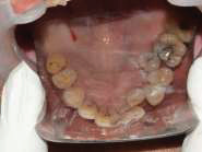
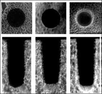
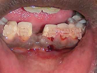
A 25 yr old systemically healthy male patient reported to the dental clinic with a chief complaint of missing upper left front tooth followed by a history of trauma 10yrs ago.[fig.1,2 and 3] Extra oral examination was within normal limits. Intraoral examination revealed missing 11 and loss of the mesio-distal width due to mesial migration of the adjacent teeth.[fig.2] Conventional 2 dimensional periapical radiograph revealed sufficient bone height but a CT scan showed a labio-lingual bone width of about 2-3mm only at the crest of the ridge (Fig. 4).
To regain the lost labio-linguo width a ridge-splitting technique in the area of 11 was planned followed by an immediate placement of an implant of 13-mm length, 3.5-mm diameter (Noble Biocare Replace Select Implant).To regain the lost mesio-distal width subsequent orthodontic treatment was planned.
At the time of the surgery, local infiltrative anesthesia was administered (lidocaine 2%, epinephrine 1:100,000). A full thickness mucoperiosteal flap was raised by giving a careful horizontal incision palatal to the crest of the ridge along with papilla preserving vertical relieving incision. Only a buccal flap is raised to maintain the palatal blood supply to the maxillary alveolar bone. The initial osteotomy was performed on midcrestal bone using a No. 15 blade and a mallet.[fig.5] Then vertical relieving incisions were given on the bone to avoid Greenstick Fracture [fig.6] as we proceed with chisels of increasing width to enlarge the osteotomy Maintenance of integrity of the small bony bridge in the apical part of the osteotomy is extremely important as it stabilizes the buccal plate in its new position and also preserves its blood supply.[fig.7,8]
After reaching a depth of 8mm, a pilot implant drill is used to mark the direction of the implant. This is followed by the use of an End pore osteotome of size 1 and 2 to make sufficient space for the placement of a 13mm long and 3.5mm wide implant. The implants presented initial primary stability (Fig. 9, 10), the cover screws were placed and the implants were submerged for a healing period of 6 months. The widened space between cortical plates was filled with a mix of bovine inorganic bone filler, Bio-Oss® which in turn was stabilized with a resorbable collagen membrane (Bioguide).The tissue was approximated with a 4-0 Prolene.[fig.11]
After two weeks of the surgical procedure, active orthodontics was planned to regain the lost mesio-distal width between the adjacent teeth. An artificial acrylic crown identical to the adjacent central incisor was prepared and bonded to the orthodontic brackets to act as a guideline to obtain to the desired space.[fig.12]
After 6 months, as we achieved the estimated msio-distal width; the 2nd stage was carried out placing a 15 degree angled abutment and restoring with a Porcelain fused to metal (PFM) crown.[fig.13,14]
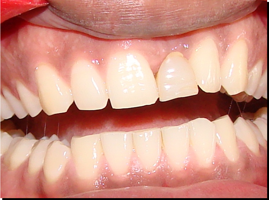
Fig.1:Pre treatment photograph , when the patient presented with an interim removable denture

Fig:2 :Pre treatment photograph
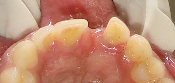
Fig:3:Pre treatment – incisal view
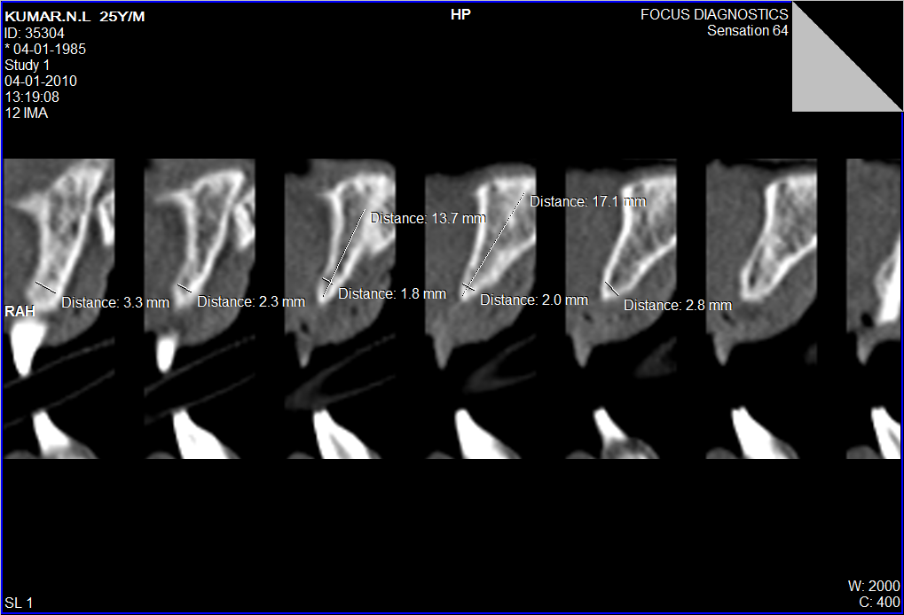
Fig.4:CT scan reveals insufficient bone at the crest

Fig.5:Initial incision in bone with a no#15 Bp blade
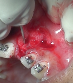
Fig.6:Vertical incisions in bone by a narrow chisel

Fig.7:The bone being split using the chisel

Fig. 8:The final drill used after expansion of the bone by osteotomes
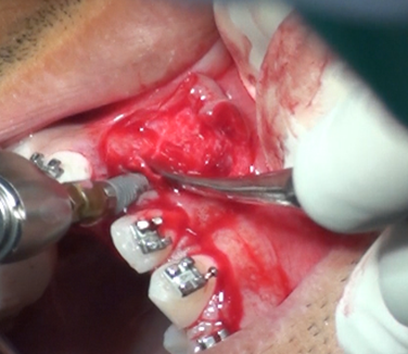
Fig.9 Root form tapered Implant fixture being driven in at low speed and 35 Ncm torque
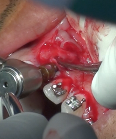
Fig.10:The implant completely driven in

Fig.11:A good passive primary closure
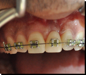
Fig . 12 :Orthodontics done to create the mesio-distal width ,with an acrylic artificial tooth ,the same size of the adjacent central incisor

Fig.13:The final crown in place
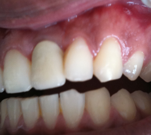
Fig. 14 Left lateral view of the same
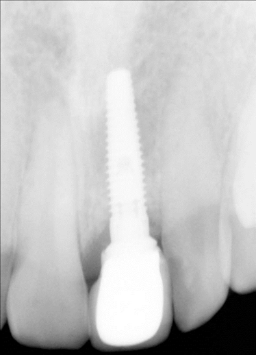
Fig.15:Rradiograph of the implant 1year post restoration
DISCUSSION
The ridge-splitting technique allows placement of implants in a narrow crestal ridge in a single procedure. The split crest technique is a suitable procedure particularly for the maxilla as the maxillary cortical plates are relatively thin and the cancellous bone is sparse. Thus, the separation between the cortical plates can be achieved
with relative ease. Conversely, the mandible demonstrates
thicker cortical plates and denser cancellous bone and separation
between the cortical plates may prove to be more difficult. Complete fusion between the buccal and lingual cortical plates, which can exist particularly in a case of a very thin and resorbed alveolar ridge, is a contraindication for this procedure. The separation between the bony plates is a delicate task. The maintenance of the integrity of the small bony bridge in the apical part of the osteotomy is extremely important as it stabilizes the buccal plate in its new position and preserves its blood supply. Therefore, the split crest approach is suitable for treatment of ridge
defect class I while class II and III represent a contraindication for this approach.
CONCLUSION
This article described implant placement in atrophic maxillary alveolar crests using the ridge-splitting technique. The correct indication associated with careful clinical maneuver of the ridge-splitting technique allows predictable placement of implants even in narrow alveolar ridges.
Disclaimer from Guident
The views and opinions expressed in articles published in Guident are solely those of the authors and do not necessarily reflect the views of the editors, the editorial board or the publisher, Ivory India. Guident does not endorse any product, technique or treatment described. Clinical content is intended for qualified dental and medical professionals and is not a substitute for individual clinical judgement. Guident, its editors and the publisher accept no liability for any consequence arising from the use of information published in the journal.
How to cite this article
Dr. Vikas Gowd, Dr. Kirti Gowd, Dr. Growd. Ridge Split Technique in the Anterior Esthetic Zone Along with Immediate Implant Placement. Guident. 2018 Sep;11(10). Available from: https://guident.in/articles/implantology/ridge-split-technique-in-the-anterior-esthetic-zone-along-with-immediate-implant-placement.html