Rationale of Cortico Basal Implants: A Review of Literature with Case Presentations
V.K Dental, Bangalore, KAR India
ABSTRACT
Purpose - To explain the rationale of the corticobasal implants.
Methods – Presented three cases of full mouth rehabilitation. 2% lignocaine with 1: 80,000 adrenaline was used to anesthetize the upper and lower arches. Extractions were carried out with immediate placement of corticobasal implants with syncrystallization of implants in one of the patients presented here.
Results- We have rehabilitated the patients using hybrid denture, shell denture and porcelain fused metal. Primary stability of the implants was achieved with good esthetics and function.
Conclusion- Quantity and quality of the bone plays a major role in the success of the implants. Cortico basal implants are anchored in the cortical bone that do not resorb during the life time. Irrespective of the implants placed in the extraction sockets or healed ridges, no significant changes were seen in the life of the implant. Cortico basal implants are a very good option and we call it a blessing as the patient’s esthetics and function can be restored in 3-4 days.
Keywords- Cortical, cortico basal implants, bone density, bone implant contact (BIC), bone multicellular unit (BMU), Syncrystallization.
BACKGROUND
Dental implants are the need of the hour for the missing teeth. Life of the implants basically depends on two factors- Quality and quantity of bone. These factors are detrimental for the early and long term success of the implant. Poor quality and quantity of bone would result in
lack of primary stability, impaired healing and failure of the implants. Cortico- basal implants have broad indications, lower degree of limitations and indicated in moderate to severe atrophic jaws. The cortical bone used for anchoring of the implants is the basal bone which is highly mineralized and is not resorbed through the course of life.1 Basal cortical bone has a much faster and stable repairing capacity. Cortical implants engage the basal cortical bone of both the jaws for achieving retention of the implant. It is the fixed unalterable framework of maxilla and mandibular bone that provides excellent bicortical anchorage, hence basal implantology is also referred to as bicortical implantology. The advantages of basal implants over conventional implants are that, it is done in a single appointment using a flapless procedure, simple, cost-effective procedure which utilizes minimum armamentarium. It provides an infection free zone of bone for implants, greater stress bearing area, lesser resorption rate and a possibility to provide immediate prosthesis, which cannot be achieved with the conventional crestal implants. Moreover, Basal implantology does not require ridge augmentation procedures and they are single piece implants that can be loaded immediately so, the strength provided by the implant is excellent.2
RATIONALE OF CORTICO BASAL IMPLANTS
The concept of basal implantology originates from the presence of two distinct parts of jaw bone. a) The tooth bearing alveolus also called the crestal bone. b) Basal bone. (figure 1)

Figure-1: Physiology of Bone
The alveolar process is defined as part of the maxilla and mandible that forms around and supports the teeth. Its volume, shape and height are determined by the shape of the teeth (crown and root) they harbour.3,4 The bony component of the alveolar process is comprised of outer plates, the facial (labial and buccal) surface and the lingual/palatal surface, these plates being made of thin cortical bone. The area enclosed by the cortical bone plates is occupied by cancellous (spongy) bone.5
The cortical surfaces are in continuity with the body of the jaw and vary in thickness from one region of the jaw to the other. Both in the mandible and in the maxilla, the facial plate is usually thin and tends to be thicker in the second and third molar area. The palatal/lingual plates of the alveolar process are usually thicker than the facial plate. Despite the basic similarity between the two jaws, the alveolar process in the mandible it is not as cancellous as that in the maxilla. The thin facial bone plate, most often seen in the anterior area of the mandible, its composed of mainly cortical dense compact bone as seen on the lingual plate 6 . When teeth are lost, the portion of the alveolar process that supported the missing tooth will be subject to atrophic reduction, where the size of the ridge will become markedly reduced in both its horizontal and vertical dimensions.7
The buccal bone plate’s original dimensions have a large effect on the degree of ridge resorption following tooth extraction.8 This clear difference in post-extraction remodelling behaviour relies on the histological characteristics of the bone plate, as the thin facial plate is predominantly composed of bundle bone that is functionally dependent on tooth presence and will thus completely resorb following tooth extraction or loss. This resorption will result in pronounced vertical reduction of the ridge height. For the wider lingual crest and more apical parts of the buccal plate, where the bone composition is lamellar, compact in nature and less affected by the bundle bone resorption, less changes are expected.9
BONE DENSITY
Carl .E. Misch10 was an American Prosthodontist, recognised for his contributions to the field of Implant dentistry. Based on the C.T. findings he classified bone density into 5 grades.
Misch’s classification (Table-1)
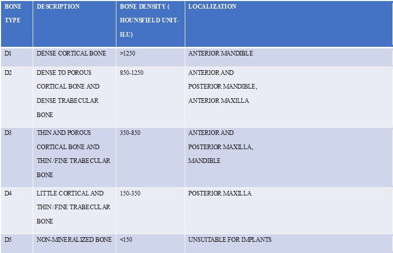
D1 BONE TYPE- This bone consists almost entirely of a dense cortical. It is found in the anterior mandible and can be compared to oak or maple with least resorption. On a Hounsfield scale, it measures 1250 units or more. It has a large bone-implant contact (BIC) area and great initial implant stability. Its nutrition depends on the periosteum due to a lower number of blood vessels. Surgery must be conservative and special care to avoid bone heat must be taken that would result in delayed bone healing. The post-surgical strongest osseointegration in this type is derived from high bone-implant contact.11
D2 BONE TYPE- It has a is a combination of dense-to-porous cortical bone on the crest and trabecular bone from 40% to 60% on the inside, most frequently in the anterior mandible, followed by the posterior mandible and also the anterior maxilla. This type of bone can be compared to white pine or spruce. Its density can be measured from 850 to 1250 on the Hounsfied scale. It provides a good bone-to-implant contact and good vascularization, therefore fewer drills are needed during the surgery and thus predictable osseointegration. Fair blood supply within the bone tissue allows bleeding during the osteotomy, which in turn is very useful in reducing overheating during surgical bed preparation.11
D3 BONE TYPE- It consists of a porous crestal layer of cortical bone and a fine trabecular bone with < 50%. Tactile analogy can be made to compressed balsa wood. It is commonly found in the anterior and posterior maxilla, but may also be found in the posterior mandible. On the Hounsfield scale, 350 to 850 units indicate D3 bone.
Its porous structure means less BIC, which cause a higher risk of implant failure. Necessitates a different drilling protocol to achieve osseointegration. It does have a great vascularization that promotes new bone formation.
Lower BIC can be enhanced by implant surface treatments such as titanium plasma spray (TPS) or hydroxyapatite (HA) coating.11
D4 BONE TYPE- This bone is trabecular without any cortical. It is located in the posterior maxilla and it can be compared to compressed styrofoam or lightweight balsa wood. It can be read from 150 to 350 units on a Hounsfield scale.
As a result of drilling on low density bone, BIC is seriously compromised and the surgical technique must be delicate and precise so as not to remove bone and instead compress it around the implant.
The most common locations for this type of bone are the posterior region of the maxilla. It is rarely observed in mandible. In addition, the bone-implant contact after initial loading is often less than 25% as they are sparse bone, initial fixation of any implant design presents a surgical challenge. Implant failure after initial loading reported the highest score in this type. Moreover, it takes the longest time to integrate with the implant after placement. Splinting is must here.11
D5 BONE TYPE- A very soft bone, with incomplete mineralization and large intertrabecular spaces. This bone type is most often immature bone. The bone density may be determined by tactile sense during surgery, the general location or by radiographic evaluation. It can be read below 150 units on a Hounsfield scale. This kind of bone is unfit for the implant placement.11
BONE MULTICELLULAR UNIT (BMU)
Since basal implants have a unique design their peri-implant healing is also unique. What conventional implantologists call as “Osseointegration” is called as “Osseoadaptation” by basal implantologists, this stems from the fact that the bone with continuous functional loads remodels and adapts over the surface of the implant.

Figure-2: Bone multicellular Unit (BMU)
According to philosophy of basal implantology the process of Osseoadaptation is carried out by a “Bone Multicellular Unit” (BMU), it is said to be like a cutting cone with a tail, the cutting cone comprises of osteoclastic cells that eat away the peri-implant bone and the tail comprises of osteoblastic cells that lay down bone, as this unit moves in the bone the osteoclastic activity is subsequently followed by osteoblastic activity.12
The formation of this BMU takes place when these implants are subject to immediate loading which leads to remodeling of bone under functional stresses leading to development of this unit, and thus initiates the healing phase and leads to formation of a dense peri-implant bone.
Gaur et al. stated that it is necessary to rigidly splint strategic implants within 3 days as the process of bone remodeling starts. They stated that it takes almost 4–6 months for bone remodeling to complete and thus achieve biological stability from mechanical stability.12
The cascade of processes13 involved is as follows (Figure 3)
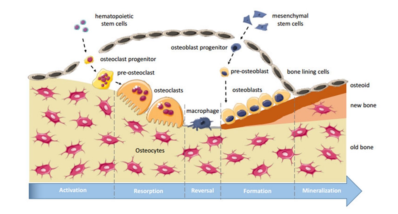
- Activation Phase- In this phase the precursor cells/human mesenchymal stem cells develop into osteoblasts and osteoclasts. This phase lasts for 3 days.
- Resorption Phase- During this phase osteoclastic activity occurs which reveals soft and porous bone. Osteoclastic activity occurs at a rate of 40µm/day.
- Reversal Phase- In this phase osteoblastic activity takes place. The osteoblasts lay down neo bone in the haversian canals at a rate of 1-2µm/day.
- Progressive Phase- This phase involves the osteoblasts forming concentric lamella in haversian canals, which leads to reduction in diameter of the canal and increase in bone density. At this stage the diameter of the haversian canal is 40- 50µm. The bone formed is a Non-Mineralized Matrix Osteoid and this phase lasts for 3 months.
- Mineralization Phase- After 10 days of osteoid formation mineralization phase begins. This phase involves two stages
- Primary Mineralization Stage- This stage imparts primary hardness to the osteoid and accounts for 60% of all mineralization.
- Secondary Mineralization Stage- This stage imparts final hardness and final morphology of bone. This phase lasts for 6-12 months.
- Dormant Phase - In this phase osteoblasts develop into osteocytes and line the haversian canals and take up mechanical, metabolic and homeostatic function.
In simple terms it can be stated that the peri-implant healing is a life-long process utilizing the concept of micro-motion and bone compression, that’s why these implants are also called as “Orthopaedic Implants” as they employ the same principles of peri-implant healing and bone densification and hence remodelling is a life-long process.
CASE PRESENTATIONS
CASE 1
A female patient aged 42 years with no relevant systemic history came to us with a chief complaint of loose teeth for 6-8 months. On clinical and radiological examination, we diagnosed this as a case of chronic generalised periodontitis. Following blood investigations, we extracted all the teeth and placed cortico basal implants immediately.Surgical procedure-Extractions and immediate implant placement in upper jaw on day 1 followed by lower jaw extractions and implant placement on the day 2. Swiss manufactured Monoimplants were used in all our cases. Flapless placement of implants were carried out through the existing extraction sockets. (figure 4,6) On day 3 trial was done (figure 5) with delivery of prosthesis on day 4 (figure-4,6). We chose to give a hybrid denture in this case. We have a follow up of this patient for two years and no complaint from the patient and clinical examination revealed a properly maintained oral hygiene with no significant changes following prosthesis restoration. 10 implants were placed in the maxilla and 10 in the mandible.
Following are the clinical and radiological pictures of the patient treated. (figures 4-7)

Figure-4: Pre-op, Intra-op & Post-op OPG

Figure-5: Hybrid Denture
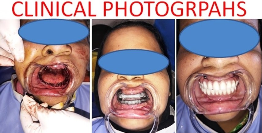
Figure-6: Immediate Implant placement, metal trial & Prosthesis Delivery
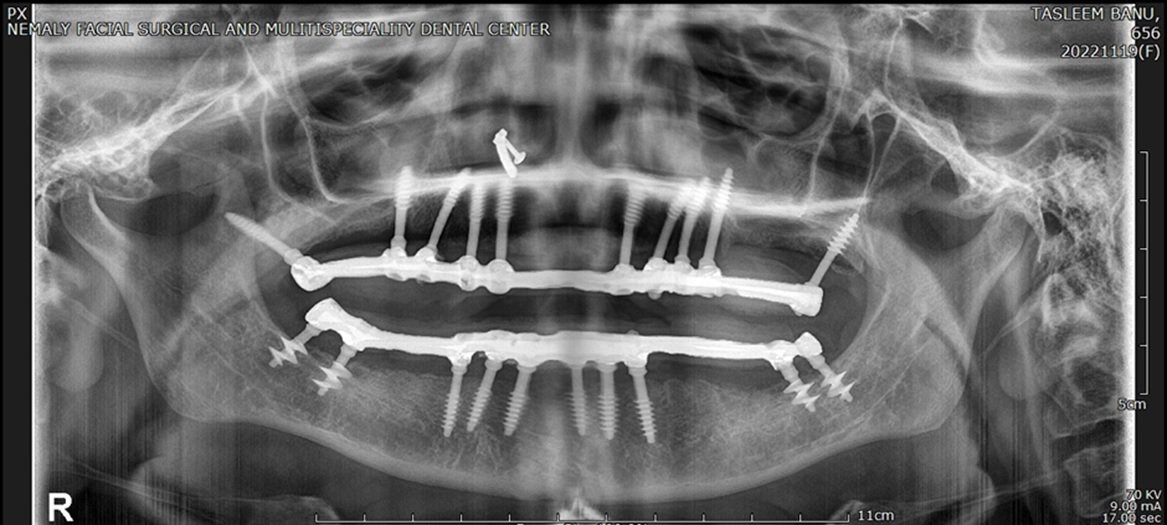
Figure-7: 2 Years Post-op
CASE 2
A male patient 76 years old came to us with a complaint of difficulty while chewing. History of diabetes and is on medication. On examination maxillary posteriors were missing with generalised mobility of existing teeth. We arrived at a diagnosis of chronic generalised periodontitis . Radiographic evaluation revealed generalised bone loss (figure 8). HBA1C revealed fair control of blood sugar.
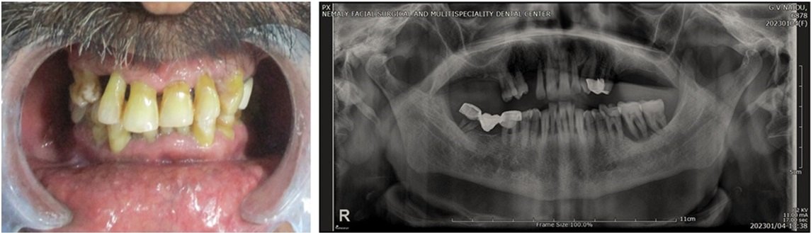
Figure-8: Pre-op
Surgical procedure- All the extractions and implant placement were carried out under local anaesthesia using 2% lignocaine and 1:80,000 Adrenaline. 3-4ml of L.A was administered per quadrant. Upper teeth were extracted on day 1 followed by immediate placement of the implants in the upper jaw. Lower teeth were extracted on day 2 followed by immediate placement of the implants in the lower jaw (Figure 9 and 10). We have used 12 implants for the maxilla and 10 implants for the mandible. Swiss made Mono Implants were used in this case. 1.5mm, 2 wires were used to syncrystallize the implants on each arch, following implants placement (Figures 9 and 10). Day 3 trial was done and Day 4 shell denture was delivered (Figure 11 ).
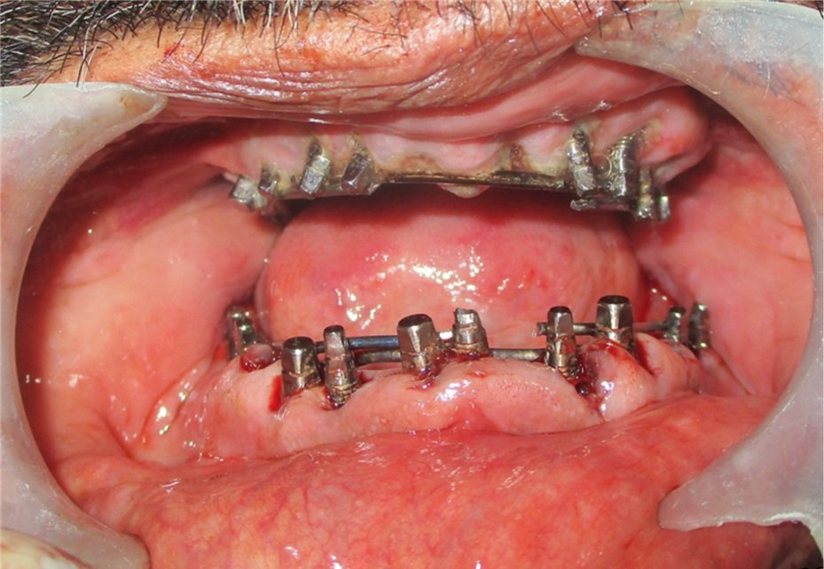
Figure-9: Implant placement

Figure-10: Post-op OPG
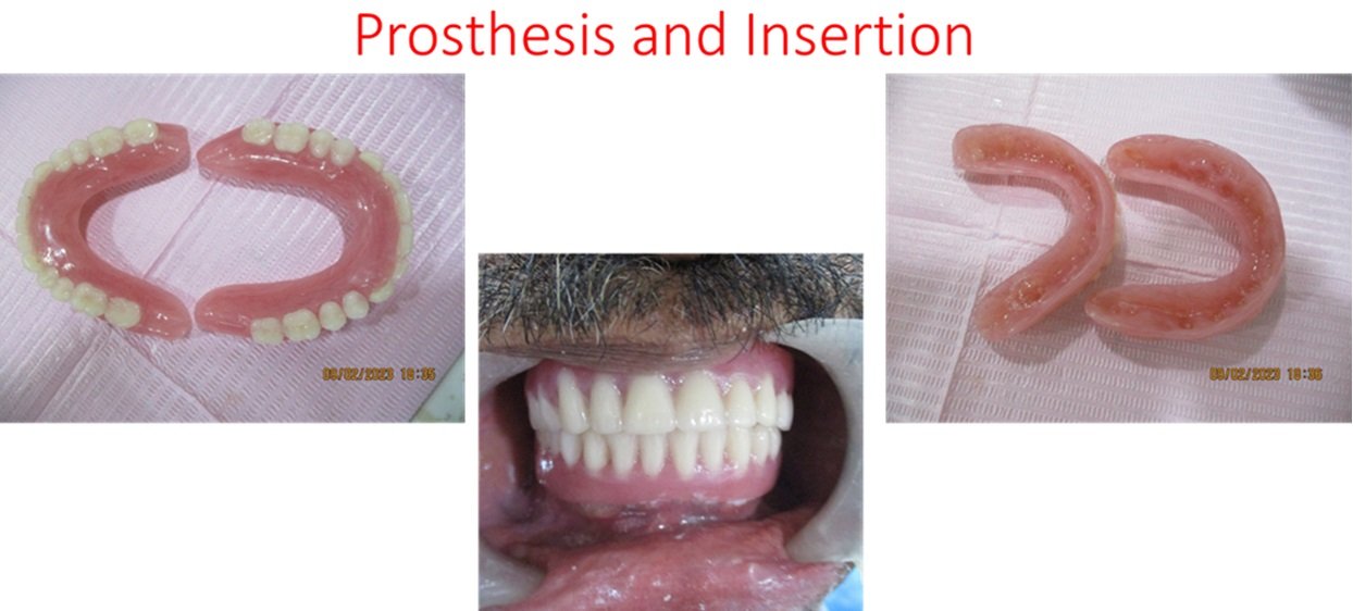
Figure-11
DISCUSSION
These three cases we have rehabilitated with different prosthesis. There are many criteria that determine what kind of prosthesis has to be given following implant placement. Quality and quantity of bone presents determines the kind of prosthesis we deliver. In case one we opted for a hybrid denture. Considering the physique, age of the patient, quality and quantity of the bone present, periodontal condition we opted for a hybrid denture. Its been two years that we have placed implants and delivered the hybrid prosthesis. The patient is happy with her function and esthetics.
In case two, owing to the poor periodontal status, we opted to give a shell denture. As it is proven scientifically that soft tissue shrinkage happens for 2-3 months following extractions, a pink prosthesis can be used in cases of gingival inflammation present at the time of extraction. Following recession of the gum line after insertion of the shell dentures, correction can be done using a reliner or composite. Esthetics and function can be maintained. If the patient desires ceramic teeth, the shell dentures can be retrieved and ceramic teeth can be delivered.
In case three, owing to the age, physique, quality and quantity of bone present, we preferred metal ceramic prosthesis. As the opposing teeth were also metal ceramic prosthesis we chose the same following implant placement in the maxillary arch and in the 3rd quadrant. Patient is extremely happy with his esthetics and function.
The modern basal implant has a sophisticated yet simple design, surgical protocol, and is a prosthetic friendly system.14 The healing of bone, i.e., osteonal remodeling, starts 3 days postoperatively. Hence, within the first 3 days, very stable bone is available. For this reason, it is recommended to equip the implant within a short period with the prosthetic splinting and to leave it in place at a minimum of 6–9 months until remodeling ceases. It should be noticed that the remodeling of the bone is not endangering the stability of implants . In 2016, S. Ihde and A. Ihde proposed the concept of “supporting polygon” to determine the position of occlusal contacts within or outside of polygon drawn up by the load transmitting part of the implants in the second or third corticals. The aim of the treatment for work on strategic basal implants should be:
- A bilateral equal and symmetrical occlusion
- A bilateral equal and symmetrical mastication, with contact on first, second premolar, and the mesial half of first molar.
- A symmetrical function of the muscles, especially tongue.14
CONCLUSION
In the span of 2 years we have placed more than 1000 cortico basal implants at our center. We have just provided 3 cases with complete rehabilitation for the sake of better understanding for our readers. Irrespective of the placement of these implants following immediate extraction or healed ridges, no significant changes are seen in an implant survival. Cortico basal implants are a very good option and we call it a blessing as the patients esthetics and function can be restored in 3-4 days. A reliable technique as it can be done in a single appointment using a flapless procedure, simple, cost-effective that utilizes minimum armamentarium.
List of abbreviations
- BIC-BONE IMPLANT CONTACT
- BMU- BONE MULTICELLULAR UNIT
- PFM-PORCEALIN FUSED METAL
Declarations
Ethics approval and consent to participate-ethics approval has been obtained
Consent for publication-consent for publication has been taken from the patients whose photographs are displayed.
Availability of data and material- The datasets used and/or analysed during the current study are available from the corresponding author on reasonable request.
Competing interests- We declare that we have no competing interests
Funding- No funding in any form was provided to this article.
Authors' contributions-
Dr. Nemaly Chaithanyaa – Operated all the cases and has authored the article.
Acknowledgements- Would like to thank my teachers Dr.Veerendra Kumar S.C. for his mentorship.
REFERENCES
- Shemtov-Yona K . Quantitative assessment of the jawbone quality classification: Ameta-analysis study. PLoS ONE 16(6): 1-19.
- Prasad DK, Mehra D, Prasad DA. Recent advances, current concepts and future trends in oral implantology. Indian Journal of Oral Sciences. 2014;5(2):55.
- Yadav R, Sangur R, Mahajan T, Rajanikant A, Singh N, Singh R. An alternative to conventional dental implants: Basal implants. Rama Univ J Dent Sci. 2015;2(2):22-8.
- Schroeder HE. The periodontium. In: Oksche A, Vollrath L, editors. Handbook of Microscopic Anatomy. berlin: Springer Berlin Heidelberg; 1986 : 233–46.
- Nelson SJ, Ash MMJ. Dento-osseous structures, Blood vessels and Nerves. ninth edit. Wheeler’s Dental Anatomy, Physiology and Occlusion. ninth edit. Saunders Elsevier; 2010; 239–51
- Merheb J, Quirynen M, Teughels W. Critical buccal bone dimensions along implants. Periodontol 2000. 2014; 66: 97–105.
- Atwood DA. Some clinical factors related to the rate of resorption of residual ridges. J Prosthet Dent. 1962; 12: 441–50.
- Cardaropoli D, Tamagnone L, Roffredo A, Gaveglio L. Relationship between the buccal bone plate thickness and the healing of postextraction sockets with/without ridge preservation. Int J Periodontics Restorative Dent. 2014; 34: 211–17.
- Arau´jo MG, Silva CO, Misawa M, Sukekava F. Alveolar socket healing: what can we learn? Periodontol 2000. 2015; 68: 122–34
- Misch CE. Contemporary Implant Dentistry-E-Book. St. Louis: Elsevier Health Sciences; 2007.
- Arriving at a definitive bone quality.Vaishnavi Rajaraman, V. Suresh, M. Dhanraj, Ashish R. Jain. Drug Invention Today : 2018; 10 (5) : 834-38.
- Ihde Stefan. Principles of BOI- Clinical, Scientific, and Practical Guidelines to 4-D Dental Implantology. Springer, Heidelberg; Germany, 2005
- Aakarshan Dayal Gupta, Aviral Verma, Tanay Dubey, Saloni Thakur. Basal osseointegrated implants:classification and review. International Journal of Contemporary Medical Research 2017;4(11):2329-35.
- Patel K, Madan S, Mehta D, Shah SP, Trivedi V, Seta H. Basal implants: An asset for rehabilitation of atrophied resorbed maxillary and mandibular jaw – A prospective study. Ann Maxillofac Surg , 2021;11:64-9.
Disclaimer from Guident
The views and opinions expressed in articles published in Guident are solely those of the authors and do not necessarily reflect the views of the editors, the editorial board or the publisher, Ivory India. Guident does not endorse any product, technique or treatment described. Clinical content is intended for qualified dental and medical professionals and is not a substitute for individual clinical judgement. Guident, its editors and the publisher accept no liability for any consequence arising from the use of information published in the journal.
How to cite this article
Dr. Veerendrakumar SC., Dr. Nemaly Chaithanyaa Oral Surgeon & Implantologist. Rationale of Cortico Basal Implants: A Review of Literature with Case Presentations. Guident. 2024 Jan;17(2). Available from: https://guident.in/articles/implantology/rationale-of-cortico-basal-implants-a-review-of-literature-with-case-presentations.html