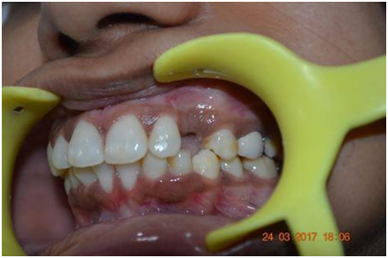
Improving the Socket Condition- Prior to Implant Placemnent
Associate Dentist Global Research Group, New Delhi Consultant Max Healthcare, New Delhi
Improving the socket condition related to a future implant site prior to implant surgery can be a safeguard for the future health of the implant and its surrounding tissues, as the presence of a sufficient zone of keratinized mucosa can minimize the soft tissue complications (especially in immediate implant placements) and stabilize the peri-implant tissue margins. This was affirmed with the fact that immediate implant placement into a freshly extracted socket might present an especially difficult challenge to achieving soft tissue coverage to many clinicians, especially with patients that have the thin scalloped tissue biotype. Achieving primary flap closure had been regarded as one of the most challenging aspects of immediate implant placement procedures. Although the implant success rates of immediate implant placement have not been shown to differ between closure and non-closure, and in many cases, the muco-gingival conditions around extraction sockets may become unfavorable for primary closure. Periosteal-releasing incisions as well as palatal rotational flaps have been introduced for complete flap closure. Nonetheless, these procedures may cause alteration of the harmony of the continuity of the keratinized tissue band, and the possible tension created in achieving primary flap closure may increase the chance of eventual bone graft or membrane exposure, leading to bone loss, infection, and even loss of implants.
Langer suggested a technique utilizing the regenerative capacity of the body to produce extra tissue around the root of a tooth that is scheduled for extraction. This technique provides sufficient keratinized mucosa for a soft tissues closure procedure on top of the implant. This new regenerated tissue subsequently minimizes surgical trauma that occurs due to attempts to achieve primary closure in immediate implant placement. In addition, the new regenerated tissue preserves the anatomical mucogingival integrity to its biological level and eliminates the chance for post extraction alveolar bone resorption. The method entails grinding the failing tooth below the free gingival margin, in order to level with the alveolar bone crest, and left in the socket for 2 to 4 weeks, allowing the surrounding gingival tissue to proliferate over the top of the remaining root. This procedure allows ridge preservation, facilitates complete tissue closure over the implant and grafts and prevents bone resorption due to the fact that the root is still in its housing. The gingival tissues proliferate, covering the remaining tooth root with keratinized gingiva, after which a muccoperiosteal flap can be elevated, the remaining root is then extracted, and an implant immediately placed.
Some distinct advantages of this technique include complete primary coverage of the implant at stage 1 surgery, reduced treatment time, and decrease the cost to the patient, when compared with other socket preservation techniques.
Drawbacks include the possibility of damaging the crestal bone during extraction and the presence of fenestration or dehiscence defects, which would necessitate further grafting procedures. The presence of residual roots of the reduced teeth will prevent epithelial down growth along the socket walls and provide scaffolding for the regeneration of keratinized tissues on top of the socket orifice. The soft tissues will take only few weeks to regenerate and fill the socket orifice as part of the healing process. Langer suggested that pulp extirpation can be performed prior to the commencement of the procedure to avoid possible pain, would be adequate for this procedure, however complete root canal treatment would ensure absence of infection in the extraction socket during implant placement surgery. With this technique, the degree of tissue closure varies and usually is time dependent. To achieve complete soft tissue closure over the root remnant, the waiting period can reach up to 3 months time, till complete soft tissue coverage and maturation occurred, however some thinning of the soft tissues can be expected on top of the roots at the time of implant placement, that warrants the possibility for soft tissue manipulations to overcome these possible fenestrations. any regenerated tissues should be done in excess of what is required so as to compensate for ensuing tissue shrinkage or remodeling .After sufficient healing time has elapsed and a stable tissue contour is established, any excessive tissue can then be trimmed or sculptured to the desired level.
Another technique that aimed at improving the soft tissue condition on top of an extraction socket, is called the Socket seal technique, it is introduced to improve the socket condition prior to implant therapy, reduce the time waiting period in the previous method for developing soft tissues on top of the socket orifice, and to augment the future peri-implant tissues. Several techniques were introduced to improve the condition of the soft tissue on top of the socket orifice via onlay grafting methods, all of them aimed to prevent post extraction bone resorption of the alveolar ridge, and to enhance the quality of bone at the future implant site. The technique of socket sealing, aims to block off the socket orifice of an extracted tooth immediately after unsalvageable tooth extraction. Socket seal surgery was first described by Landsberg and Bichacho as a treatment alternative for preserving the integrity of the alveolar ridge and inhibiting apical epithelial migration into the socket thus improving the overall socket condition to optimally house an implant body. The procedure involves a-traumatic, flapless tooth extraction using either the periotomes (Storz am Markt GMBH, Emmingen-Liptingen, Germany), followed by debridement and decortication of the socket walls to enhance osteogenic activity by providing a pool of cells into the site through the blood stream. The free gingival margins surrounding the socket orifice should then be circumferentially de-epithelialized with a water-cooled, round coarse diamond bur or with a sharp scalpel in order to provide vascular bed for the future free gingival graft and to promote clot formation inside the socket. The socket is then packed with the bone graft of choice; the type of grafting materials can range from allografts to alloplasts with or without growth factors mix. A fairly thick free gingival graft is then harvested from the palate to cover the bone-grafting material and protect it from the hostile oral environment. The free gingival graft is then adapted and fitted to the freshly de-epithelialized gingival margins on top of the bone graft and carefully sutured, to completely seal the socket orifice. The purpose of the socket sealing technique is to prevent physical, chemical, or bacterial contamination of the organizing blood clot and bone-grafting material until healing occurs. The technique is becoming more popular and considered a simple clinical procedure that can be performed along with tooth extraction in preparation for optimizing a future implant site. According to the author’s clinical experience, the soft tissue graft lies on the bone graft particles and not on a periosteal or connective tissue bed, therefore, the surrounding gingival tissue located on the rim of the socket becomes the only source of vascularity to the free gingival graft. As a result, the soft tissue graft might be prone to thinning, necrosis, and subsequent infection, due to the poor blood supply to the graft. And if the soft tissue graft survives; it generally does not come to possess the same texture or color as the surrounding soft tissues. All socket sealing techniques should be approached with caution as the post operative results did not yet confirm a highly predictable success rate.
REFERENCES
- Becker W, Becker BE, Polizzi G,Berstrom C. Autogeneous bone grafting of bone defects adjacent to implants placed into immediate extraction sockets in patients: a prospective study. Int J Oral Maxillofac Implants 1994; 9:389–396.
- Langer B. Spontaneous in situ gingival augmentation. Int J Periodontics Restorative Dent 1994(14): 525-535.
- Pietrokovski J and Massler M. Alveolar ridge resorption following tooth extraction, J Prosthet Dent 1967(17): 21-27.
- Lam RV. Contour changes of the alveolar process following extraction. J Prosthet Dent 1960(10): 25-32.
- Atwood DA. Postextraction changes in the adult mandible as illustrated by microradiographs of midsagittal sections and serial cephalometric roentgenograms. J Prosthet Dent 1963(13): 810-842.
- Roberts EW,Turley PK, Brezneak N, et al. Bone physiology and metabolism. J Calif Dent Assoc 1987(15): 54-61.
- Garretto LP, Chen J, Parr JA, et al.Remodeling dynamoics of bone supporting rigidly fixed titanium implants. A histomorphometric comparison in four species including human, Implant Dent 1995(4): 235-243.
- Landsberg CJ.Socket seal surgery combined with mediate implant placement: A novel approach for single-tooth replacement. Int J Periodontics Restorative Dent 1997(17):141-49.
- Landsberg CJ, Bichacho N.A modified surgical/prosthetic approach for optimal single implant supported crown. Part I–The socket seal surgery. Pract Periodont Aesthet Dent 1994(6): 11-17.
- Dahlin C, Lindhe A, Gottlow J, and Nyman S. Healing of bone defects by guided tissue regeneration. Plast Reconstr Surg 1988(8)1:672.
- Misch CE, Misch FD, and Misch CM. A modified socket seal surgery with composite graft approach. J Oral Implantol 1999(4): 244-250.
- Dahlin C, Alberius P, and Linde A. Osteopromotion for cranioplasty. An experimental study in rats using a membrane technique. J Neurosurg 1991(74):487.
- Hammack BL and Enneking WF. Comparative vascularization of utogenous and homogenous bone transplants, J Bone Joint Surg 1960(42A): 811.
- Male AJ, Gasser J, Fonseca RJ, et al Comparison of only autogous and allogenic bone grafts to the maxilla in primates, J Oral Maxillofac Surg 1983(42): 487-499.
- Howes R, Bowness JM, Grotendorst GR, Martin GR, and Reddi AH. Platelet derived growth factor enhances demineralized bone matrix and induces cartilage and bone formation. Calcif Tissue Int 1988(42): 34-38.
- Becker W and Becker B. Flap designs for minimization of recession adjacent to maxillary anterior sites, a clinical study, Int J Oral Maxillofac Implants 1996(11):46-54
Disclaimer from Guident
The views and opinions expressed in articles published in Guident are solely those of the authors and do not necessarily reflect the views of the editors, the editorial board or the publisher, Ivory India. Guident does not endorse any product, technique or treatment described. Clinical content is intended for qualified dental and medical professionals and is not a substitute for individual clinical judgement. Guident, its editors and the publisher accept no liability for any consequence arising from the use of information published in the journal.
How to cite this article
Dr.prerna Kaushik. Improving the Socket Condition- Prior to Implant Placemnent. Guident. 2018 Jul;11(8). Available from: https://guident.in/articles/implantology/improving-the-socket-condition-prior-to-implant-placemnent.html