
Alternative Management of Impacted Maxillary Canine - a Case Report
Wisdom Dental Clinics, Jaipur, India
ABSTRACT
Nowadays esthetic treatment have been in a great demand, especially in patients for restoration of anterior teeth. Implant treatment being esthetic, represents a valid therapeutic option to replace lost teeth due to various causes with high success and survival rates at medium-long term. The success rate of dental implants is based upon its primary and secondary stability, which is determined by insertion torque and implant stability quotient. This case report presents an alternative treatment of impacted canine, by inserting implant in canine region after extraction, without bone graft being used and getting good stability. The success of implant supported prosthesis was also attained in very short period of time.
Keywords- Impacted Canine, Implant stability Quotient, Secondary Stability
INTRODUCTION
According to literature, after third molars, canines are the next most frequently found impacted tooth.1 Impacted canines can be brought into occlusion with orthodontic therapy and reimplantation,2,3 which depends upon the position of the impacted canine within the maxilla. But if the patient does not desire orthodontic treatment or the above mentioned procedures are not clinically feasible,then surgical removal of the impacted canine is recommended, else will result in occlusal asymmetry and poor esthetics. Thus, implant placement may become necessary to replace the missing tooth.4
CASE REPORT
A 24 year old female was considered for replacement of her missing left maxillary canine (Figure 1a). The patient has no relevant past medical history. OPG revealed that she underwent restoration at left maxillary molar and there was a retained upper left deciduous canine root stump and maxillary left permanent canine was impacted, no alveolar height loss was detected. Mandibular both deciduous canines were also retained and both permanent mandibular canines were impacted but asymptomatic (Figure 1b).
Clinical examination showed that there was decayed root stump of left maxillary deciduous canine. Treatment options given to the patient were -
1) Traction of impacted canine by orthodontic treatment
2)Trans alveolar extraction of impacted tooth and placement of an implant or crown and bridge.
Because of time limitation and preference regarding the procedure, patient gave consent for surgical extraction of impacted canine and implant retained rehabilitation.
Important parameters, such as quality and quantity of soft and hard tissues were evaluated. Transalveolar extraction of impacted maxillary canine was performed in a as atraumatic manner as possible under local anesthesia. After 1 month site preparation was made at the missing maxillary canine position using osteotomy drills with a surgical handpiece along with irrigation. Next, a Nobel Bio-care implant fixture with a 3.5-mm diameter and 13-mm length was placed and 45 N cm of insertion torque was achieved that is ( i.e.), good primary stability was attained. A healing abutment was placed over the implant so to avoid second stage surgery and reducing number of appointment. Radiograph was taken to document the implant position . (Figure 2a,b).
Patient was recalled after 3 months, upon which stability of implant was measured by Osstell ISQ (Implant Stability Quotient) meter and 74 reading was obtained which states it to be highly stable (Figure 3).
Implant level impressions were made (Figure 4) and screw retained prosthesis was delivered( Figure 5 a,b,c) . Patient was advised for routine follow up after every six months.
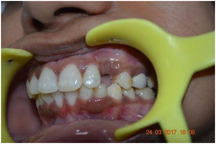
(Figure 1a Missing upper left canine)

Figure 1b OPG revealed impacted permanent upper left canine and lower permanent canines)
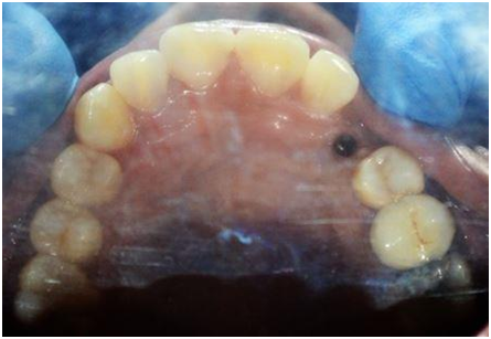
(Figure 2a Implant placed)

(Figure 2b OPG after implant placement)
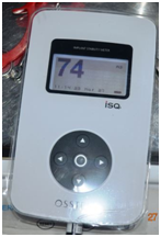
(Figure 3 Implant stability Quotient -Osstell)

(Figure 4a Implant level impression made)

Figure 4 b,c,d:Screw retained crow
DISCUSSION
Impacted teeth can form cysts around the tooth and resorption of the roots of the neighboring permanent teeth which can be a complication. Depending upon the case, there are various treatment methods for treating an impacted canine. Orthodontically, maxillary impacted canines can be treated, otherwise surgical removal remains the alternative treatment option5
Three basic protocols for implant placement were defined according to the time between tooth extraction and implant installation.6 In the type-1 protocol (immediate implant installation), implants are placed in fresh extraction sockets, with the aim to engage the remaining socket walls with the implant. In the type-2 protocol (early implant placement), implants are placed approximately 4–8 weeks after tooth extraction. The main objective of this protocol is to ensure the lack of pathology when placing the implant and, at the same time, to optimize the availability of soft tissue for primary healing and probable lateral bone augmentation. In the type-3 protocol (early delayed/conventional implant placement) the implants are placed once most of the dimensional changes in the alveolar ridge have occurred (12–16 weeks).
Chen et al. analyzed immediate and early postextraction implants on the basis of survival rates and clinical outcomes. He concluded that there are no statistically significant differences in terms of implant survival in the short term (12 months of follow-up).7 Even the Quirynen’s group concluded the same due to the absence of long term data i.e., the clinical outcomes of postextraction implants and implants placed in healed sites are practically the same.8 In the present scenario , type -2 protocol was followed.
The fixture placement should be based upon an adequate understanding of the structural changes that occur in the alveolar process after tooth loss( dental extraction) .9It is important to extract teeth atraumatically to preserve available bone for placement of immediate implants, otherwise in such cases bone grafts should be used to provide primary stability to the implant and speed up the site for filling/healing.5
Therefore, the clinician must know and predict the changes of postextraction ridge to plan the timing of implant placement and the management of the site in the best way possible in order to get predictable results.
In the present case , implant stability ( ISQ value) was determined by the Osstell apparatus A substantial increase or decrease in implant stability could be detected with this method that otherwise could not be clinically perceived. The factors affecting the readings are effective implant length, bone quality and quantity, implant length, diameter and shape. Effective implant length is the length of the exposed threads and abutment height.10
CONCLUSION
Treatment of impacted maxillary cuspids can be challenging when implants are considered in that area. The present case took around 4 months months to complete, from extraction of impacted canine to delivering of implant supported final prosthesis. Due to time limitation, Impacted canine was removed in least invasive manner and insertion torque level >45 Ncm was achieved i.e. primary stability of implant was obtained. Hence, use of bone graft was not required. At the time of delivering crown, implant stability was determined by ISQ value which indicated high stability.
Declaration Of Patient Consent
The authors certify that they have obtained all appropriate patient consent forms. In the form the patient(s) has/have given his/her/their consent for his/her/their images and other clinical information to be reported in the journal. Due efforts will be made to conceal patients name and identity, but anonymity cannot be guaranteed.
Financial Support And Sponsorship
Nil
Conflict Of Interest
There are no conflicts of interest
REFERENCES
- Cooke J, Wang HL. Canine impactions: incidence and management. Int J Periodontics Restorative Dent 2006;26:483-91.
- Sagne S, Thilander B. Transalveolar transplantation of maxillary canines. A follow-up study. Eur J Orthod 1990;12:140-47.
- Ioannidou E, Makris GP. Twelve-year follow-up of an autogenous mandible canine transplant. Oral Surg Oral Med Oral Pathol Oral RadiolEndod 2003;96:582-90.
- Lanka Mahesh, Maurice Salama, Gregori M. Kurtzman. Extraction of an impacted maxillary canine with immediate implant placement. Inside Dentistr2012;8(3).
- Garcia B, Boronat A, Larrazabal C, Penarrocha M, Penarrocha M. Immediate implants after the removal of maxillary impacted canines: a clinical series of nine patients. Int J Oral Maxillofac Implants 2009;24:348-52.
- Hammerle CH, Chen ST, Wilson Jr TG. Consensus statements and recommended clinical procedures regarding the placement of implants in extraction sockets.Int J Oral Maxillofac Implants 2004;19(Suppl):26-8.
- Chen ST, Wilson Jr TG, Hammerle CH. Immediate or early placement of implants following tooth extraction: review of biologic basis, clinical procedures, and outcomes. Int J Oral Maxillofac Implants 2004;19:12-25.
- Quirynen M, Van Assche N, Botticelli D, Berglundh T. How does the timing of implant placement to extraction affect outcome?. International Journal of Oral & Maxillofacial Implants 2007;22:203-23.
- Hämmerle CH, Araújo MG, Simion M, Osteology Consensus Group 2011. Evidenceâeuro;based knowledge on the biology and treatment of extraction sockets. Clinical oral implants research 2012;23:80-2.
- Meredith N, Alleyne D, Cawley P. Quantitative determination of the stability of the implant-tissue interface using resonance frequency analysis. Clin Oral Implants Res 1996;7:261-7.
Disclaimer from Guident
The views and opinions expressed in articles published in Guident are solely those of the authors and do not necessarily reflect the views of the editors, the editorial board or the publisher, Ivory India. Guident does not endorse any product, technique or treatment described. Clinical content is intended for qualified dental and medical professionals and is not a substitute for individual clinical judgement. Guident, its editors and the publisher accept no liability for any consequence arising from the use of information published in the journal.
How to cite this article
Dr. Gaurav Gupta Jaipur Dental College, Dr. D.K Gupta Dr. Neelija Gupta Department of Pedodontics and Preventive Dentistry. Alternative Management of Impacted Maxillary Canine - a Case Report. Guident. 2018 Jul;11(8). Available from: https://guident.in/articles/implantology/alternative-management-of-impacted-maxillary-canine-a-case-report.html
