Anteriors with Two Root Canals - a Report of 2 Cases
Department of Dentistry Burhani Hospital, Surat, India
INTRODUCTION
A root canal is the naturally occurring anatomic space within the root of a tooth. It is composed of pulp chamber (within the crown part) and main canal and its accessory canals in the surface of the root.
ACCESSORY CANALS
- Minute canals that extend in a horizontal, vertical or lateral direction from the pulp to the peridontium.
- Contains connective tissue and vessels but do not supply the pulp with collateral circulation.
- Formed by the entrapment of periodontal vessels in Hertwig’s Epithelial Root Sheath {HERS} during calcification- GROSSMAN.
- These accessory canals are usually located in areas where the roots of multi-rooted teeth join, or around the last third of the root, but can also be found anywhere along the length of a main root canal.
According to Cohen, incidence of accessory canals-
73.5%- Apical 1/3rd
11.4%- Middle 1/3rd
15.1%- Cervical 1/3rd
LATERAL CANALS
An accessory canal that branches to the lateral surface of the root.

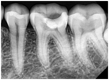
An x-ray showing multiple accessory canals successfully treated
X-ray provided by Dr. Michael Scianamblo
CASE-1
A female patient of age 49 years reported with a complaint of slow growing swelling on lower left side of jaw since 2 months which was associated with mild localised dull, aching pain with difficulty in chewing. Extra orally swelling over the lateral surface of the chin over the left side. IOPA revealed a large radiolucency around the apical area of canine.
Treatment done was non-surgical root canal treatment exploration, cleaning, shaping and filling of root canal with ceramic crown placement in relation to lower left canine.
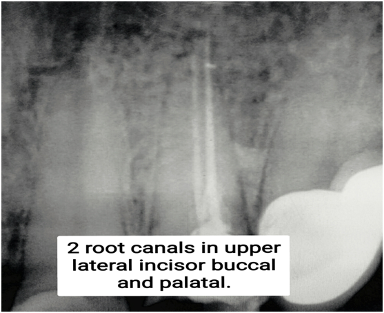
CASE 1 IOPA- showing 2 distinct root canals in upper lateral incisor
CASE-2
A female patient 53years old present with a chief complaint of swelling on upper lip on the left side of the mouth since 1 month. Patient gave history of swelling in the same region 2 years back which regressed after root canal treatment {RCT} and later recurred.
On intraoral examination, a small solitary swelling was evident on palatal aspect of lateral incisor. Tooth was tender on percussion. Suggestive of root canal failure. IOPA showed apical radiolucency in the endodontically treated tooth.
Treatment done was – Re-Root Canal Treatment with evident lateral canal in Lateral incisor and later on replacement with a ceramic crown.
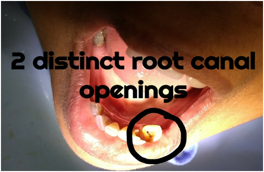
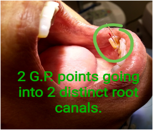
Patient's intraoral images showing 2 root canals
DISCUSSION
Disturbances in normal development of HERS may lead to few dental anomalies like grooves formation, dens in dente, fusion or germination.
Careful examination of tooth before and during access cavity preparation is essential along with thorough probing of pulp chamber. A common reason for not locating a second canal is inadequate access opening into the tooth which leaves a lingual shelf of dentine over the second (usually the lingual canal).
Accurate pre-operative radiographs, straight and angled, using a parallel technique and use of dental microscope can add on to providing clued of multiple number of root canals.
Thus these two cases demonstrates that conventional root canal therapy can be an acceptable treatment modality for the teeth with anatomical irregularities provided all canals are located, cleaned and shaped, disinfected and filled adequately.
CONCLUSION
These case reports highlight the clinical importance and its management of lateral canals. One should be aware of the anatomical root morphology, variability such as lateral canals and apical ramification to determine proper endodontic treatment success.
Based upon, follow-up of these 2 cases we can state that infection via lateral or accessory canals can be successfully managed by non-surgical endodontic treatment with beautiful results.
Many times C-shaped canal system tends to vary considerably in their configuration and therefore may lead to complexities in root canal debridement, cleaning and shaping, filling and restoration.
REFERENCES
- Vertucci FJ root canal morphology and its relationship to endodontic treatment.
- Cooke and Cox C-shaped canal configuration.
- 3.Grossman’s Endodontic Practice 12th edition chapter 9, page181-189.
Disclaimer from Guident
The views and opinions expressed in articles published in Guident are solely those of the authors and do not necessarily reflect the views of the editors, the editorial board or the publisher, Ivory India. Guident does not endorse any product, technique or treatment described. Clinical content is intended for qualified dental and medical professionals and is not a substitute for individual clinical judgement. Guident, its editors and the publisher accept no liability for any consequence arising from the use of information published in the journal.
How to cite this article
Dr. Kalpana Sinsinbar. Anteriors with Two Root Canals - a Report of 2 Cases. Guident. 2018 Jul;11(8). Available from: https://guident.in/articles/endodontics/anteriors-with-two-root-canals-a-report-of-2-cases.html