Implant Placement with GBR in the Mandible: Case Study
School of Dental Medicine, University of Berne, Switzerland
Circumferential bone anchorage in the alveolar ridge is key to the long-term success of an implant. Therefore, bone defects at the implant site have to be corrected by means of GBR.
The same surgical technique with simultaneous GBR therapy presented in this case report has been used at our clinic since the turn of the century. Inaddition, the long-term result after
11.5 years is presented.
GBR WITH BONE, BIOMATERIAL AND MEMBRANE
The patient was referred after the extraction of two molars(36 and 37). The result was a local bone defect in region 36.The implant surgery took place about four months later. This involved a 10 mm long tissue level implant (Straumann AG,Basel) inserted in the correct prosthetic position with goodprimary stability. There was a medium-sized buccal bone defect with a favourable 2-wall defect morphology. This defect wasfilled initially with locally harvested, autologous bone chips andthen covered with Geistlich Bio-Oss® granules. The graft was covered with a Geistlich Bio- Gide® membrane applied in a double layer. After the periosteal incision, the tension-free primary wound closure concluded the procedure. Five months later the implant was uncovered and prosthetic restoration was performed by the referring colleague.The clinical examination after 11.5 years revealed a peri-implant mucosa that was free of inflammation, and the radiograph showed stable bone crest levels. A Cone Beam CT confirmed the existence of an intact buccal bone wall.
WHAT MAKES THE METHOD WORK SO WELL?
Two long-term studies with CBCT imaging showed excellent results with regeneration of a buccal bone wall. These resultscan be attributed primarily to the applied biomaterials that complement one another optimally. The locally harveste dautologous bone chips stimulate new bone formation across the defect area in the early phase of healing. This stimulation is caused by growth factors in the bone matrix that are passedinto the surrounding blood clot. The Geistlich Bio-Oss® granules applied have the critical function of preserving the regeneratedbone volume long-term.Various histological studies have indicated that Geistlich Bio-Oss® has a very low substitution rate. Geistlich Bio-Gide® membrane made from noncross- linked collagen is simple to apply, has few complications, fulfils the vital barrier function for four to eight weeks and stabilizes the applied bone filler. And it does not have to be removed, because it is slowly absorbed by tissue.
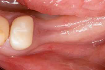
Situation after the extraction of tooth 36 and 37.
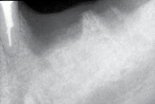
Local bone defect in region 36.
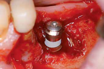
Buccal bone defect with dual-walled morphology.
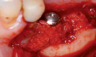
Situation after implantation and filling the defect with autologous bone
chips.
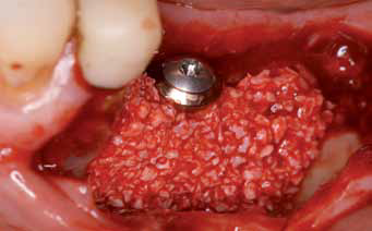
Geistlich Bio-Oss® Granulate is applied to the bone.
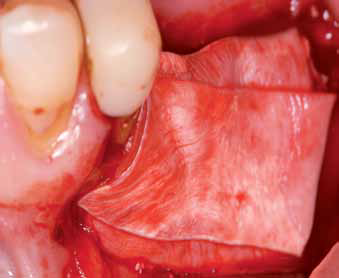
A double layer of Geistlich Bio-Gide® covers the graft.
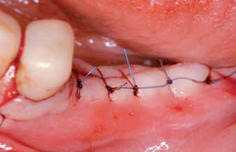
Primary wound closure after periosteal incision.
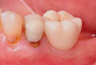
Prosthetic restoration 1-year after implantation.
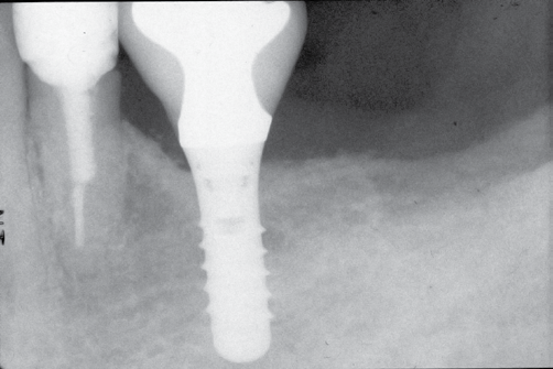
Radiograph findings 1-year after implantation.
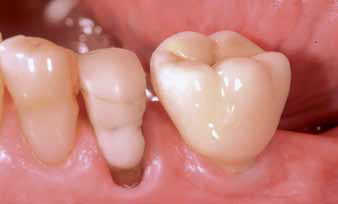
Stable clinical situation after 11.5 years.
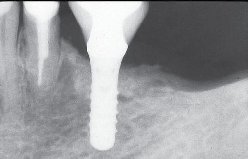
The radiograph shows a stable bone situation after 11.5 years.
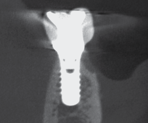
The CBCT image shows an intact buccal bone wall after 11.5 years.
LITERATURE
- Buser D, et al.: J Periodontol 2013; 84: 1517–27.
- Buser D, et al.: J Dent Res 2013; 92: 176S–82S.
- Buser D, et al.: Int J Periodont Rest Dent 2008; 28: 440–51.
- Caballé-Serrano J, et al.: Clin Oral Invest (2016, accepted)
- Caballé-Serrano J, et al.: Clin Oral Implants Res. 2016 (e-pub)
- Jensen SS, et al.: Clin Oral Impl Res 2006 ; 17: 237–43.
- Jensen SS, et al.: J Periodontol 2014; 85: 1549–56.
DISCLAIMER
This Article was first published in Geistlich News Volume (Issue 1/2016). Article reprint with the permission of the Author and Geistlich Pharma India Pvt. Ltd.
Disclaimer from Guident
The views and opinions expressed in articles published in Guident are solely those of the authors and do not necessarily reflect the views of the editors, the editorial board or the publisher, Ivory India. Guident does not endorse any product, technique or treatment described. Clinical content is intended for qualified dental and medical professionals and is not a substitute for individual clinical judgement. Guident, its editors and the publisher accept no liability for any consequence arising from the use of information published in the journal.
How to cite this article
Dr. Daniel Buser Department of Oral Surgery and Stomatology. Implant Placement with GBR in the Mandible: Case Study. Guident. 2016 Sep;9(10). Available from: https://guident.in/articles/implantology/implant-placement-with-gbr-in-the-mandible-case-study.html