Minimal Intervention Slot Preparations
Victoria, Australia
Over the millennia teeth have adapted to managing carious lesions so that progression is often slow and tooth damage is contained. The advent of the air rotor has dramatically increased pulpal response to the extent that tooth preparation can damage pulps more than the pre existing lesion.
There is a need to review the manner in which dentists approach the removal of caries, particularly from proximal lesions where extensive tooth structure needs to be removed to gain access.
John McLean was talking about slot preparations in the early 80’s although it was a technique that didn’t seem to take of, probably due to the difficulties in determining the extent of the lesion and uncertainties about complete caries removal.
Technologies such as KaVo’s HealOzone, laser photo activated dye disinfection (PAD) and AgF/KI are emerging to treat caries in situ by disinfection and changing demineralized tooth structure to resist further caries attack.
With these technologies, access is still needed to the caries but complete caries removal may not be required, thus changing the whole dynamics of restorative dentistry.
The patient is a young adult male whose changing lifestyle had initiated a series of quite rapidly advancing carious lesions. Conventional access to the mesial lesion in 17 would have required removal of a large amount of healthy tooth structure and the distal inclination of the tooth challenged the standard tunnel preparation technique.


Fig 1 Mesial lesion on requiring extensive tooth removal to gain access.
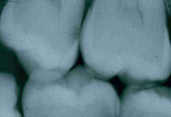
Fig 2. Radiograph of lesion.
Access was achieved using an end cutting high speed diamond inclined in a slightly apical direction. The preparation must be deep enough to avoid cutting the proximal surface of the adjacent tooth and preparation ceases when the operator’s sensory perception of the bur recognized that the tip has reached the lesion.
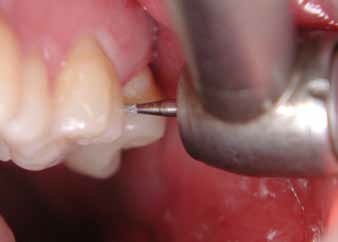
Fig 3. Initial access using high speed diamond bur.
This is followed by gentle preparation of the cavity using a #6 round steel bur, again relying upon the operator’s tactile sensation of the lesion.
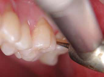
Fig 4. Further preparation using a slow speed #6 round steel bur.
Uncertain that all caries had been removed the preparation was etched for 5 seconds with 35% phosphoric acid, washed and dried. A solution of Diammine Silver Fluoride (Saforide, Halas) was placed into the preparation with a micro brush followed by several applications of Potassium Iodide (KI generic) to reduce the staining effects of the AgF. The preparation was flushed with water and dried with oil free air.
A stainless steel sectional matrix was placed into the interproximal space and lightly wedged from the facial and lingual to secure the matrix and prevent overhanging margins.
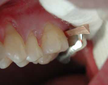
Fig 5. Sectional matrix secured with buccal and lingual wooden wedges.
A fast set capsulated GIC (Fuji IX, GC Corp. Ketac Molar, 3M ESPE. Riva, SDI) was placed to slightly overfill the preparation and allowed to set. Figure 6.
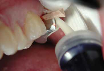
Fig 6. Placement of fast set GIC from capsule.
After setting, the matrix was removed and the GIC contoured to the tooth.
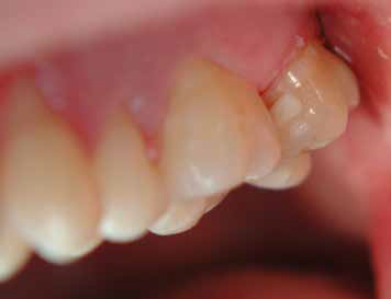
Fig 7. Inserted GIC restoration.
A radiograph of the completed restoration shows to GIC restoration in place requiring much less tooth preparation than either a standard Class II preparation or tunnel preparation.

Fig 8. Radiograph of the completed restoration.
The transilluminated bicuspid shows a carious lesion in the proximal surface.

Fig 9. Trans- illuminataed lesion on proximal surface.
Cutting a slot to the depth of the DEJ removes most of the caries. Figure 10. Treatment of the remaining caries with AgF/KI will disinfect the preparation and prevent caries developing at the restorative margins.
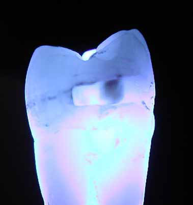
Fig 10. Caries remaining for AgF/Kl tratment.
Over the last few years there has been an emerging interest using a non surgical approach to the management of dental caries. It is early days but clearly pointing to the future direction of caries management.
The next generation of dentists may wonder why it took today’s profession over 150 years to apply non surgical modalities to the management of caries and no doubt somewhat confused why we found it necessary remove healthy tooth structure to achieve temporary changes in oral cosmetics.
DISCLAIMER
Published in the Austrailion Dental Association News bulletin May 2004. Reprinted with permission of editor and author.
Disclaimer from Guident
The views and opinions expressed in articles published in Guident are solely those of the authors and do not necessarily reflect the views of the editors, the editorial board or the publisher, Ivory India. Guident does not endorse any product, technique or treatment described. Clinical content is intended for qualified dental and medical professionals and is not a substitute for individual clinical judgement. Guident, its editors and the publisher accept no liability for any consequence arising from the use of information published in the journal.
How to cite this article
Dr. Geoffrey M Knight. Minimal Intervention Slot Preparations. Guident. 2016 Sep;9(10). Available from: https://guident.in/articles/aesthetic-dentistry/minimal-intervention-slot-preparations.html