Application of PRF in Alveolar Ridge Augmentation
Department of Prosthodontics, K.D. Dental College & Hospital, Mathura
ABSTRACT
BACKGROUND: Different barrier membranes and augmentation techniques are used to recover lost bone structures with varied success. Recently, a combination between bone graft materials and Platelet-Rich-Fibrin (PRF) is implemented in implantology.
AIM: Aim of this study is to assess the possibility of bone augmentation using PRF
CONCLUSION: using A-PRF and i-PRF can be beneficial for bone augmentation of the alveolar ridge before implant placement.
INTRODUCTION
Extraction of teeth will be followed undoubtedly by loss in height and width of the alveolar process. It results in narrowing and shortening of the alveolar ridge.
In the past partial dentures or FPD could camouflage the alveolar defects and the resorption process could be counterattacked by repair of the partial dentures.
However there is paradigm shift from partial dentures to implant supported dentures. Implants are an aid for prosthetic devices and therefore need to be placed in a three dimensionally perfect location.
Especially in the esthetic zone it is important to simulate the natural contours even if there is no tooth or root in the alveolar process.
Different barrier membranes and augmentation techniques are used to recover lost bone structures with varied success .The majority of authors recognize the autogenous bone as the ‘golden standard’. The autogenous bone is the only one that has osteogenic, osteoinductive and osteoconductive qualities, while other materiaals possess only osteoconductive properties. It is well known that PRF is able to stimulate integration and remodulation of bone structure.
BIOLOGICAL ASPECT
Developed in France by Choukroun et al. the PRF production protocol attempts to accumulate platelets and released cytokines in a fibrin clot. Granules present in platelet contain many proteins, which may be platelet specific (eg. beta-thromboglobulins) or non platelet specific (fibronectin, thrombospondin, fibrinogen, and other coagulation, growth promoters, fibrinolysis inhibitors, immunoglobulins etc) calcium and serotonin etc. Also phospholipids double layer of platelet membrane constitute many receptors for other molecules. Growth factors released by alpha-granules encompass a group of cytokine polypeptides with relatively low molecular weight ranging from 6 to 45 kDa. Activation and degranulation is important to initiate and support aggregation at the healing site and the release of the cytokines (IL-1 beta, IL-6, TNF-alpha) 14 and growth factors (TGF beta 1, PDGF, VEGF, EGF) that stimulates cell migration and proliferation within the fibrin matrix and thus begins the first stage of healing.
A soluble fibrillary molecule, fibrin is an activated form of plasmatic molecule fibrinogen that is massively present both in plasma and in the platelet alpha granules that plays potential role in platelet aggregation during homeostasis. Fibrinogen is the final substrate of all coagulation reactions which is transformed into an insoluble fibrin by thrombin while the polymerized fibrin gel constitutes the first cicatricial marix of the injured site. Characteristic of polymerisation naturally and slowly during centrifugation, along with physiologic thrombin action on collected autologus fibrinogen is crucial to determine the three dimensional organization of fibrin network that will give great elasticity and very strong PRF membrane.
METHOD FOR OBTAINING PLATELET RICH FIBRIN (PRF)
A blood sample is taken without anticoagulant in 10 ml tubes and immediately centrifuged at 2700-3000 rpm for 10-12 minutes. The resultant product consists of following three layers: (a) RBC at the bottom, (b) PRF clot in middle and (c) upper most layer consisting of platelet poor plasma (PPP). The outcome of this technique entirely depends upon the speed of blood collection and transfer to the centrifuge. As the blood sample starts to coagulate almost immediately upon contact with the tube-glass and it takes a minimum of few minutes of centrifugation to concentrate fibrinogen in middle and upper part of the tube, rapid handling is the only way to obtain a clinical usable PRF clot and any delay to collect blood and starts centrifugation may be the cause of failure resulting in polymerisation of fibrin in a diffuse way in the tube with small blood clot without consistency. Driving out the fluids trapped in the fibrin matrix by squeezing the PRF clot between the sterile dry gauze, practitioners will obtain a highly resistant autologous PRF membrane (a highly promising biomaterial) for multiple clinical usage.
PRF clot obtained after centrifugation is removed from the test tube and attached red blood cells scraped off and discarded, 27 but it should be remembered that PRF membranes are inhomogeneous and platelets and leukocytes are concentrated within one end (towards RBCs) of membrane.
ADVANTAGES OF PRF OVER PRP
PRP requires large amount of blood (450 ml) and is done in hospital settings. For platelet concentrating systems a double spin or single spin technique is utilised, 31 on other hand PRF can be procured by using a table top centrifuged in a matter of 10 minutes.
The most important advantage of PRF over PRP has been the deletion of any additive constituent such as bovine thrombin which is mandatory for making PRP.
APPLICATION OF PRF IN RIDGE AUGMENTATION
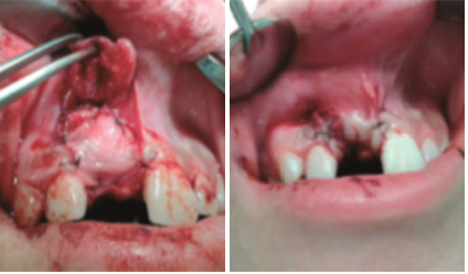
PRF membranes protect the surgical site and promote soft tissue healing and cut few milimeters PRF fragments mixed with graft material functioned as a “biological connector” between the different graft elements, and as a matrix that supports neo-angiogenesis, capture of stem cells and migration of osteoprogenitor cells to the center of the graft.
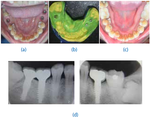
Immediate bone augmentation after infected tooth extraction using titanium membranes applied to the socket walls followed by socket filling with autologous platelet-rich fibrin and primary closure was found to be feasible and safe and yielded adequate bone filling to support implant fixation at 8 weeks or above.


DISCUSSION
Placement of dental implants is directly related to the volume of the remaining alveolar bone. It is necessary to create new bone structure when there is an absence of a sufficient volume, in order to place a dental implant. To form new bone structure, the bone graft material must be biocompatible and able to withstand the biomechanical stress. Using bone grafts is very successful when it is performed in an environment with good blood supply. This depends on the primary wound closure and the condition of the adjacent bone. Good blood flow assures the necessary cells, growth factors and promoters for the osteogenic-biomineralisation cascade. A biological connector between the different parts of the bone graft material is obtained when it is mixed with A-PRF and i-PRF. PRF assists angiogenesis, engagement of the stem cells and migration of osteogeneous cells in the central part of the graft. According to Toffler M et al. using PRF membrane in augmentation procedures is beneficial for better wound closure and faster recovery of the soft tissues. Other authors concluded that the use of PRF reduces postoperative pain, swelling and minimizes the infection phenomenon.
CONCLUSION
PRF increases the amount of growth factors inside the wound and improves the qualities of the bone graft material. Further research and more clinical cases are needed to confirm the achieved results.
REFERENCES
Chenchev IL, Ivanova VV et al. Application of Platelet-Rich Fibrin and Injectable Platelet-Rich Fibrin in Combination of Bone Substitute Material for Alveolar Ridge Augmentation - a Case Folia Medica 2017; 59(3):362-366
Heggeler Ten JMAG , Slot DE et al. Effect of socket preservation therapies following tooth extraction in non-molar regions in humans: a systematic review. Clin. Oral Impl. Res. 22, 2011; 779–788
Wang HL, Boyapati L. “PASS” Principles for Predictable Bone Regeneration Implant Dent 2006; 15:8–17
Gupta V, Mathur A et al. Regenerative Potential of Platelet Rich Fibrin In Dentistry: Literature Review. Asian J Oral Health Allied Sci. 2011;1(1)
Disclaimer from Guident
The views and opinions expressed in articles published in Guident are solely those of the authors and do not necessarily reflect the views of the editors, the editorial board or the publisher, Ivory India. Guident does not endorse any product, technique or treatment described. Clinical content is intended for qualified dental and medical professionals and is not a substitute for individual clinical judgement. Guident, its editors and the publisher accept no liability for any consequence arising from the use of information published in the journal.
How to cite this article
Dr. Shaivy Ambuj, Dr. Neha Srivastava, Dr. Manesh Lahori. Application of PRF in Alveolar Ridge Augmentation. Guident. 2019 Jun;12(7). Available from: https://guident.in/articles/implantology/application-of-prf-in-alveolar-ridge-augmentation.html