
Management of a Large Periapical Cyst
Department of Conservative Dentistry & Endodontics, Darshan Dental College & Hospital, Udaipur Dr. Jitender Lohar (Reader), Department of Conservative Dentistry & Endodontics, Pacific Dental College & Research Centre, Udaipur
ABSTRACT
Traumatic injuries commonly affect the anterior teeth leading to slow death of the pulp which result to the formation of periapical cyst by the proliferation of cell rests of Malassez following pulpal necrosis. Most of the conditions are usually asymptomatic but can result in a slow-growth tumefaction in the affected region which can be painful. Radiographic examination shows round or oval, well circumscribed radiolucent area involving the apex of the infected tooth. The first line of treatment choice is Nonsurgical management of a periapical cyst. However, if lesion does not respond to a nonsurgical approach periapical surgery can be considered.
Key Words: Non-Vital Tooth, Radicular Cyst, Periapical Surgery, Platelet Rich Fibrin (Prf)
INTRODUCTION
Radicular cysts are the most common (52%-68%) cystic lesions affecting the jaws. They are most commonly found at the apices of the involved teeth, however they may also be found on the lateral aspects of the roots in relation to lateral accessory root canals.1 Radicular cysts are direct sequel to chronic apical periodontitis but not every chronic lesion develops into a cyst. These cysts can occur in the periapical area of any teeth, at any age but are seldom seen associated with the primary dentition.2 It is more frequent in maxillary than mandibular teeth.
Most of the radicular cyst are symptom less and are discovered when periapical radiograph are taken of teeth with non-vital pulps. Patient often complains of slowly enlarging swellings.3 Radiographically most radicular cyst appears as round or pear shaped unilocular radiolucent lesion in the periapical region. The cyst may displace adjacent teeth or cause mild root resorption.4 The treatment options for radicular cyst can be conventional nonsurgical root canal therapy when lesion is localized or surgical treatment like enucleation,
marsupialization or decompression when lesion is large.5 This case report presents the successful surgical management of large infected radicular cyst.
CASE REPORT
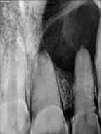
A 28 year old male patient reported to the Department of Conservative Dentistry & Endodontics, Darshan dental College and hospital udaipur with the chief complaint of pain and swelling in the upper palatal region of jaw. He gave history of trauma 12 years back in upper anterior tooth. Post trauma, he had pain in relation to maxillary central and lateral incisor which subsided after 1 week on its own. He gave a history of swelling along with pus discharge from gumssince last 3 month. On clinical examination, 21,22 was found to be discolored and a diffuse palatal swelling of approximately 3 x 2 cms in size, extended 2 cms on the left and 1 cm on the right from the midline of the palate. The swelling was tender and fluctuant on palpation. Vitality test gave a negative response in relation to 21 and 22. An Intra Oral Periapical Radiograph (IOPA) in relation to 21 & 22 (Figure. 1) revealed large well-defined periapical radiolucency involving central and lateral incisor.
On FNAC, about 0.8 ml of straw colored, shimmering fluid, serous in consistency was aspirated and sent for cytological examination which revealed numerous cholesterol crystals and RBCs along with few inflammatory cells. On the basis of clinical, radiological and cytological examination, a provisional diagnosis of Radicular cyst was made. Differential
diagnosis Included Nasopalatine cyst, Median palatal cyst, Traumatic bone cyst, and Globulomaxillary cyst.
An endodontic therapy was planned with respect to 21 & 22 (Figure. II). Patient became extremely apprehensive whenever an effort was made to apply rubber dam, which contraindicated its usage. Repeated calcium hydroxide dressings were given at an interval of 5 days for 1 month.

Figure. II (a) Figure .II (b) Figure. II (c)
However, there was continuous fluid drainage through the canal of 21; hence surgical enucleation of the cyst (Figure. III) through a palatal approach was adopted, full thickness flap was raised.

Cystic fluid was clearly visible through the perforation. Cyst enucleation was carried out into Enucleated cystic lining (Fig. IVa)
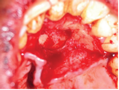
was sent for histopathological examination which revealed 6-7 cell layered thick, non keratinized stratified squamous epithelium. Connective tissue capsule showed the presence of cholesterol crystals along with acute & chronic inflammatory cells which confirmed the diagnosis of an Infected Radicular cyst (Fig. IVb).

and prf placed in the cystic cavity and flap was secured in position with sutures (Figure. V) and after 1 week the sutures were removed. A marked reduction in palatal swelling could be appreciated after one month( Figure. VI)
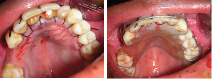
Figure. V Figure. VI
DISCUSSION
The periapical cyst is the most common odontogenic cyst (52.3-70.7 %) followed by the dentigerous cyst (16.6-21.3 %) and odontogenic keratocyst(5.4 - 17.4 %).2 Treatment planning depend open determined by factors such as the extension of the lesion, relation with noble structures, evolution, origin, clinical characteristic of the lesion such as Discolored,Diffuse palatal swelling,Preoperative IOPA and systemic condition and cooperation of the patient.3 The treatment of these cysts is still under discussion and manyprofessionals opt for a conservative treatment by means of an endodontic technique.4,5 However, in large lesions the endodontic treatment alone is not sufficient and it should beza associated with decompression or marsupialisation or enucleation.6 In this case, endodontic treatment was carried out in multiple visits with calcium hydroxide dressings. The placement of root canal medicament between appointment in root canal treatment of teeth with chronic periapical lesions is important for reducing bacteria better than that obtained with mechanical preparation, particularly by penetration of areas that are unreachable by instruments or irrigation solutions, such as dentinal tubules and ramifications. Calcium hydroxide has also shown clinical efficiency in reducing exudate due to its hygroscopic properties. Studies have shown that at least 2 weeks are necessary for calcium hydroxide bactericidal activity7 Theperiapical lesion did not respond to the intracanal medicament because of the causative factor being located beyond the root canal system, viz., within the inflamed periapical tissue, thus requiring surgical intervention. After the surgery a plan end of 6 month follow-up, patient was free from all symptoms and signs with optimum tissue healing. Radiographic examination revealed a decrease in the size of periapical radiolucency bilaterally. Frank 10 reported the treatment of 3 cases based on the normal physiologic pattern of root development that brings about the resumption of apical development so that the root canal can be obliterated by conventional lateral condensation techniques. Considering patient’s young age and good health, bone graft was not used. This case is a good example of a large lesion been treated with conventional RCT and cyst enucleation without the use of materials which enhance bone regeneration like Platelet Rich Fibrin (PRF) G-bone (Synthetic Granules and blocks made of Multiphasic Calcium Hydroxyapatite in low crystalline form).
CONCLUSION
The clinical case in this article was managed successfully by root canal therapy followed by surgery. It is suggested that the treatment of the periapical lesion should be defined according to the clinical and radiographic evaluations according to each case.
REFERENCES
- Massive residual dental cyst: Case report George Dimitroulis, John Curtin, Australian Dental Journal1998; 43: (4)
- Ali M, Baughman RA: Maxillary odontogenic keratocyst: A common and serious clinical misdiagnosis. JADA, 2003; 134 (7): 877-883.
- Ribeiro PD (Jr.) Gonçalves ES, Neto ES, Pacenko MR: Surgical approaches of extensive Periapicalcyst. Considerations about Surgical technique. Salusvita,
- Hoen MM, LaBounty GL, Strittmatter EJ: Conservative treatment of persistent periradicular lesions using aspiration and irrigation. J Endod, 1990; 16 (4): 182- 186.
- Rees JS: Conservative management of a large maxillary cyst. Int Endo J, 1997; 30 (1): 64-67.
- Caliskan MK: Prognosis of large cyst-like periapical lesions following nonsurgical root canal treatment: a clinical review. Int Endod J, 2004;37(6):408-416.
- Leonardo MR, Silveira FF, Silva LA, Tanomaru Filho
- Frank AL: Therapy for divergent pulpless tooth by continued apical formation. JADA, 1966; 72(1):87-93.
Disclaimer from Guident
The views and opinions expressed in articles published in Guident are solely those of the authors and do not necessarily reflect the views of the editors, the editorial board or the publisher, Ivory India. Guident does not endorse any product, technique or treatment described. Clinical content is intended for qualified dental and medical professionals and is not a substitute for individual clinical judgement. Guident, its editors and the publisher accept no liability for any consequence arising from the use of information published in the journal.
How to cite this article
Dr. Manisha Bhati, Dr. Tariq Mohd. Shekh. Management of a Large Periapical Cyst. Guident. 2019 Jun;12(7). Available from: https://guident.in/articles/endodontics/management-of-a-large-periapical-cyst.html


