Autogeneous Blood Derived Products
Department of Oral & Maxillofacial Surgery, Seema Dental College & Hospital, Rishikesh (249203)
ABSTRACT
The utility of platelet-rich plasma (PRP) has spanned various fields of dentistry, due to its role in wound healing. Though PRP is being used over a long time, there is still confusion over proper terminology to define, classify and describe the different variations of platelet concentrates. There is also a wide variation in the reported protocols for standardization and preparation of PRP, in addition to lack of accurate characterization of the tested products in most articles on the topic. Additionally, the high cost of commercially available PRP kits, precludes its use over a larger population. This article reviews in detail about various blood products prepared and various method of preparation of PRP.
Key words PRP, Autogenous, Blood, Platelets.
Introduction
Blood is a complex tissue based on its derivation from mesenchyme cells and its structure. Blood contains formed elements like RBC, WBC and platelets. Plasma contains organic/inorganic substances, plasma proteins, albumin, globulin and fibrinogen.
Blood when allowed to clot with anti-coagulant it seperates Serum a straw colored fluid. When mixed with anti-coagulant and centrifuged then seperates RBC and plasma.
Pic A: Pre-Operative OPG
PREPARATION OF PRP
PRP is obtained from a sample of patients’ blood drawn at the time of treatment. A 30 cc venous blood draw will yield 3-5 cc of PRP depending on the baseline platelet count of an individual, the device used, and the technique employed. The blood draw occurs with the addition of an anticoagulant, such as citrate dextrose A to prevent platelet activation prior to its use. Preparation-related costs are significantly lower than with commercial kits.
PRINCIPLES OF PRP PREPARATION
PRP is prepared by a process known as differential centrifugation. In differential centrifugation, acceleration force is adjusted to sediment certain cellular constituents based on different specific gravity.
There are many ways of preparing PRP. It can be prepared by the PRP method or by the buffy-coat method.
In the PRP method, an initial centrifugation to separate red blood cells (RBC) is followed by a second centrifugation to concentrate platelets, which are suspended in the smallest final plasma volume.
WB (whole blood) is initially collected in tubes that contain anticoagulants. The first spin step is performed at constant acceleration to separate RBCs from the remaining WB volume. After the first spin step, the WB separates into three layers: an upper layer that contains mostly platelets and WBC, an intermediate thin layer that is known as the buffy coat and that is rich in WBCs, and a bottom layer that consists mostly of RBCs. For the production of pure PRP (P-PRP), upper layer and superficial buffy coat are transferred to an empty sterile tube. For the production of leucocyte rich PRP (L-PRP), the entire layer of buffy coat and few RBCs are transferred. The second spin step is then performed. ‘g’ for second spin should be just adequate to aid in formation of soft pellets (erythrocyte-platelet) at the bottom of the tube. The upper portion of the volume that is composed mostly of PPP (platelet-poor plasma) is removed. Pellets are homogenized in lower 1/3rd (5 ml of plasma) to create the PRP (Platelet-Rich Plasma).
Buffy coat method
In the buffy coat method, whole blood (WB) is centrifuged at a ‘high speed’ with subsequent collection of the buffy coat. A buffy coat contains high concentration of leucocytes.From small volume of WB (10 mL), a very thin layer of a buffy coat can be produced. The difficulty lies in separating this thin buffy coat layer that contains mainly white blood cells (WBCs) and platelets, from the underlying RBC layer.
Procedure 1, 2 PRP method
- Obtain WB by venipuncture in acid citrate dextrose (ACD) tubes
- Do not chill the blood at any time before or during platelet separation.
- Centrifuge the blood using a ‘soft’ spin.
- Transfer the supernatant plasma containing platelets into another sterile tube (without anticoagulant).
- Centrifuge tube at a higher speed (a hard spin) to obtain a platelet concentrate. >
- The lower 1/3rd is PRP and upper 2/3rd is plateletpoor plasma (PPP). At the bottom of the tube, platelet pellets are formed.
- Remove PPP and suspend the platelet pellets in a minimum quantity of plasma (2-4 mL) by gently shaking the tube.
Buffy coat method
- WB should be stored at 20°C to 24°C before centrifugation.
- Centrifuge WB at a ‘high’ speed.
- Three layers are formed because of its density: The bottom layer consisting of RBCs, the middle layer consisting of platelets and WBCs and the top PPP layer.
- Remove supernatant plasma from the top of the container.
- Transfer the buffy-coat layer to another sterile tube.
- Centrifuge at low speed to separate WBCs or use leucocyte filtration filter.
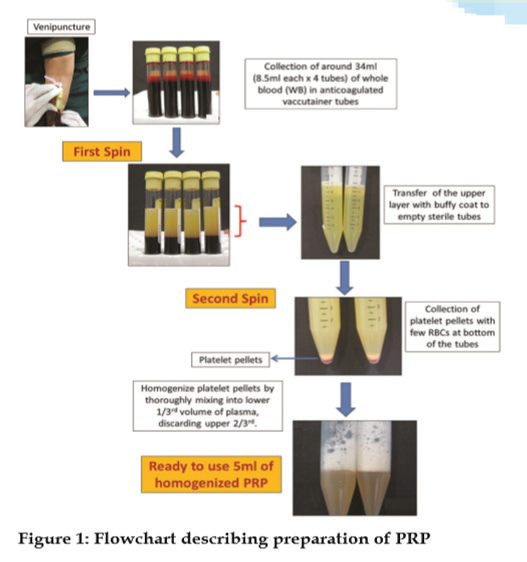
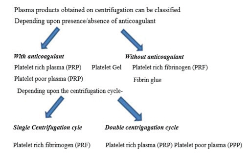
Preparation of PRP

Preparation of platelet rich fibrin

Platelet deficient products

Platelet deficient products
COMMERCIALLY AVAILABLE PRP KITS
There are many PRP systems commercially marketed, which facilitate the preparation of ready to apply platelet rich suspensions in a reproducible manner. All operate on a small volume of drawn blood (20-60 mL) and on the principle of centrifugation. These systems differ widely in their ability to collect and concentrate platelets depending on the method and time of its centrifugation. As a result, suspensions of different concentration of platelets and leucocytes are obtained.
Differences in the concentrations in platelets and WBCs influence the diversity of growth factors concentration. It is difficult to assess which kit for PRP preparation is better and which is worse PRP devices can be usually divided into lower (2.5-3 times baseline concentration) and higher (5-9 times baseline concentration) systems.
The high-yielding devices include Biomet GPS II and III (platelet count 3-8×); Harvest SmartPRep 2 APC+ (4-6×); ArterioCyteMedtronic Magellan (3-7×). The lower concentration systems include Arthrex ACP (2-3×), Cascade PPR therapy (1-1.5×), and PRGF by Boitech Institute Vitoria, Spain (2-3×).Regen PRP (Regen Laboratory, Mollens, Switzerland).4-8
Uses of PRP
PRP provides an immediate surgical hemostatic agent that is biocompatible, safe, and effective. PRP accelerates endothelial, epithelial, and epidermal regeneration, stimulates angiogenesis, enhances collagen synthesis, promotes soft tissue healing, decreases dermal scarring, enhances the hemostatic response to injury, and reverses the inhibition of wound healing caused by glucocorticoids. The high leukocyte concentration of PRP has an added antimicrobial effect. Since PRP is an autologous blood product, it carries no risk of transmitting infectious disease.
PRP has an extremely broad range of clinical healing applications in head and neck surgery, otolaryngology, cardiovascular surgery, burns and wound healing, oral and maxillofacial surgery, cosmetic surgery, and periodontics. In addition to its effectiveness for patients with chronic non-healing wounds, it has also been used as an antiangiogenic agent and as a carrier for growth factors.
References
- Sweeny J, Grossman BJ. Blood collection, storage and component preparation methods. In: Brecher M, editor. Technical Manual. 14th ed. Bethesda MD: American Association of Blood Banks (AABB) 2002;955-8.
- Welsh WJ. Autologous platelet gel: Clinical function and usage in plastic surgery. Cosmetic Derm2000; 11:13-9.
- Scherer SS, Tobalem M, Vigato E, Heit Y, Modarressi A, Hinz B, et al. Non-activated versus thrombin-activated platelets on wound healing and fibroblast-to-myofibroblast differentiation in vivo and in vitro. Plast Reconstr Surg 2012; 129: 46e-54e.
- Platelet Rich Plasma (PRP) Guidelines,© International Cellular Medicine Society - 2011. Available from: http://www.cellmedicinesociety.org. [Last accessed 2014 Dec 5].
- Marlovits S, Mousavi M, Gabler C, Erdös J, Vécsei V, et al. A newsimplified technique for producing platelet-rich plasma: A short technical note. Eur Spine J 13:102-06.
- Waters JH, Roberts KC. Database review of possible factors infuencing point of care platelet gel manufacture. J Extra Corpor Technol 2004; 36:250-4.
- Lozada JL, Caplanis N, Proussaefs P, Willardsen J, Kammeyer G. Plateletrich plasma application in sinus graft surgery: Part I--background and processing techniques. J Oral Implantol 2001; 27:38-42
- Kevy SV, Jacobson MS. Comparison of methods for point of care preparation of autologous platelet gel. J Extra Corpor Technol 2004; 36:28-35.
Disclaimer from Guident
The views and opinions expressed in articles published in Guident are solely those of the authors and do not necessarily reflect the views of the editors, the editorial board or the publisher, Ivory India. Guident does not endorse any product, technique or treatment described. Clinical content is intended for qualified dental and medical professionals and is not a substitute for individual clinical judgement. Guident, its editors and the publisher accept no liability for any consequence arising from the use of information published in the journal.
How to cite this article
Dr. Mudit Agarwal, Dr. R. Muthunagai, Dr. R. Sivasankary. Autogeneous Blood Derived Products. Guident. 2020 Jan;13(2). Available from: https://guident.in/articles/general/autogeneous-blood-derived-products.html