Management of C Shaped Root Canal in the Mandibular Second Molar an Endodontic Challenge
Department of Conservative Dentistry and Endodontics, Faculty of Dental Sciences, BHU, Varanasi
ABSTRACT
The primary goal of root canal treatment is to achieve complete obturation of the canals by thorough cleaning and shaping which eliminates microorganism, debris from all the root canals to avoid recurrence of infection. The presence of unusual complex anatomies always poses a difficulty in performing endodontic therapy. Early negotiation and management of these complex anatomies is important for successful endodontic treatment. The commonest teeth to get unusual internal canal configuration are the mandibular molars. This case report deals with the successful management of single C shaped canal in the mandibular second molar with the help of conventional periapical radiograph and cold lateral condensation technique for obturation.
INTRODUCTION
Normally, mandibular molar have two roots, mesial and distal and three roots canals, but sometimes mandibular molars presents with variations in its internal anatomy of canal configuration. The commonest among the unusual anatomy is C shaped canals, The C shaped canal configuration presents a ribbon shaped orifices looks like 180 0 arcs, usually starts from mesiolingual line angle of the chamber sweeps around buccally and finally ends at the distal aspect. The occurrence of C shaped canal in the mandibular molar was first documented by Cooke and Cox in 1979.1 In mandibular second molars prevalence of C shaped canals ranges from 2.7 % to 44.5 %.3 Apart from high occurrence rate in mandibular second molar, it is also observed in maxillary third molar [4.7 %], mandibular third molar [3.5 to 4 %], mandibular first and second premolar [1%], mandibular and maxillary first molar [0.12 %] and rare occurrence in maxillary lateral incisor.4,7-11 The bilateral occurrence of C configuration canal ranges from 70 to 81%.15,16 It is believed that failure of fusion of Hertwigs Epithelial Root Sheath on the buccal or lingual root surfaces results in the formation of C shaped canals.2 Another reason stated by Manning is that deposition of cementum due to aging results in the formation of C shaped canal.5,6 Incomplete debridement of this complex anatomy results in treatment failure. This case report presents the successful detection and management of C shaped canal by conventional radiographic and obturation techniques.
CASE REPORT
A 22 year old male patient referred to department of conservative dentistry and endodontics with the chief complaint of pain in his lower right back tooth region for past 2 days. His medical history was non contributory and clinical examination of tooth 47 revealed dental caries and tooth was tender to percussion. Radiographic examination revealed dental caries involving pulp with widening of periodontal ligament space. It is also noted that single root outline till the apex. Thermal test and electric pulp test gives exaggerated response in comparison with control tooth. Based on clinical and radiographic evaluation diagnosis was made as symptomatic apical periodontitis and root canal therapy was initiated.
After administration of local anaesthesia, tooth was isolated with rubber dam. Access cavity was prepared with Endo access and Endo Z bur [DENTSPLY, Switzerland]. The floor of the pulp chamber was examined carefully. The presence of single canal, curved in a semicircular manner without separate orifice was seen. The canal was negotiated with 10 no K file. Working length was determined using electronic apex locator [Canalpro, Coltene] and confirmed with intraoral periapical radiograph, which showed single fused root canal system. The cleaning and shaping was done using protaper rotary files up to F3. Irrigation was done using 5.25 % NAOCL and then with copious amount of normal saline. The activation of NAOCL by endoactivator for better removal of debris from the canal was used. The canals were dried with paper points and obturation was done by cold lateral condensation technique using sealapex sealer. The cavity was restored with permanent restoration.
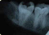
PREOPERATIVE RADIOGRAPH
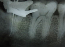
WORKING LENGTH
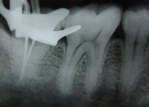
MASTERCONE RADIOGRAPH
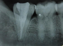
POSTOPERATIVE RADIOGRAPH
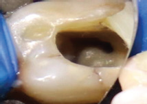
CLINICAL IMAGE
DISCUSSION
The commonest anatomical variation in the mandibular second molar is C shaped canal and its occurrence ranges from 2.7 % to 44.5 %. The management of this complex root canal needs proper clinical and radiographic evaluation, advanced techniques for cleaning, shaping and obturation. Magnification devices such as dental loupes, dental operating microscope is helpful in managing these complex structures. Conventional periapical radiographs is useful to know the root outline, but main disadvantage is, it’s a two dimensional view. An advanced imaging technique includes Computed Tomography and Cone Beam Computed Tomography is more accurate in diagnosing C shaped canal.3 Proper access cavity preparation and careful clinical evaluation of the pulp chamber gives clue in identifying the different types of C shaped canal, which was classified by Melton and Fan et al. Fan et al concluded that most of the C shaped canal is the orifice with an uninterrupted semicircle C shaped configuration.18 Continuous C shaped canal [MB-D] should be negotiated with three or more initial K files No 8 and 10, which decreases the chance of missing canal. The cleaning and shaping of the canal includes Niti rotary files using anticurvature filing method, which prevents strip perforations. The use of sonic and ultrasonic activators for irrigation provides better removal of debris from these irregular complex root canals.20 Obturation techniques include lateral compaction of gutta percha starting from distal side to mesial gives better result. Thermo plasticized gutta percha technique provides effective obturation of the root canals. By having proper knowledge about common occurrence of this abnormal internal anatomy and use of proper instruments and techniques facilitates long term success of endodontic treatment.
CONCLUSION
The treatment of mandibular second molar with this complex C shaped anatomy poses an endodontic challenge to every clinician. Thorough knowledge about abnormal anatomic variations and proper clinical and preoperative radiographic evaluation helps in early detection of C shaped root canals. Early detection of these canals and use of advanced instruments and proper application of cleaning and obturation techniques will results in successful management of C shaped canals.
REFERENCES:
- Cooke III HG Cox FL. C-shaped canal configurations in mandibular molars. J Am Dent Assoc. 1979;99(5):836-9.
- Orban B, Mueller E. The development of the bifurcation of multirooted teeth. J Am Dent Assoc. 1922;16(2):297-319.
- Seo DG, Gu Y, Yi YA, Lee SJ, Jeong JS, Lee Y, et al. A biometric study of C-shaped root canal systems in mandibular second molars using cone-beam computed tomography. Int Endod J 2012;45:807-14.
- Jerome CE. C-shaped root canal systems: diagnosis, treatment, and restoration .Gen Dent 1994;42(5):424.
- Manning SA. Root canal anatomy of mandibular second molars. Int Endod J 1990;23(1):40-5.
- Al-Fouzan KS. C-shaped root canals in mandibular second molars in a Saudi Arabian population. Int Endod J 2002;35(6):499-504.
- Bolger WL, Schindler WG. A mandibular first molar with a C-shaped root configuration. J Endod 1998;14(10):515-9.
- Yilmaz Z, Tuncel B, Serper A, Calt S. C-shaped root canal in a maxillary first molar: a case report. Int Endod J 2006;39(2);162-6.
- Dankner E, Friedman S, Stabholz A. Bilateral C shape configuration in maxillary first molars. J Endod 1990;16(12):601-3.
- LuTY, Yang SF, Pai SF. Complicated root canal morphology of mandibular first premolar in a Chinese population using the cross section method. J Endod 2006;32(10):932-6.
- Sidow SJ, West LA, Liewehr FR, Loushine RJ. Root canal morphology of human maxillary and mandibular third molars. J Endod 2000;26(11):675-8.
- Kuzekanani M, Haghani J, Nosrati H. Root and canal morphology of mandibular third molars in an Iranian population. J Dent Res Dent Clin Dent Prospects 2012;6(3):85.
- Cleghorn BM, Christie WH, Dong CC. Root and root canal morphology of the human permanent maxillary first molar: a literature review. J Endod 2006;32(9):813-21.
- Boveda C, Fajardo M, Millan B. Root canal treatment of an invaginated maxillary lateral incisor with a C-shaped canal. Quintessence Int Engl Ed. 1999;30:707-11.
- Zheng Q, Zhang L, Zhou X, Wang Q, Wang Y, Tang L, et al. C-shaped root canal system in mandibular second molars in a Chinese population evaluated by cone-beam computed tomography. Int Endod J 2011;44(9):857-62.
- Sabala CL, Benenati FW, Neas BR. Bilateral root or root canal aberrations in a dental school patient population. J Endod 1994;20(1):38-42.
- Neelam Mittal, Suraj Arora. Root Banking: A Case Report. Clinical dentistry. 2009;3:20-4.
- Fan B, Cheung GS, Fan M, Gutmann JL, Bian Z, C-shaped canal system in mandibular second molars: Part 1- Anatomical features. J Endod 2004;30:899-903.
- Mittal N, Narang I. 3-D diagnosis-assisted management of anomalous mandibular molar. Contemp Clin Dent. 2012;3(Suppl 1):S51-S54.
- Jafarzadeh H, Wu YN. The C-shaped root canal configuration: A review. J Endod 2007;33:517-23.
Disclaimer from Guident
The views and opinions expressed in articles published in Guident are solely those of the authors and do not necessarily reflect the views of the editors, the editorial board or the publisher, Ivory India. Guident does not endorse any product, technique or treatment described. Clinical content is intended for qualified dental and medical professionals and is not a substitute for individual clinical judgement. Guident, its editors and the publisher accept no liability for any consequence arising from the use of information published in the journal.
How to cite this article
Dr. Neelam Mittal, Dr. Praveen Kumar. Management of C Shaped Root Canal in the Mandibular Second Molar an Endodontic Challenge. Guident. 2020 Jan;13(2). Available from: https://guident.in/articles/endodontics/management-of-c-shaped-root-canal-in-the-mandibular-second-molar-an-endodontic-challenge.html