Endodontic Management of Radix Entomolaris and Paramolaris-a Case Report
Department of Conservative Dentistry and Endodontics, Seema Dental College And Hospital, Rishikesh
INTRODUCTION
The main aim of endodontic therapy is the elimination of bacteria from the infected root canals and prevention of further reinfection which is mainly achieved by thorough cleaning and shaping of root canals followed by tri-dimensional obturation with a coronal fluid tigh seal.6 Root canal anatomy is highly complex and unpredictable. An awareness and understanding of the presence of additional root and unusual root canal morphology are essential, as it determines the successful outcome of endodontic treatment.1
The majority of mandibular first molars have two roots, mesial and distal with two mesial and one distal canal. A major variant is the presence of three roots in mandibular first molar, first mentioned in the literature by Carabelli known as radix entomolaris, when located in distolingual position. When located on mesiobuccal surface, the anomaly is known as radix paramolaris. The external morphology of this anomaly having additional lingual or buccal root, are described by Carlsen and Alexandersen.2
Radiographic diagnosis plays a pivotal role in successful endodontic treatment of tooth. One of the main reasons for failure of endodontic treatment is incomplete removal of pulp tissue and microorganisms from all the root canals. So, radiographs taken at different angulations give information about extra canals or roots.2
Radix Entomolaris has a frequency of less than 5% in white Caucasian, African, Eurasian and Indian populations while it appears to be commonly present in races of Mongoloid traits such as the Chinese, Eskimos, and Native American populations with a frequency of 5-30%.
Radix Paramolaris is very rare and occurs less frequently than the Radix Entomolaris. Radix Paramolaris is commonly observed in 1.5–3% of the African population whereas it is less frequent, and frequency of existence is around 2% in the Indian population.1 In this report three such cases are presented.
CASE REPORTS
CASE 1
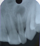
26-year-old male patient reported with the chief complaint of pain in lower right back tooth region since two weeks. The pain aggravated upon biting. Clinical examination revealed a faulty restoration and tenderness to percussion w.r.t 46. Intraoral periapical radiograph revealed secondary carious lesion involving the pulp with widening of PDL space and presence of an extra root in 46. Based on clinical and radiographic examination, diagnosis of Symptomatic Irreversible Pulpitis with Acute Apical Periodontitis w.rt 46 was established. After adequate anesthesia and isolation with rubber dam, access cavity was prepared. Working length was determined using #10 k file followed by shaping and cleaning till #25/0.06 in all canals. All the canals were irrigated with 2.5% sodium hypochlorite and 17% EDTA after use of each file. In the subsequent appointment canals were dried using paper points and obturation was done using MTA Fillapex sealer. Later, the access cavity was sealed with composite.

Pre-Operative Radiograph
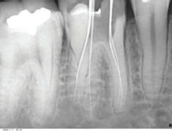
Working Length
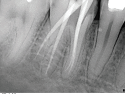
Master Cone
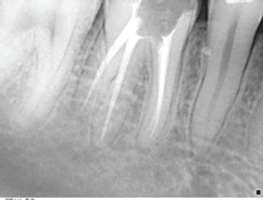
Obturation
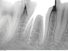
Post Endodontic Restoration
CASE 2
38-year-old male patient reported to the department with the chief complaint of dislodged restoration, food lodgement and pain upon biting in lower right back tooth region since a month. Clinical examination revealed Faulty Restoration and tenderness to percussion w.r.t 46. Intraoral periapical radiograph revealed Root Canal Treated tooth with short obturation and missed canal and widening of the PDL space w.r.t 46. Based on clinical and radiographic examination, diagnosis of Acute Apical Periodontitis w.r.t 46 was established. Treatment plan was Re-RCT w.r.t 46. After adequate anesthesia and isolation with rubber dam, old restoration and gutta percha was removed. New working length was determined using #15 k files followed by shaping and cleaning till #25/0.06 in all the canals. All the canals were irrigated with 2.5% sodium hypochlorite, 2% Chlorhexidine and 17% EDTA after use of each file. In the subsequent appointment canals were dried using paper points and obturation was done using guttapercha and MTA Fillapex sealer. Later, the access cavity was sealed with composite.
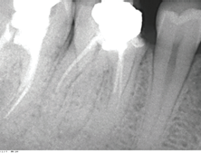
Pre-Operative Radiograph
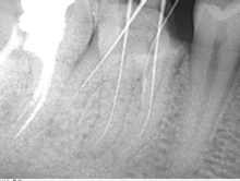
Working Length
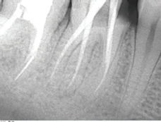
Master Cone
Obturation
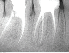
Post Endodontic Restoration
CASE 3
24-year-old female patient reported with the chief complaint of pain in lower left back tooth region since 10-15 days. Pain was mild, continuous, non-radiating in nature, aggravated upon biting. Clinical examination revealed deep proximal caries and tenderness to percussion w.r.t 36. Intraoral periapical radiograph revealed radioleucency on distal aspect involving the pulp with widening of PDL space and presence of an extra root in 36. Based on clinical and radiographic examination, diagnosis of Symptomatic Irreversible Pulpitis with Acute Apical Periodontitis w.rt 36 was established. After adequate anesthesia and isolation with rubber dam, access cavity was prepared. Working length was determined using #10 k file followed by shaping and cleaning till #25/0.06 in all canals. All the canals were irrigated with 2.5% sodium hypochlorite and 17% EDTA after use of each file. In the subsequent appointment canals were dried using paper points and obturation was done using MTA Fillapex sealer. Later, the access cavity was sealed with composite.
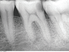
Pre-Operative Radiograph
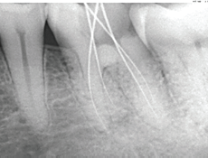
Working Length
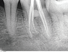
Master Cone
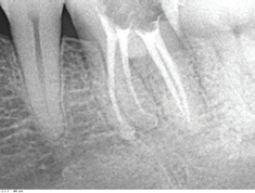
Obturation
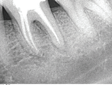
Post Endodontic Restoration
DISCUSSION
A thorough knowledge of the root canal anatomy is very important for the clinician. With the advent of newer technologies and recent concept on access preparation, it is very easy to locate the aberrant canals. Inadequate knowledge of anatomic variations resulting in missed canals is one of the critical reasons for failure of endodontic therapy.
The etiology behind the formation of the radix entomolaris is still unclear. In dysmorphic, supernumerary roots, its formation could be related to external factors during odontogenesis, or to penetrance of an atavistic gene or polygenetic system. In eumorphic roots, racial genetic factors influence the more profound expression of a particular gene that results in the more pronounced phenotypic manifestation. An RE can be found on the first, second and third mandibular molar, occurring least frequently on the second molar. Some studies report a bilateral occurrence of the radix entomolaris from 50 to 67%.3
MORPHOLOGY
Carlsen and Alexanderson classified RE based on the location of its cervical part into four types.
Type A–Distally located cervical part with two normal distal root components
Type B–Same as Type A; however, only one normal distal root component
Type C–Mesially located cervical part
Type AC–Central location between mesial and distal root components.
De Moor et al. classified RE‑based on the curvature in buccolingual orientation into three types.
Type I–Refers to a straight root/root canal
Type II–Refers to an initially curved entrance which continues as a straight root/root canal
Type III–Refers to an initial curve in the coronal third of the root canal, and a second buccally oriented curve starting from the middle to apical third.
Recently, Wang et al. gave another classification for RE depending on its radiographic appearance.
Type 1: Presents the most identifiable radiographic image
Type 2: A large beam angulation is necessary mesially or distally for their identification
Type 3: Identification becomes extremely difficult because of the overlap of the adjacent distobuccal root.
Carlsen and Alexanderson classified RP based on the location of its cervical part into two types.
Type A–Refers to an RP in which the cervical part is located on the mesial root complex
Type B–Refers to an RP in which the cervical part is located centrally, between the mesial and distal root complexes.1
CLINICAL IMPLICATIONS
Accurate clinical and radiographic diagnosis can avoid failure of root canal treatment because of missed canal. There are various methods to locate additional canals such as, knowledge of law of symmetry, law of orifice location, visualizing the dentinal map and canal bleeding points. The tactile sensation using hand instrument like Endodontic explorer, Pathfinder, DG 16 probe and Micro‑openers are also helpful.
Advanced imaging techniques can aid to locate and confirm additional canals in case of multirooted teeth, especially molars. These techniques include digital radiography, fiber‑optic illumination, dental endoscopy and orascopy, surgical loupes, operating microscope, micro‑computed tomography (CT), visualization endograph using Ruddle’s solution, and magnetic resonance microscopy.4
In the present case series, the variations in distal root anatomy were identified through careful reading of angled IOPA radiographs. The first radiograph was taken with conventional angulation, and the second one with a mesial shift of approximately 20°. This buccal object rule has also been called same lingual, opposite buccal rule/Clark’s rule/Walton’s projection. An additional root appears as a shadow or a thin radiolucent line in the radiographs. In order to confirm the location of this additional root, an H‑file was inserted in other canals, and a K‑file was introduced into the additional canal before taking the radiograph. The existence of an accessory cusp (tuberculum paramolare) and periodontal probing for cervical prominences can also provide an indication of an additional root.
Sometimes, radix canal orifice could be occluded by secondary or calcified dentine. Therefore, the visual access and superior control during access procedures make a convenient tool in such cases. Most important basic principle for successful root canal treatment is the principle of ‘straight-line access’. Straight‑line access in this respect has to be emphasized, as the majority of the radix root is curved. With the advent of rotary instruments, access burs such as Endo Z, SS white access burs, access can be modified to get a straight line access.
As the orifice of radix entomolaris is distolingually located, the shape of access cavity should be modified from classical triangular form to trapezoidal or rectangular form in order to better locate the orifice of distolingual root.3
When the third root is detected, it should be cleaned and shaped like every other canal. A severe root inclination or canal curvature, particularly in the apical third of the root (as in a type III RE), can cause shaping aberrations such as straightening of the root canal or a ledge, with root canal transportation and loss of working length resulting. The use of flexible nickel-titanium rotary files allows a more centered preparation shape with restricted enlargement of the coronal canal third and orifice relocation.5
Nevertheless, unexpected complications such as instrument separation do occur, and are more likely to happen in an RE with severe curvature or narrow root canals. Therefore, after relocation and enlargement of the orifice of the RE, initial root canal exploration with small files (size 10 or less) together with radiographical root canal length and curvature determination, and the creation of a glide path before preparation, are step-by-step actions that should be taken to avoid procedural errors.
CONCLUSION
Clinicians should be aware of these unusual root morphologies in the mandibular first molars in Caucasian people. The initial diagnosis of a radix entomolaris or paramolaris before root canal treatment is important to facilitate the endodontic procedure, and to avoid ‘missed’ canals. Preoperative periapical radiographs exposed at two different horizontal angles are required to identify these additional roots. Knowledge of the location of the additional root and its root canal orifice will result in a modified opening cavity with extension to the distolingual. The morphological variations of the radix entomolaris or paramolaris in terms of root inclination and root canal curvature demand a careful and adapted clinical approach to avoid or overcome procedural errors during endodontic therapy.
REFERENCES
- Arun J, Mayuri BS, Nandini TN, Mallikarjun Goud K. Radix entomolaris and paramolaris: A case series. International Journal of Oral Health Sciences, Jan-Jun 2015 ; 5(1): 57-62.
- Amit Parashar, Shikha Gupta, Abhishek Zingade and Shashi Parashar. The Radix Entomolaris and Paramolaris: A Review and Case Reports with Clinical Implications. J Interdiscipl Med Dent Sci 2015, 3:1.
- Filip L. Calberson, Roeland J. De Moor and Christophe A. Deroose. The Radix Entomolaris and Paramolaris: Clinical Approach in Endodontics. JOE — Volume 33, Number 1, January 2007.
- A. R. Vivekananda Pai, Rachit Jain, Ashwini S. Colaco. Detection and endodontic management of radix entomolaris: Report of case series. Saudi Endodontic Journal • May-Aug 2014;Vol 4:Issue 2.
- Parupalli Karunakar, Umrana Faizuddin, Madanala Nagarjun, M. S. Ranga Reddy. Endodontic Management of Radix Entomolaris in Second Molar Contemporary Clinical Dentistry,Volume 9;Issue 1:January - March 2018.
Disclaimer from Guident
The views and opinions expressed in articles published in Guident are solely those of the authors and do not necessarily reflect the views of the editors, the editorial board or the publisher, Ivory India. Guident does not endorse any product, technique or treatment described. Clinical content is intended for qualified dental and medical professionals and is not a substitute for individual clinical judgement. Guident, its editors and the publisher accept no liability for any consequence arising from the use of information published in the journal.
How to cite this article
Dr. Annil Dhingra, Dr. Sneha. Endodontic Management of Radix Entomolaris and Paramolaris-a Case Report. Guident. 2020 Feb;13(3). Available from: https://guident.in/articles/endodontics/endodontic-management-of-radix-entomolaris-and-paramolaris-a-case-report.html