Healing of a Periapical Lesion Non Surgical Approach by Using Calcium Hydroxide
Department of Endodontics and Conservative Dentistry Saifee Dental Clinic, Masjid, Mumbai
ABSTRACT
Micro-organisms within the root canal should be eradicated for successful outcome of root canal treatment. Periapical lesions of endodontic origin usually heal by conservative therapy alone. This article presents a non surgical resolution of an extensive periapical lesion of endodontic origin associated with maxillary left lateral incisor. The tooth was treated by conventional endodontic therapy employing calcium hydroxide as intracanal medicament.
INTRODUCTION
Traumatic injuries generally disrupt the pulpal blood supply causing pulp necrosis and leading to anaerobic conditions favourable for the growth of opportunistic microorganisms, which may subsequently result in the development of periapical lesions.
As a consequence of pathologic changes in the dental pulp, the root canal system can harbour numerous irritants. Egress of these irritants from infected root canal into the periapical tissues can initiate formation and perpetuation of the periapical lesion.
Elimination of the microorganisms, in case of large periapical lesions, has been a challenge for the clinician. The polymicrobial infection makes sterilization of the root canal system difficult.
Both chemical and mechanical debridement procedures are essential in effectively disrupting and removing the microbial ecosystem that is associated with the disease process. Thus, whenever a pulp is removed and the canal treated and filled in a manner favorable to a physiological reaction, we can expect a satisfactory percentage of endodontic success.
Case Report
A 38-year-old male patient, with no relevant medical history, presented with a painful swelling, in the anterior portion of the upper jaw. The patient gave a history of trauma to the maxillary anterior teeth ten years ago.
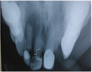
Extra oral examination was unremarkable. Intraoral examination revealed painful swelling of the palatal mucosa, adjacent to tooth 22. A radiographic evaluation demonstrated periapical lesion associated with 22 [Fig 1]. There was a large radiolucent lesion with a uniform radiolucency and well-defined margins involving the apex of the tooth.
Vitality test was negative with tooth 22.
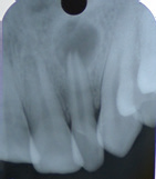
Figure 1: Pre Operative IOPA showing large periapical lesion associated with 22.
Taking into account the medical and dental history of the patient, a nonsurgical endodontic treatment was planned, to treat the involved tooth.
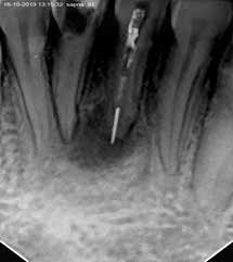
The access cavity was prepared without local anaesthesia. The root canal was negotiated with a size 10 hand file. After achieving apical patency, the canal was negotiated with a size 15 hand file and a working length radiograph was recorded using the same [Fig 2] Subsequently biomechanical preparation was performed by the crown down technique using pro taper gold system of rotary files.
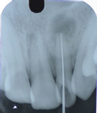
Figure 2: Working Length IOPA recorded using No. 15 K File
The tooth was irrigated with NaOCl 5.2%, EDTA 17% and final irrigation was done with Chlorhexidine 2%.
After drying the canal with sterile paper points Calcium hydroxide ( Cal Excel by Ammdent) dressing was placed.[Fig 3]

Figure 3: Calcium Hydroxide dressing placed
The paste was changed every month for a period of 3-months until the teeth displayed no symptoms.
A master cone radiograph was recorded and root canal obturation was performed with AH plus sealer (Dentsply) using lateral condensation followed by a coronal restoration with composite resin. [Fig 4 and Fig 5]
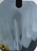
Figure 4: Master Cone Radiograph

Figure 5: Post Operative IOPA recorded on the day of obturation appointment
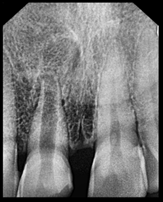
After a two year follow-up period the patient was asymptomatic. Moreover radiographic examination revealed resolution and adequate healing of the periapical radiolucency. [Fig 6]

Figure 6: Follow up radiograph recorded 2 years after procedure. Resolution of periapical apical lesion can be seen with adequate healing.
Discussion
The development and progression of endodontic induced lesion is directly associated with the presence of micro-organisms in root canal. The infected root canal is considered to be a poly-microbial infection consisting of aerobic and anaerobic bacteria. The periapical and periradicular lesions are treated by either non-surgically or surgically.
But the non- surgical method should be done initially. The success of the non-surgical endodontic treatment method is based on appropriate cleaning,shaping and filling of root canal.
Adequate biomechanical preparation with the aid of 5.25% NaOCl is currently recommended for the above purpose followed by placement of calcium hydroxide dressing. The favourable clinical and radiographic and histological responses obtained with calcium hydroxide are attributed to the involvement of Calcium and hydroxyl ions in several cellular and molecular mechanisms leading to regeneration of periapical connective tissues.
The benefits of calcium hydroxide include anti-inflammatory action, neutralisation of acidic products, activation of alkaline phosphatase and anti bacterial action. The anti bacterial action is attributed to its ph of 12.5 which results in damage to bacterial cytoplasmic membrane, protein denaturation and damage to the bacterial DNA
Apical patency provides decompression of the periapical lesion. Moreover, the removal of the source of antigen, associated with regular renewal of the calcium hydroxide root canal dressing eliminated the exudate and provided significant resolution of the periapical radiolucency as seen in the follow up radiograph.
REFERENCES
- Soares J, Santos S, Silveira F, Non surgical treatment of extensive cyst like periapical lesion of endodontic origin. Int. Endod J 2006; 39:566-75
- Ross, MH, Histology: A text and Atlas, 3rd edition, Philadelphia: Lippincott, Williams and Wilkins. 1995, pg. 315
- Castelluci: Periapical disease, Las Vegas institute, pg 160
- Blayney: JADA 15: 1217-1221,1928, quoted by Lovdahl, PE, Gutmann
- Caliskan MK, Sen BH (1996) Endodontic treatment of teeth with apical periodontitis using Calcium hydroxide: A long term study, Endod Dent Traumatology 12,215-221
- Nair, PNR: Pathobiology of the Periapex. In Cohen, S.Burns:Pathways of the pulp, 8th edition, pg 470
- Nair PNR (1998) New perspectives on radicular cysts: Do they heal? Int Endodon J 31.155-160
- Weine Franklin S, Endodontic Therapy, 6th edition, pg 252
- Baugmarten, JC, Hutter, JW: Endodontic Microbiology and Treatment of Infections in Cohen: Pathways of the Pulp, 8th edition, pg 501
- Soares JA, Leonardo MR, Silva LAB, Tanomaru FM (2006) Elimination of intracanal infection in dogs teeth with induced periapical lesions after rotary instrumentation: influence of different Calcium Hydroxide Pastes. J Appl Oral Sci 14, 172-177
- Antony DR, Gordon, TM, Del Rio CE(1982) The effect of three vehicles on the pH of Calcium Hydroxide, Oral Surg Oral Med Pathol 54, 560-565
Disclaimer from Guident
The views and opinions expressed in articles published in Guident are solely those of the authors and do not necessarily reflect the views of the editors, the editorial board or the publisher, Ivory India. Guident does not endorse any product, technique or treatment described. Clinical content is intended for qualified dental and medical professionals and is not a substitute for individual clinical judgement. Guident, its editors and the publisher accept no liability for any consequence arising from the use of information published in the journal.
How to cite this article
Dr. Abbas Ali Rangwala, Dr. Bareen J. Tramboo, Dr. Tasneem Bandukwala. Healing of a Periapical Lesion Non Surgical Approach by Using Calcium Hydroxide. Guident. 2020 Mar;13(4). Available from: https://guident.in/articles/endodontics/healing-of-a-periapical-lesion-non-surgical-approach-by-using-calcium-hydroxide.html