Root End Resection or Apicoectomy-a Novel Approach in Rehabilitation: A Report of Two Cases
Department of Conservative Dentistry and Endodontics, Seema Dental College And Hospital, Rishikesh
ABSTRACT
Apicoectomy is the excision of the apical portion of the tooth and the attached soft tissues during periapical surgery.1 The surgery goal is periapical lesion removal and the apical third sealing, allowing soft and hard tissue regeneration. According to the meta-analysis of Tsesis et al, paraendodontic surgery success rate is 91.6%, while failure rate is 4.7%.2 In periradicular surgery with retrograde root-canal obturation, several surgical steps are essential to achieve healing: surgical exposure of the root-end, debridement of pathological tissue, root-endresection, root-end cavity preparation, root-end filling, and wound closure.3
INTRODUCTION
An apicoectomy, a surgical procedure involving the root apex to treat apical infections, was defined in 1884 by J. Farrar as a means of removing the root cause of disease and achieving a permanent cure. This method, also known as root resection or endodontic surgery, emerged in the late 19th century as an alternative to dental extraction, particularly for cases of dental abscesses related to "pyorrhea alveolaris." Despite successful conventional endodontic treatment, apical lesions may persist, prompting consideration for apical surgery.
Conventional endodontic treatment has a success rate ranging from 53% to 98% for initial procedures, but it can be lower for cases with persistent periapical lesions. The nature of periapical lesions, whether granulomas or cysts, is not always clear during treatment. While granulomas tend to heal after endodontic therapy, there has been debate among dentists regarding the healing of periapical cysts. Issues like draining fistulas, pain during chewing, or radiolucent areas increasing in size suggest problems with the initial treatment, and in such cases, surgical intervention becomes necessary. The decision to opt for surgery or consider retreatment from the coronal part of the tooth depends on various clinical and anatomical factors.
Literature suggests that mineral trioxide aggregate (MTA), due to its high biocompatibility and sealing properties, promotes better tissue healing when in contact with dental pulp or periradicular tissues compared to other root end filling materials like amalgam, gutta percha, composite resin, and others. MTA's biocompatibility and potential to induce the growth of cement oblasts and osteoblasts make it capable of supporting complete histological repair after periapical surgery and retrofilling.
The article describes a clinical case where root canal treatment was combined with apicectomy surgery and retrofilling using MTA, showcasing the application of this approach.4,5
CASE REPORT-1
A 26-year-old female patient reported to Department of Conservative dentistry And Endodontics with a chief complaint of discolored tooth with pus in upper front tooth region for 1 months. Patient also gave history of trauma as she fell down while riding bicycle 5 years back for which she had not consulted any doctor and she noticed discolouration of tooth 3 years back . On clinical examination Discoloration i.r.t. 11 and sinus opening noted on labial aspect wrt 11 , grade I mobility present wrt to 11,12 was noted. 11,12 gave no response to Cold Test and EPT. On radiographical examination Diffuse Periapical Radiolucency was noted i.r.t 11,12
On the basis of clinical and radiographic examination root canal treatment was advised which was followed by root end resection.
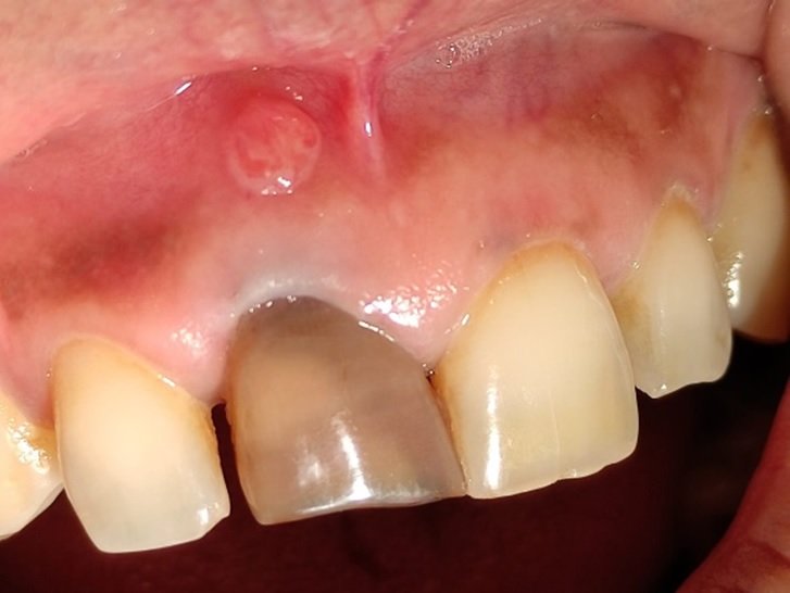
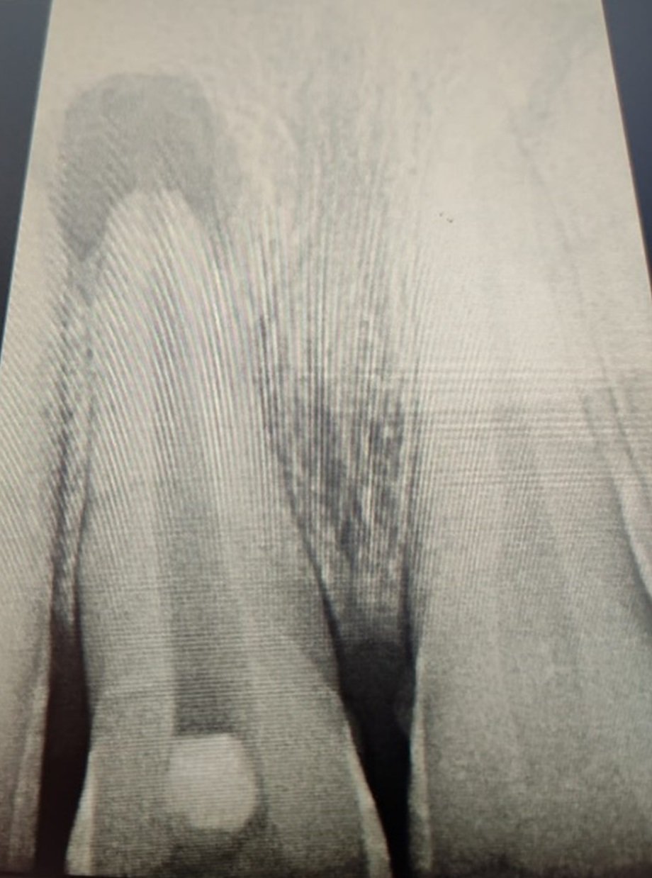
PRE OPERATIVE IMAGE AND RADIOGRAPH
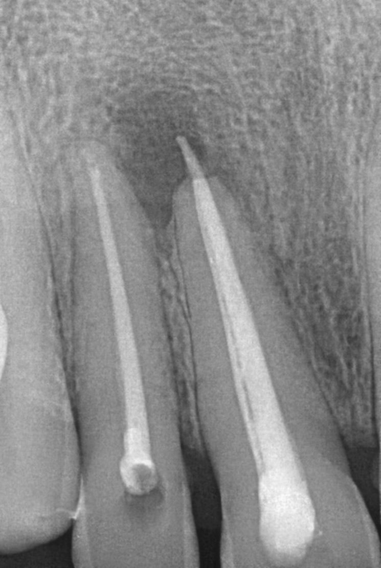
OBTURATION

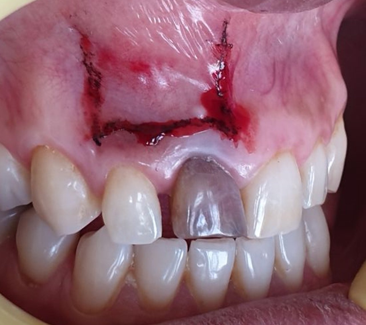
FLAP-MARKED INCISION

ROOT END RESECTION
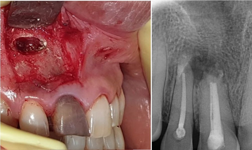
RETROGRADE FILLING WITH MTA

SUTURES PLACED
CASE REPORT-2
A 28 year old patient reported to the department with a chief complaint of pain and pus discharge in the upper front tooth region for 1 month. There was a history of falls during childhood.
On clinical examination, previously initiated root canal treatment irt 11 was noted, grade I mobility irt 11,12. radiographic examination revealed radiolucency with respect to periapical region irt 11,12,21,22. Cold test was negative i.r.t 11 and lingering response i.r.t 12,21,22 Teeth presented a no response irt 11 and delayed response to electric pulp testing i.r.t 12,21,22. The treatment planned was root end resection w.r.t 11,21 with retrograde filling of MTA. It was explained to the patient and consent was obtained. After the access opening of 11,21,12,22 was done, the working length was determined using K file and thorough shaping and cleaning was done. The teeth were then obturated using cold lateral condensation technique. Raising Ochsenbein – Luebke (O-L) Flap, a periapical surgery was performed and periapical pathosis was enucleated. Root end resection was done and retrograde cavity was prepared using ultrasonic tip. MTA was placed and was confirmed using a radiograph and the sutures were placed. The patient was recalled after 10 days for suture removal.


PREOPERATIVE RADIOGRAPH AND IMAGE

POST OBTURATION

FLAP INCISION MARKED

FLAP RAISED

BONY WINDOWS
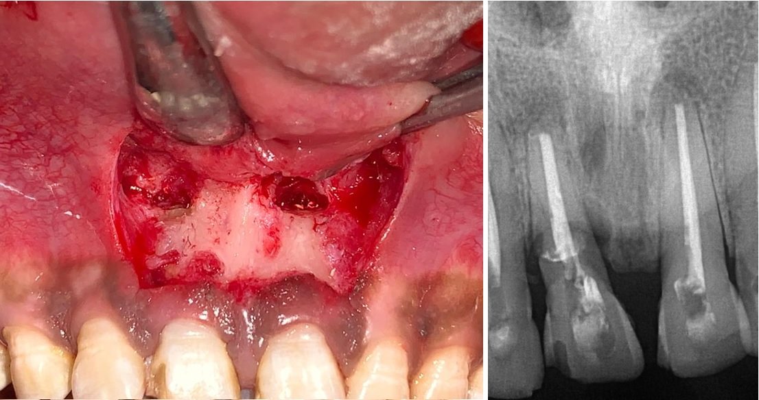
ROOT END RESECTED
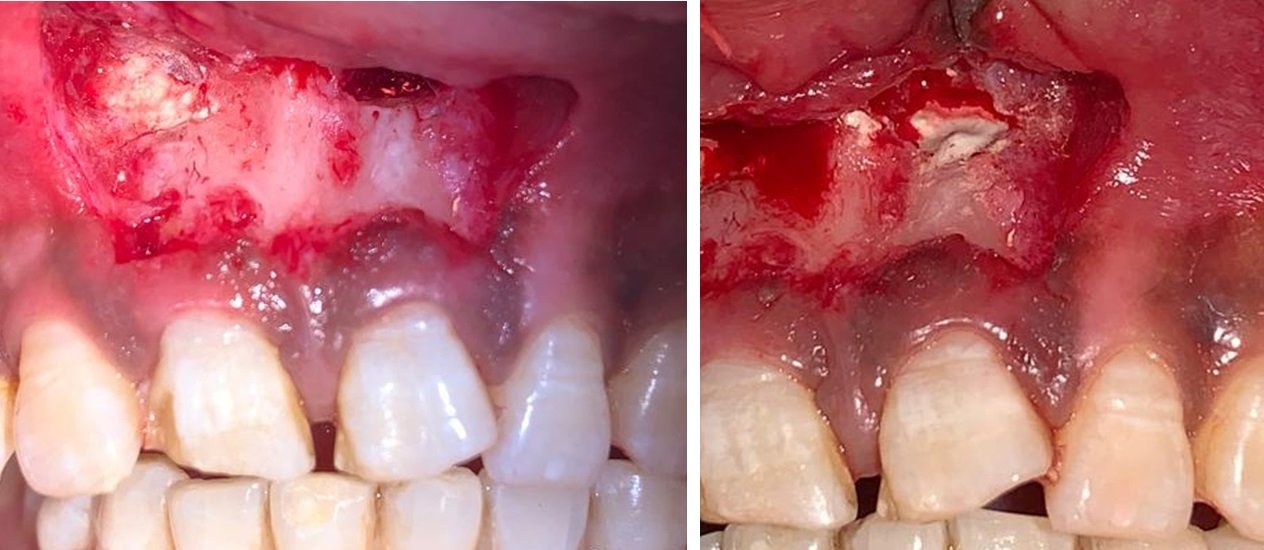

ROOT-END FILLING

SUTURE PLACEMENT

SUTURE REMOVAL AND FOLLOW-UP AFTER 10 DAYS
DISCUSSION
Trauma can result in varying responses in dental pulp. While some pulps remain normal, others become necrotic. Necrotic pulps can serve as a nutrient source for pathogenic bacteria, leading to the formation of periapical lesions. In this particular case, the affected maxillary incisors had nonvital pulp tissue and a radiolucent lesion beyond the apex, prompting the initiation of root canal treatment.
Root canal treatment primarily aims to remove microbial infection from the complex root canal system. Irrigants are used to reduce microbial flora and dissolve necrotic tissue if a tissue-solvent solution is employed. In this case, 2.5% hypochlorite was chosen as an irrigating solution due to its tissue-dissolving property.
For large cystic lesions, treatment options vary from conventional nonsurgical root canal treatment with calcium hydroxide therapy to surgical interventions. In this instance, the large size of the lesion, cystic appearance on radiographs, prolonged infection, and patient's time constraints led to surgical intervention.6
The surgical procedure involved root-end resection and preparation, followed by placement of root canal filling to seal the communication between the infected root canal and periradicular tissues. An ideal root canal filling material should possess various properties, including non-absorbability, biocompatibility, adhesion to dentinal walls, and the ability to induce cementogenesis. Among the materials used, mineral trioxide aggregate (MTA), Biodentine, and IRM are common choices. MTA was chosen due to its excellent physical properties, biocompatibility, potential to induce cellular response, and non-mutagenic nature.
While MTA has benefits, it also has drawbacks such as difficulty in controlling filling length, potential for voids, absence of solvent for removal, challenging handling, long setting time, and higher material cost. However, follow-up examinations of the case showed the absence of symptoms, preservation of tooth function, and no signs of pain or swelling.4,5
CONCLUSION
With a high percentage of successful treatment outcomes of conventional endodontics together with high success of surgical endodontics almost all teeth with endodontic lesions can be successfully treated. The surgical technique applied in this case, apicoectomy, was appropriate. This was proven by both clinical and radiographic results.
REFERENCES
- Locurcio LL, Leeson R. A case of periradicular surgery: apicoectomy and obturation of the apex, a bold act. Stomatological Dis Sci 2017;1:76-80.
- A Textbook of Advanced Oral and Maxillofacial Surgery Volume 2 Chapter 12
- KimS and Kratchman S. Modern Endodontic Surgery Concepts and Practice: A Review. J Endod 2006;32:601– 623.
- Torabinejad M, Parirokh M. Mineral Trioxide Aggregate: a comprehensive literature review - Part II: leakage and biocompatibility investigations. J Endod. 2010; 36(2):190-202.
- Parirokh M, Torabinejad M. Mineral Trioxide Aggregate: a comprehensive literature review - Part III: clinical applications, drawbacks, and mechanism of action. J Endod. 2010; 36(3): 400-13.
- Pawar AM, Kokate S R, Shah RA. Management of a large periapical lesion using Biodentine as retrograde restoration with eighteen months evident followup. Journal of Conservative Dentistry 2013; 16 (6)
Disclaimer from Guident
The views and opinions expressed in articles published in Guident are solely those of the authors and do not necessarily reflect the views of the editors, the editorial board or the publisher, Ivory India. Guident does not endorse any product, technique or treatment described. Clinical content is intended for qualified dental and medical professionals and is not a substitute for individual clinical judgement. Guident, its editors and the publisher accept no liability for any consequence arising from the use of information published in the journal.
How to cite this article
Dr. Rohit Babu, Dr. Siddhi Goyal, Dr. Anil Dhingra. Root End Resection or Apicoectomy-a Novel Approach in Rehabilitation: A Report of Two Cases. Guident. 2023 Sep;16(10). Available from: https://guident.in/articles/endodontics/root-end-resection-or-apicoectomy-a-novel-approach-in-rehabilitation-a-report-of-two-cases.html