Benign Fibro-Epithelial Polyp: A Case Report
Department of Oral Medicine and Radiology, K.D. Dental College and Hospital, Mathura India
ABSTRACT
The most typical epithelial benign tumour of the oral cavity is a fibro-epithelial polyp. Such a polyp is a sessile or pedunculated growth that resembles a pink, red, or white knob and is of mesodermal origin. On the gingiva, the tongue, or the buccal mucosa, fibro-epithelial polyps are frequently found. An inflammatory hyperplastic lesion known as a fibro-epithelial polyp develops in response to persistent irritation brought on by calculus, sharp tooth edges, unnatural denture borders, or overhanging restorations. A polyp of this type is uncommon before the fourth decade of life and is common in both sexes. Despite careful surgical excision, the lesion may return as long as the ongoing irritation source exists.
Keywords-Fibro-epithelial polyp, traumatic fibroma, benign tumor
INTRODUCTION
The most frequent benign tumour of the oral cavity is a fibroma of the oral mucosa, which develops from fibrous connective tissues.1 With a prevalence of 1-2%, oral mucosal fibromas are most frequently found in older persons, while they can happen to anyone. An inflammatory hyperplastic connective tissue lesion is known as a fibroma. Due to the hyperplasia of the constituent cells, the local response to tissue damage causes a rise in the size of an organ or tissue.2
A fibroma in the oral cavity typically develops as a result of persistent irritation brought on by factors including lip- or cheek-biting, uneven denture borders, overhanging restorations, calculus, sharp tooth edges, or other oral prosthesis.3
Fibrous hyperplasia is the result of an inflammatory hyperplastic lesion that has healed.4
The phrase "focal fibrous hyperplasia," which denotes a reactive tissue response, was recommended by Daley et al. as an alternative to the term "fibroma".5
There are several other names for fibroma of the oral mucosa, including irritation fibroma (IF), traumatic fibroma, a fibrous nodule, and fibro-epithelial polyp.6
Usually, a fibroma of the oral mucosa clinically presents as painless swelling that is sessile or occasionally pedunculated; the affected site can be firm and resilient or soft with a spongy consistency.7
Any pedunculated lesion on the mucosal surface is referred to by Cooke as a "polyp" (fibro-epithelial polyp) and any pedunculated or sessile lesion in the gingiva is referred to as a "epulides." 8
Epulides typically arise in the maxillary anterior region, whereas fibro-epithelial polyps most frequently occur in the buccal mucosa along the line of occlusion.9
CASE REPORT
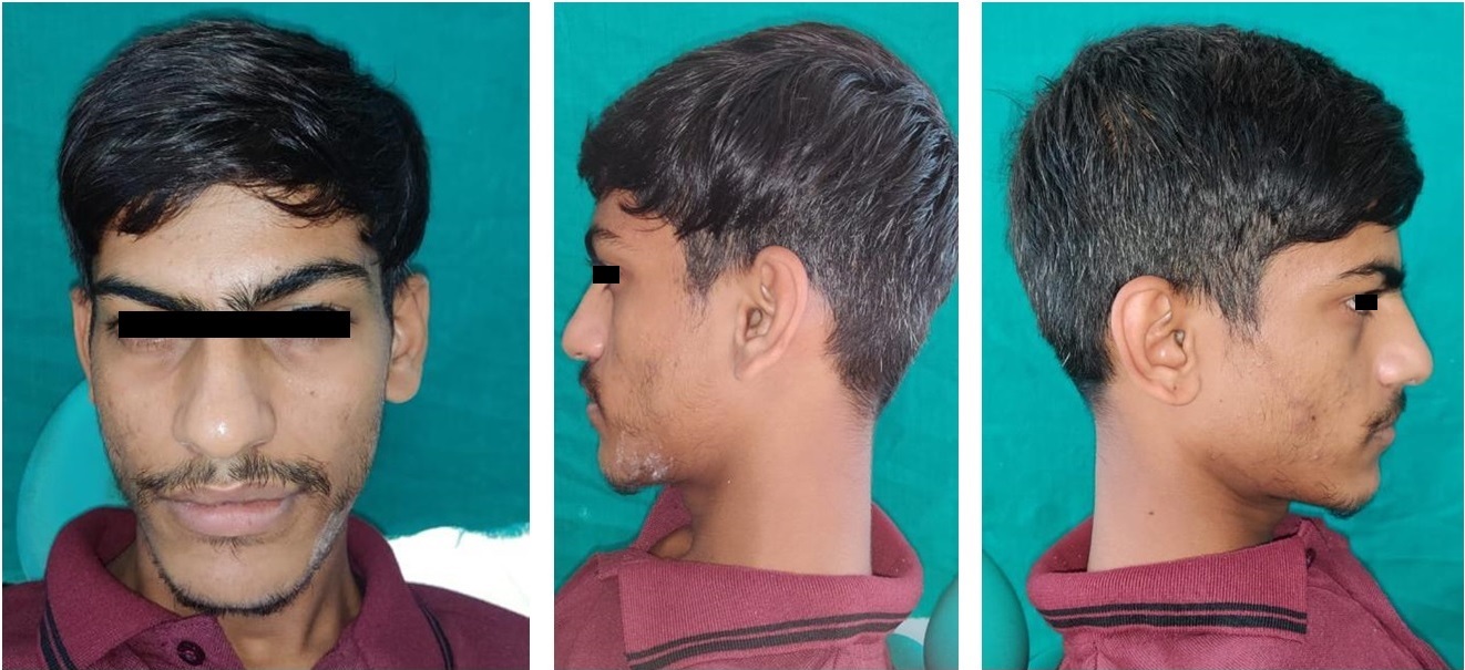
A 46 year old female patient reported to our outpatient department with a chief complain of growth in lower left front region of mouth since 4-5 months. History elicited that the a solitary, painless growth had been observed in her left mandibular anterior region which was initially small in size and then it gradually enlarged to present size of oval shape, well-defined, pedunculated lesion. Patient gave the similar history of the lesion at the same area which was surgically excised 1 year ago. Also, there was no relevant dental, medical and family history reported by the patient. On systemic examination all vital signs were within normal limits. On extraoral examination, no abnormality was detected.
On intraoral examination patient revealed a firm, solitary, irregular, painless, well defined, exophytic and pedunculated growth between 31 and 32 with both labial and lingual extension. The growth was smooth and shiny in appearance. The colour of the lesion was reddish pink measuring about 0.5x0.4x0.4mm in dimensions. Patient had a poor oral hygiene with a generalized periodontal condition.

Figure 1- Buccal and Lingual image of the lesion
An intraoral periapical radiograph was taken to rule out any associated bone changes. No significant findings was noted on complete hemogram analysis.
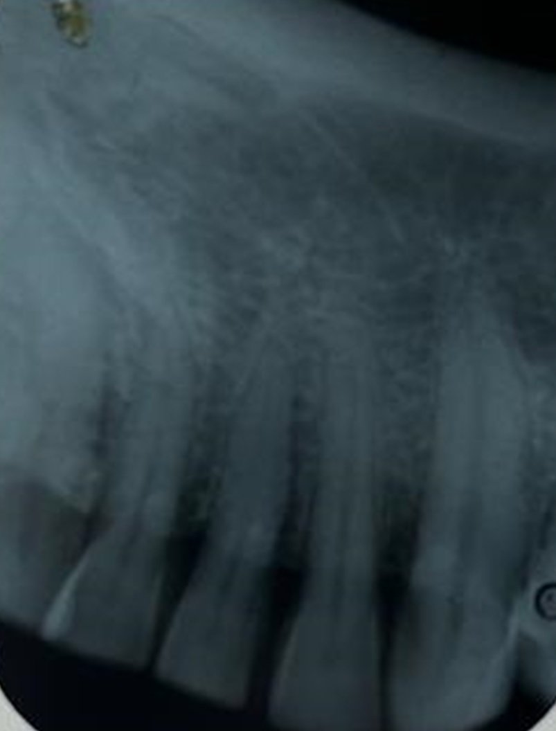
Figure 2- Periapical radiograph showing no bony changes
Treatment- Initial phase treatment included scaling & root planning and emphasizing oral hygiene.
An excisional biopsy was done under local anaesthesia using diode laser. Irrigation with normal saline and betadine was done. Gauze pack was kept in the operated site. The specimen was stored in formalin bulb and was sent to Histopathology laboratory. Post-operative antibiotics and analgesics were prescribed the patient. Post-op instructions to the patient were given.
Histopathological examination of H&E stained specimen showed polypodal tissue lined by intact squamous epithelium. Subepithelium shows oedematous fibrocollagenous tissue with moderately dense chronic inflammation. There is no cellular atypia. Overall features are suggestive of benign fibro-epithelial polyp.

Figure 3- Specimen and photomicrograph.
DISCUSSION
The most typical benign soft tissue tumour found in the oral cavity is a fibro-epithelial polyp, also known as fibrous hyperplasia.10 A fibroma develops as a result of a protracted healing process that includes the creation of scar tissue and granulation tissue, resulting in a fibrous mass underneath the mucous membrane.11
According to Axell's research, the prevalence of fibromas among Swedish adults is 3.25%. Fibromas are uncommon until the fourth decade of life and are equally prevalent in both sexes.12
A fibro-epithelial polyp's clinical characteristics are not universal, and the disease must be distinguished from peripheral ossifying fibroma and peripheral giant cell granuloma. A peripheral ossifying fibroma only develops on the gingiva and can be distinguished from a fibro-epithelial polyp by its firmer texture due to calcified material in the stroma.
When palpated, a pyogenic granuloma and a peripheral giant cell granuloma typically have a more vascular appearance and may bleed.13
A fibro-epithelial polyp is diagnosed based on the area of edoema in the soft tissues.
If there is swelling on the tongue, a neurofibroma, neurilemmoma, or granular cell tumour must be taken into account. A mucocele, lipoma, or salivary gland tumour could be the cause of swelling on the lower lip or buccal mucosa. A traumatic fibroma has two alternative collagen arrangement patterns, a radiating pattern and a circular pattern, depending on the severity of the irritation and the location of the lesion. This is a key characteristic that sets them apart from other types of fibromas.
A true fibroma does not display either of those patterns; instead, an irritation fibroma with a radiating pattern is associated with sites that are flexible (such as the cheeks) and less severe trauma, while an irritation fibroma with a circular pattern is associated with sites that are immobile in nature (such as the hard palate). Malignancy is not a threat posed by fibroepithelial polyps.14
Recurrence rates are low, and it typically results from repeated trauma at the lesion site.
Electrocautery, a Nd:YAG laser, a flashlamp-pumped pulsed dye laser, cryosurgery, intralesional injection of ethanol or corticosteroids, and sodium tetradecyl sulphate sclerotherapy are other therapeutic options in addition to surgery. However, because fibro-epithelial polyps can mimic the clinical characteristics of a real fibroma, it is essential to perform a histological examination of the tissue in order to differentiate between a benign tumour and a fibro-epithelial polyp.15
CONCLUSION
In situations of reactive hyperplastic lesions of the oral cavity, traumatic fibroma, one of the most frequent benign soft tissue fibrous lesions, should always be taken into consideration. To prevent recurrence, immediate surgical intervention should be performed together with the removal of the irritating source since it makes it difficult to chew and eat normally.16
REFERENCES
- Mishra A, Pandey RK. Fibro-epithelial polyps in children: A report of two cases with a literature review. Intractable Rare Dis Res. 2016 May;5(2):129-32. doi: 10.5582/irdr.2016.01015. PMID: 27195199; PMCID: PMC4869581.
- Neville BW, Damm DD, Allen CM, Bouquot JE. Soft tissue tumors. In: Oral and Maxillofacial Pathology. 3ed. Philadelphia, Saunders, PA, USA, 2002; pp. 507-512
- Convissar R. Ablation/Vaporization Techniques and Procedures- Clinical Scenarios. In: Principles and Practices of Laser Dentistry. Elsevier, St. Louis, Mosby, 2011; pp. 107-108.
- Wood NK, Goaz PW. Differential diagnosis of oral and maxillofacial lesions. 5th ed. Missouri: Mosby; 2006; pp. 136-138
- Rangeeth BN, Moses J, Reddy VK. A rare presentation of mucocele and irritation fibroma of the lower lip. Contemp Clin Dent. 2010; 1:111-114
- Toida M, Murakami T, Kato K, Kusunoki Y, Yasuda S, Fujitsuka H. Irritation fibroma of the oral mucosa: A clinicopathological study of 129 lesions in 124 cases. Oral Med Pathol. 2001; 6:91-94.
- Yeatts D, Burns JC. Common oral mucosal lesions in adults. Am Fam Physician. 1991; 44:2043-2050
- Cooke BE. The fibrous epulis & the fibro epithelial polyp: Their histogenesis & natural history. Br Dent J. 1952; 93:305-309
- Prasanna J, Sehrawat S. Fibroepithelial hyperplasia: Rare, self-limiting condition – Two case reports. J Adv Oral Res. 2011; 2:63-70
- Throndson RR, Johnson JM. Spontaneous regeneration of bone after resection of central giant cell lesion: A case report. Tex Dent J. 2013; 130:1201-1209.
- Regezi JA, Sciubba JJ, Jordan RC, Abrahams PH. Oral Pathology: Clinical Pathologic Correlations. 5th ed. St. Louis, MO: WB Saunders; 2003; pp.165-166.
- Axell T. A prevalence study of oral mucosal lesions in an adult Swedish population. Odontol Revy. 1976; 36:1-103
- Coleman GC, Nelson JF. Principles of Oral Diagnosis. St. Louis: Mosby; 1993.
- Patil S, Rao RS, Sharath S, Agarwal A. True fibroma of alveolar mucosa. Case Rep Dent. 2014; 2014:904098.
- Halim DS, Pohchi A, Pang EE. The prevalence of fibroma in oral mucosa among patient attending USM dental clinic year 2006-2010. Indonesian J Dent Res. 2010; 1:61-66.
- Samudrawar, Ratna & Mazhar, Heena & Kashyap, Mukesh & Gupta, Rubi. (2017). Fibro-Epithelial Polyp: Case Report with Literature Review. International Healthcare Research Journal. 1. 14-17. 10.26440/IHRJ/01_07/116.
Disclaimer from Guident
The views and opinions expressed in articles published in Guident are solely those of the authors and do not necessarily reflect the views of the editors, the editorial board or the publisher, Ivory India. Guident does not endorse any product, technique or treatment described. Clinical content is intended for qualified dental and medical professionals and is not a substitute for individual clinical judgement. Guident, its editors and the publisher accept no liability for any consequence arising from the use of information published in the journal.
How to cite this article
Dr. Sameeksha Bhardwaj, Dr. Anuj Gaur. Benign Fibro-Epithelial Polyp: A Case Report. Guident. 2023 Sep;16(10). Available from: https://guident.in/articles/oral-medicine/benign-fibro-epithelial-polyp-a-case-report.html