Splinting Lower Incisors
Victoria, Australia
The management of mobile lower incisors are a problem for patients and dentists alike. Traditionally these teeth have been removed and replaced with a denture that may well improve the aesthetics but spends more time out of the mouth than in. There are many techniques available for splitting lower incisors and many types of fibres commercially available. Most procedures involve bonding the fibres onto the lingual aspect of these teeth however this interferes with the tongue and is difficult to maintain adhesion because there is no mechanical retention. The following splinting technique is originally written up in the March 2000 Aesthetic Update. Since then the procedure has evolved to reduce the placement time and improving predictability.
The fibres of choice for this procedure are “Fly Line Backing” manufactured by 3M Scientific Anglers. Figure 1. (Originally the idea of retired dentist and angler John Mathieson of Bunbury WA)
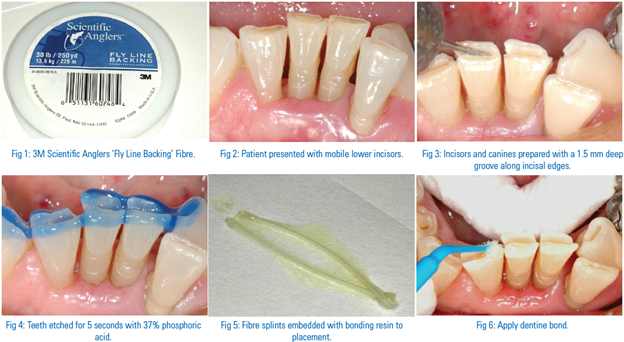
These fibres are well known to fly fishermen and available in most angling shops throughout Australia. They are a woven polyester fibre that is readily impregnated with resin and with a breaking strain of over 16 Kg are extremely strong. These fibres have no internal memory and are easily bent in any direction along the incisal edge of irregular lower incisors.
CLINICAL CASE
The patient presented with #3 level mobility of lower incisors 31 and 41. Figure 2. There was significant bone loss however both teeth were vital. . Teeth 32 and 42 were less mobile and as 32 was well outside the arch the treatment plan was to splint the teeth between the canines and extract tooth 32 to facilitate oral hygiene and improve aesthetics.
- A 1.5 mm deep grove was prepared two thirds of the way along the incisal edges of the canines and along the incisal edges of teeth 42, 41, and 31 using a narrow high speed flat fissure bur. Figure 3. A 1.5mm deep groove is required to maintain stability of the splint. Irreversible tooth preparation has to be weighed against the doubtful prognosis of these teeth if no intervention was undertaken. The age of these patients is generally such that sclerotic changes within the pulp reduce the chance of an exposure however it is prudent warm patients beforehand of this possibility.
- The teeth were etched for 5 secs. with 37% phosphoric acid, washed and dried with oil free air. Figure 4.
- Following isolation with cotton rolls, two lengths of fibres were cut so they would closely fit into the length of the preparation. They were them embedded in resin bond. Figure 5. Two lengths of fibres are required to improve strength and resist rotation of the splint.
- A dentine bonding agent of choice was placed into the preparation. Figure 6.
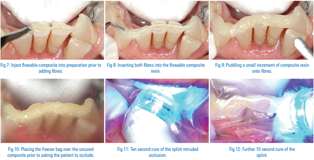
- Following this, a small amount of flowable composite resin was injected into the preparation (Figure 7) into which both impregnated fibres were placed with the aid of a periodontal probe. Figure 8.
- A small amount of composite resin was next puddle into the preparation above the fibres. It is useful to remember that placing too much resin extends the time required to contour the restoration after curing. Figure 9.
- A 3cm square piece of “freezer bag” was placed over the uncured restoration (Figure 10) and prior to closing the patient was instructed to place his tongue at the back of his hard palate to assure a retruded bite. Having the patient occlude in a retruded position prior to curing assures that the mobile incisors are in a stable position within the bite. The “freezer bag” also acts as an occlusal matrix significantly reducing the time required to contour the restoration.
- With the patient closed in a retruded position an initial cure of 10 seconds was carried out (Figure11) followed by another 10 second cure after the patient had opened and the “freezer bag” removed. Figure 12.
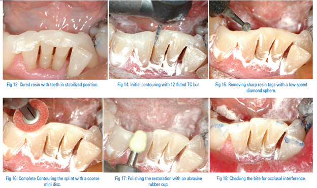
- This slide shows the splint immediately after curing, demonstrating how the incisors are correctly positioned and the minimal amount of contouring required to complete the restoration. Figure 13.
- Initial contouring was carried out on labial and lingual surfaces using a 12 fluted high speed TC bur. These burs rapidly remove composite resin but are not as aggressive on tooth enamel. Figure 14.
- A slow speed diamond sphere was used to remove any sharp tags left by the 12 fluted TC bur, this is particularly important on the lingual surfaces. Figure 15.
- A small abrasive disc was used to smooth over the restoration (Figure 16) prior to polishing with an abrasive rubber cup. Figure 17.
- Following checking the occlusion with articulating paper (Figure 18) tooth 32 was extracted under local anaesthetic to complete the case. Figure 19.
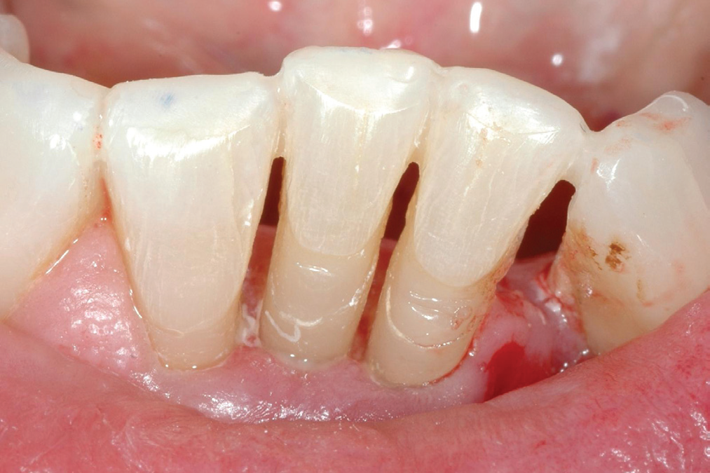
Fig 19: Removal of tooth 32 and the completed splint.
These splints are relatively easy to fabricate, take little clinical time and have proved to be a reliable clinical procedure. Patients are delighted with the outcome as they are able to eat again without discomfort or fear of losing a tooth.
Occasionally an abscess may develop under one of the splinted teeth. As the cause is usually periodontal in origin the problem can be resolved by sectioning the affected tooth at the gingival margin and extracting the root beneath. The patient may be recalled in a couple of weeks to place a RMGIC restoration at the gingival margin using a section matrix positioned on the gingivae to form the base of the restoration.
The “Fly Line Backing” fibres have multiple clinical applications, they are easy to place and do not swell if exposed to the oral environment as some fibres do.
DISCLAIMER
“Published in the Australian Dental Association News Bulletin Sept- 2012 Reprinted with permission of the editor and Author.”
Disclaimer from Guident
The views and opinions expressed in articles published in Guident are solely those of the authors and do not necessarily reflect the views of the editors, the editorial board or the publisher, Ivory India. Guident does not endorse any product, technique or treatment described. Clinical content is intended for qualified dental and medical professionals and is not a substitute for individual clinical judgement. Guident, its editors and the publisher accept no liability for any consequence arising from the use of information published in the journal.
How to cite this article
Dr. Geoffrey M Knight. Splinting Lower Incisors. Guident. 2013 Mar;6(5). Available from: https://guident.in/articles/aesthetic-dentistry/splinting-lower-incisors.html