A Comparative Analysis of Effect of Cortical Bone Thickness, Implant Diameter and Two Different Implant Designs on Stress Distribution: A Three Dimensional Finite Element Analysis Study
Department of Prosthodontics & Crown / Bridge, Sree Sai Dental College, Srikakulam, AP, India
ABSTRACT
Purpose: There are many no.of implant designs in market. Various force magnitude subjected on implants during function. Thus, a finite element analysis helps to access the biomechanics of implant. So, FEA was performed to investigate the effect of mandibular cortical bone thickness, implant design and diameter on stress around implants.
Aim & Objective : Compare the effect of mandibular cortical bone thickness, varying implant diameters, and two different implant designs on stress distribution using a three-dimensional finite element analysis.
Material and Methods: Eighteen 3D models of the posterior mandible were created, varying cortical bone thickness (0.5 mm, 1.0 mm, and 1.5 mm), implant diameters (3.3 mm, 4.1 mm, and 4.8 mm), and two implant designs (threaded and press-fit). An axial load of 100 N was applied on models, and von Mises stress values were calculated.
Results: Implant design and diameter stress distribution varies on cortical bone. As stresses increased with the decrease of crestal cortical bone thickness & less stresses are observed on threaded design implant.
Conclusion: In this study, we found that threaded implant designs reduce stress better. Implants with a larger diameter and thicker cortical bone helps spreading stress more evenly. So, while choosing an implant, proper preoperative evaluation of the cortical bone is recommended.
Keywords: Threaded implant, Press fit implant, Stress distribution
INTRODUCTION
Alveolar bone loss is a common challenge in dentistry today. Bone resorption is a natural process, but it plays a key role in many dental problems. It's important to identify the cause of bone loss and choose the right treatment. One of the most common causes is periodontitis. Studies show that bone loss is often seen in the lower jaw (mandibular arch).1
There are several treatment options to manage bone loss, such as:
Guided Bone Regeneration (GBR) after tooth extraction
Socket Shield Technique
Ridge Augmentation2
These methods can be effective, but they are technique-sensitive and require careful handling. To improve outcomes, newer treatment approaches have been developed.
One of the most effective modern solutions is dental implants. They replace missing teeth and offer strength, function, and a natural look. Implants are usually made from titanium or zirconia and are placed into the jawbone to support crowns, bridges, or dentures.
Some implant treatment options include :
Implant-supported overdentures
"Jaw in a Day" (JIAD) or All-on-4 technique
These methods have shown high success rates, especially in the front lower jaw, where the bone is dense (D1 or D2), allowing for good stability and even immediate loading. They also reduce the need for complex surgeries and help avoid issues like nerve damage.
MATERIAL AND METHODS
An analysis of the scenario was conducted using a three-dimensional finite element research to generate models featured as various implant designs, diameters & cortical bone thickness. In order to predict the stress distribution on the 3 different cortical bone thickness with 3 different implant diameters of 2 implant designs, using finite element analysis.
MATERIALS:
- Metal ceramic (Feldspathic porcelain) - premolar region
- Straight abutment - 6mm length,4mm diameter
- Implant - abutment connecting screw
- D1 type Bone models - cortical bone thickness 0.5,1.0,1.5.
-
IMPLANT DESIGNS
Press fit Implant design models - 8mm length with diameters of 3.3mm, 4.1mm, and 4.8mm. (Fig.1)
Fig. 1: Treatment options for bone loss -A . GBR graft after extraction B. Socket shield technique C. Ridge augmentation techniques

Reverse Buttress threaded Implant design models - 8mm length with diameters of 3.3mm, 4.1mm, 4.8mm.( Fig.2)
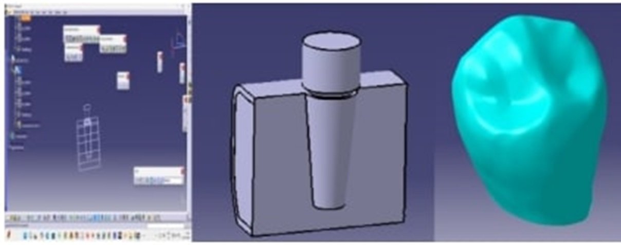
Fig . 2: Press fit implant design - abutment length 6mm,diameter 4mm
METHODOLOGY:
Step 1: Collecting information and Data related to press fit and threaded implants and study the various types of compact bones, cancelous bones & different abutments with various parameters.
Step 2: A fully parametric models of the various threaded and press fit implants which are Cylindrical Shape implants are created. Compact Bone 0.5mm Thickness, Compact Bone 1mm Thickness, Compact Bone 1.5mm Thickness & implants with 3.3 Diameter, 4.1 Diameter, 4.8 Diameter created in catia software.
Step 3: Model obtained in IGS. Analyzed using ANSYS 16.2(work bench), to obtain Von- misses stresses.
Step 4: Taking an average biting force of mandibular teeth I.e. premolar an 100N Axial load was given.
Step 5: Finally, we compare the results obtained from ANSYS and compared various geometries in static analysis finally compared the von-misses stresses.
Load Applied: An axial load of 100N was applied on the central fossa of the premolar of these final element models (18 models).
Stress distribution shall be analyzed using ANSYS of finite elemental analysis created on a computer with the following configurations.
Software
CBCT - SCANNING
MIMIC SOFTWARE
MEASURING DIMENSIONS
CATIA
DESIGN
ANSYS 15.0
FEA
CPU unit
Intel i7
Windows
windows 7
Processor
i7 processor
Memory
4GB RAM
Disk Drive
1 trillion hard disc
Display
LED monitor
Graphic Adapter
4GBRAM VGA
A Three dimensional models of two different implants were designed those are threaded & press fit. Along with abutment, metal ceramic crown was designed and 3 different cortical bone thickness were designs.
The following configurations were the subjects of the construction of three-dimensional finite element models which are 18 variables:
Model 1
Threaded design with 3.3 mm diameter implant on 0.5 mm cortical bone
Model 2
Threaded design with 3.3 mm diameter implant on 1.0 mm cortical bone
Model 3
Threaded design with 3.3 mm diameter implant on 1.5 mm cortical bone
Model 4
Threaded design with 4.1 mm diameter implant on 0.5 mm cortical bone
Model 5
Threaded design with 4.1 mm diameter implant on 1.0 mm cortical bone
Model 6
Threaded design with 4.1 mm diameter implant on 1.5 mm cortical bone
Model 7
Threaded design with 4.8 mm diameter implant on 0.5 mm cortical bone
Model 8
Threaded design with 4.8 mm diameter implant on 1.0 mm cortical bone
Model 9
Threaded design with 4.8 mm diameter implant on 1.5 mm cortical bone
Model 10
Press fit design with 3.3 mm diameter implant on 0.5 mm cortical bone
Model 11
Press fit design with 3.3 mm diameter implant on 1.0 mm cortical bone
Model 12
Press fit design with 3.3 mm diameter implant on 1.5 mm cortical bone
Model 13
Press fit design with 4.1 mm diameter implant on 0.5 mm cortical bone
Model 14
Press fit design with 4.1 mm diameter implant on 1.0 mm cortical bone
Model 15
Press fit design with 4.1 mm diameter implant on 1.5 mm cortical bone
Model 16
Press fit design with 4.8 mm diameter implant on 0.5 mm cortical bone
Model 17
Press fit design with 4.8 mm diameter implant on 1.0 mm cortical bone
Model 18
Press fit design with 4.8 mm diameter implant on 1.5 mm cortical bone
RESULTS
VON-MISSES STRESS GRAPH OF THREADED MODEL:
We can observe that in case of Equivalent von-misses stress D1 bone A fully parametric models of the Artificial various implants Cylindrical threaded and Compact Bone 0.5mm Thickness, Compact Bone 1mm Thickness, Compact Bone 1.5mm Thickness 3.3 Diameter, 4.1 Diameter, 4.8 Diameter created in catia software and performed the static analysis apply the axial load is 100 top of the 1st premolar teeth finally observed the 1.5mm thickness. 4.8 diameter implant have less von-misses stresses 18.252Mpa compared to the remaining designs . ( Graph . 1 Overall stress on threaded models )

Graph 1: Overall stresses on Threaded models
VON-MISSES STRESS GRAPH OF PRESS FIT MODEL:
We can observe that in case of Equivalent von-misses stress D1 Denture A fully parametric models of the Artificial various press fit implants Cylindrical shape and Compact Bone 0.5mm Thickness, Compact Bone 1mm Thickness, Compact Bone 1.5mm Thickness 3.3 Diameter, 4.1 Diameter, 4.8 Diameter created in catia software and performed the static analysis apply the axial load is 100 top of the 1st premolar teeth finally observed the 1.5mm thickness bone with 4.8 diameter implant have less von-misses stresses 26.102Mpa compared to the remaining designs. (Graph . 2 Overall stress on pressfit models)

Graph 2: Overall stresses on Press fit models
DISCUSSION
Cortical Bone Thickness and Implant Stress
The highest stress around a dental implant usually occurs at the implant neck, no matter the implant design, load direction, or bone thickness. This is because cortical bone is much stiffer than cancellous bone, so it takes most of the load when the implant is placed.
During activities like chewing, the implant moves slightly. The stiff cortical bone resists this movement, causing stress to build up at the neck of the implant.
When the cortical bone is thinner, it can't support the implant as well. This leads to higher stress at the implant neck and more pressure on the cancellous bone below the implant.
For example:
With 1.5 mm thick cortical bone, stress is mainly seen at the neck.
With 0.5 mm thick cortical bone, compressive forces spread more, and both bone types show higher stress.
Thinner cortical bone bends more under load, which increases stress in both the cortical and cancellous bone. This can raise the risk of bone resorption (bone loss) and implant failure.
Also, cancellous bone is much weaker — its yield stress is about 60–80 times lower than that of cortical bone. So, when stress transfers to cancellous bone, it’s more likely to be damaged.
IMPLANT DIAMETER
A wider diameter implant distributes occlusal forces over a larger surface area, reducing stress concentrations on the surrounding cortical and trabecular bone. But Narrow implants tend to cause higher stress on the crestal bone, increasing the risk of marginal bone loss and implant failure.
Increasing implant diameter improves primary stability and reduces micro-motion, especially in areas with low bone density areas. Wider implants engage more bone so, distributing functional loads more effectively & reducing stress peaks.
Wider implants reduce cortical bone stress at the crestal level, potentially lowering the risk of peri-implant bone resorption. A larger diameter implant provides better support for prosthetic components, reducing the risk of screw loosening and component fractures.
In this study also as diameter increases the stress values are reduced irrespective of design of implant.
In comparision of diameter of implant vs cortical bone thickness as increase in cortical bone thickness & increase of diameter the stress is decreased.(Fig. 3)

Fig. 3: Threaded implant design - abutment length 6mm,diameter 4mm
IMPLANT DESIGN
In threaded implants the bending tendency is more than cylindrical implants because The narrower structure of the screw-type implant makes it more susceptible to bending forces & this bending effect results in higher stress concentration on the cortical bone near the neck of the implant so, increase the risk of marginal bone loss.3,4,5
The cortical layer acts as the primary load-bearing structure, resisting implant displacement and preventing excessive micro-motion.6 so, in this study also stresses are seen at the neck area in all threaded models.
In threaded implants the surface have different types of threads i.e V shaped, square, buttress, reverse buttress. Among all the types reverse buttress is best one for mechanical stability.7 Because it resist the shear force which reduce the stress in the neck area. So, in this article we took reverese buttress type threaded design.8,9
Smaller pitch has increases bone contact and stress distribution but requires higher insertion torque.10 Deeper threads has enhance anchorage in low-density bone but can create stress concentration in cortical bone.These type of implants can be placed even in D3 & D4.
In this study Ti grade 5 threaded implant is used with small pitch & reverse buttress single threaded with a constant implant length with 8mm. The abutment length is 6mm & 4mm diameter. As cylindrical implants are smooth or slightly roughened surface these are friction based implants. These implants reduce micro-strain of bone because there is an even stress distribution due to large surface area.

Fig .4: Comparision of threaded & pressfit design with 4.8 mm implant in 1.5 mm cortical bone (least stress distributed models - 9 & 18)
According to Nandini et al. the immediate primary stability is less when compared to screw type but after one month the stability is more than screw type. After 3 months screw form is slightly greater.11
Basically, if the initial stability is good enough the dental implant load can give immediately after placement within the bone. But if it is not fair enough we have to wait for secondary stability because it is crucial for successful osseointegration. Secondary stability is achieved as additional bone is remodeled at the implant interface.12
Usually in cylindrical initial primary stability is high & stress also equally transmitted throughout the bone so, these are preferred in conditions of immediate loading. As these are friction based implants where reduced mechanical interlocking is seen.13 So , these can only used in D1 & D2 bone types.
In this study Ti grade 5 smooth surface (non-threaded) implant is used with a constant implant length with 8mm & The abutment length is 6mm & 4mm diameter. A 3D FEA model is prepared by using young’s modulus, Poisson’s ratio, density & tensile strength . Based on these values models are prepared & placed virtually on to D1 type bone finally stresses was analyzed.
As discussed above press fit implant design has more stress when compare to threaded type because it transmitted stress throughout the surface of implant. As diameter of implant & thickness of cortical bone is increases stress was decreases.
CONCLUSION
Increasing implant diameter reduces stress where the stress are less in 4.8 diameter implant designs than other diameters. But it is not a complete solution because wider implants need more bone volume especially for replacement of posterior tooth. So, while selection of implant diameter, cortical bone thickness has to analyse.Therefore among threaded designs the least stress are observed in the model 9 (4.8 threaded implant placed in 1.5mm cortical bone) & for press fit implants the least stress observed in model 18 (4.8 press fit implant placed in 1.5mm cortical bone.
A cylindrical implant has slightly more stress concentration values than threaded implants due to its compressive forces towards bone. But its advantage is minimal osteotomy preparation. So, Osseo integration will be faster. It mostly preferred in D1 & D2 bone, where immediate load can also give.
When it comes to threaded implant it has good mechanical interlocking efficiency, good primary stability & less stress distribution when compare to cylindrical implants. So, it can place even in D3 & D4. But the prognosis is depends on cortical bone around implant especially around the neck of implant.
Finally based on this study results we conclude that threaded implant design has less stress concentration. Increased diameter of implants & increased cortical bone thickness has least stress distribution. So, before selection of implants the bone quality, thickness of cortical bone, area of tooth replacement has to analyzed based on this maximum implant diameter & design is selected for better prognosis of implant prosthesis.
Funding sources - NO
Conflict of Interest - NO
REFERENCES
- Hienz SA, Paliwal S, Ivanovski S. Mechanisms of Bone Resorption in Periodontitis. J Immunol Res. 2015;2015:615486.
- Sverzut CE, Trivellato AE, Sverzut AT, Luna AH, dos Santos TI, Pontes Cde B. Rehabilitation of severely resorbed edentulous mandible using the modified visor osteotomy technique. Braz Dent J. 2009;20(5):419-23.
- Okumura N, Stegaroiu R, Kitamura E, Kurokawa K, Nomura S. Influence of maxillary cortical bone thickness, implant design and implant diameter on stress around implants: a three-dimensional finite element analysis. J Prosthodontic Res. 2010 Jul;54(3):133-42.doi: 10.1016/j.jpor.2009.12.004. Epub 2010 Feb 11. PMID: 20153281.
- Sennerby L, Roos J: Surgical determinants of clinical success of osseointegrated oral implants: a review of the literature. Int J Prosthodont. 1998, 11:408-20.
- Wilson TG Jr, Miller RJ, Trushkowsky R, Dard M. Tapered implants in dentistry: revitalizing concepts with technology: a review. Adv Dent Res 2016;28(1):4–9.
- Zarei, Jafari. et.al Assessing the Effect of Dental Implants Thread Design on Distribution of Stress in Impact Loadings Using Three Dimensional Finite Element Method. J Dent Biomater. 2016 Jun;3(2):233-240.
- Toyoshima T, Tanaka H, Ayukawa Y, et al. Primary stability of a hybrid implant compared with tapered and cylindrical implants in an ex vivo model. Clin Implant Dent Relat Res. 2015;17:950–956.
- Abuhussein H, Pagni G, Rebaudi A, Wang HL. The effect of thread pattern upon implant osseointegration. Clin Oral Implants Res 2010;21(2):129–136.
- Orsini E, Giavaresi G, Trirè A, Ottani V, Salgarello S. Dental implant thread pitch and its influence on the osseointegration process: an in vivo comparison study. Int J Oral Maxillofac Implants. 2012;27:383–392.
- Samieirad S, Mianbandi V, Shiezadeh F, Hosseini-Abrishami M, Tohidi E: Tapered versus cylindrical implant: which shape inflicts less pain after dental implant surgery? A clinical trial. J Oral Maxillofacial Surg. 2019, 77:1381-8.
- Nandini N, Kunusoth R, Alwala AM, Prakash R, Sampreethi S, Katkuri S. Cylindrical Implant Versus Tapered Implant: A Comparative Study. Cureus. 2022 Sep 28;14(9):e29675.
- Atsumi M, Park SH, Wang HL: Methods used to assess implant stability: current status . Int J Oral Maxillofacial Implants. 2007, 22:743-54.
- Szmukler-Moncler S, Salama H, Reingewirtz Y, Dubruille JH: Timing of loading and effect of micromotion on bone-dental implant interface: review of experimental literature. J Biomed Mater Res. 1998, 43:192-203.
Disclaimer from Guident
The views and opinions expressed in articles published in Guident are solely those of the authors and do not necessarily reflect the views of the editors, the editorial board or the publisher, Ivory India. Guident does not endorse any product, technique or treatment described. Clinical content is intended for qualified dental and medical professionals and is not a substitute for individual clinical judgement. Guident, its editors and the publisher accept no liability for any consequence arising from the use of information published in the journal.
How to cite this article
Dr. Macharla Juhi, Dr. CH. Siddesh Kumar, Dr. K. Jagadish. A Comparative Analysis of Effect of Cortical Bone Thickness, Implant Diameter and Two Different Implant Designs on Stress Distribution: A Three Dimensional Finite Element Analysis Study. Guident. 2025 Aug;18(9). Available from: https://guident.in/articles/research/a-comparative-analysis-of-effect-of-cortical-bone-thickness-implant-diameter-and-two-different-implant.html