Reattachment of Tooth Fragment Embedded in Lower Lip Following Dental Trauma Using Fiber Post
Sankapur, Gopinath Nagar, Bye lane-4, House no-22, Guwahati-16, Assam
ABSTRACT
Dental trauma is a common occurrence in routine dental practice. However rarely, after the trauma tooth fragments may get lodged in oral tissues such as the lip and may lead to complications if left undiscovered. This particular case report describes a 22 year old man who was diagnosed with a fragment of a fractured permanent central incisor lodged in the lower lip. The fragment was surgically retrieved and after a week endodontic treatment was performed. Later a fiber post was placed in the root canal and the fragment reattached using composite resin. The patient was followed up for a period of 6 months.
Keywords
Crown fracture, lip , tooth fragment, reattachment, fiber post
INTRODUCTION
One of the most frequent facial traumas corresponds to the damage caused to the teeth and their support structures. These traumas may be the result of many factors such as: falls, assaults, traffic, sports, work accidents and others. In regards to these factors, falls account for the most frequent incident.1
The occurrence of dental trauma depends on the impact energy and direction of the causal agent, on the location of the incidence and on the health of the supporting structures from the involved teeth. Many studies have suggested the higher prevalence of the incisor traumas among childhood and adolescnce2 which can range from 10-20%, according to sex and age of the patients.3 The reason for increased vulnerability of maxillary incisors is because of the projection of anterior teeth and to the short labial lip that do not adequately protect these teeth. These risk factors should be considered in the incidence and severity of these traumas, as they can range from concussion to tooth loss.4
Fractured incisors often cause injury to the soft tissues. In such event, considerable attention has to be spared to the tooth fragment resulting from the accident.5 In many cases the fragments may be lost or badly destroyed, however in certain cases the patient may have preserved the fragment or it may be lodged in the soft tissues and maybe amenable to surgical removal. In such cases reattachment of the fractured fragment serves as a feasible option.
Tennery was the first to report the re-attachment of a fractured fragment using acid-etch technique.6 The introduction of composite in combination with the use of acid-etch technique to bond composite to enamel, made restoration possible for the fractured incisor, with little or no additional tooth preparation .7 However, composite resins have the disadvantage of poor abrasion resistance in comparison to enamel8; water absorption and staining of the composite are further drawbacks.
Reattachment of fragment may offer following advantages: 1. Better aesthetics and achievement of lifelike translucency, 2. Incisal edge wear at a rate similar to that of the adjacent teeth, 3. Replacement of fractured portion involving less time, 4. A positive emotional and social response from the patient9, 5. Relatively inexpensive procedure.
Resin based restorative materials are frequently used in restoration of the fractured teeth. Because of the poor mechanical resistance of these materials, different approaches have been developed to strengthen resistance of composite resin, such as fiber posts. Tooth-colored fiber posts were introduced in the 1990’s and has several advantages, such as esthetics, bond to tooth structure, have a modulus of elasticity similar to that of dentin, but still require dentin preparation to fit into the canal.10
This article presents the description of a case with fractured incisor fragment lodged in lower lip, it’s surgical removal and subsequent reattachment following endodontic treatment
CASE REPORT
A 22 year old male patient reported to the Dept of Conservative and Endodontics College of Dental Sciences, Davangere ,Karnataka, India with a chief complaint of fractured teeth in his upper front tooth region. The fracture had occurred 3 weeks before due to a road traffic accident .Patient did not give any significant relevant medical history.
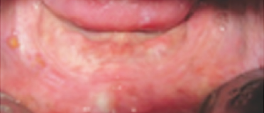
On inspection intra orally Ellis Class III fracture with tooth no 11 and 21 was seen(Figs. 1 & 2). A detailed clinical examination revealed a scar present on lower lip , with a nodule which was firm to palpation.The gingival and alveolar mucosa was intact without any tears. Periodontal probing did not reveal presence of pocket or bone loss.
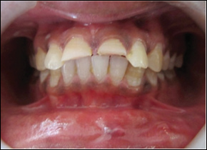
Fig 1: Pre operative intra-oral view
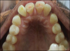
Fig 2: Showing Ellis’ Class III fracture of 11 and 21
No extra oral swelling or lacerations were present (Fig. 3). The patient revealed that the fractured incisors fragment had been lodged in the lower lip. Pre operative radiograph of the lower lip showed a radiopaque fragment lodged in it, resembling the fractured tooth( Fig. 4). Periapical radiographs of the upper central incisors showed a radiolucency involving the pulp space without any root fractures or pathological alterations in the periapical region.
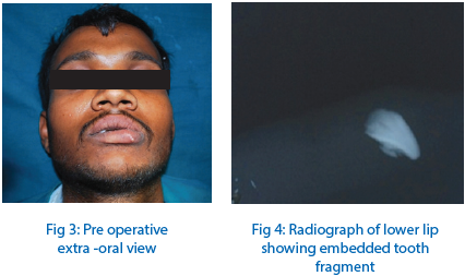
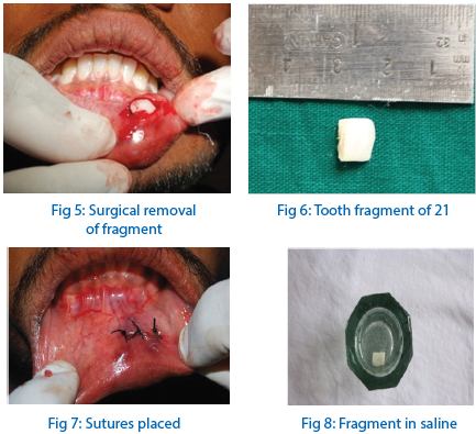
The patient was submitted to a surgical excision of the fragment under local anesthesia. For this procedure 1.0 cc of lidocaine in a 2% solution with 1:1,00,000 epinephrine was administered in the area of lip tumefaction. The lower lip was incised and the dental fragment was identified and gently removed (Fig. 5). The fragment was cleaned and checked in the oral cavity and was found to be the fractured segment of 21(Fig. 6). A 4-0 black silk suture was placed to re-approximate the tissue (Fig 7). The patient was prescribed antibiotics and analgesics post operatively. The patient was recalled after 1 week postoperatively for suture removal and healing was uneventful. The fragment was stored in a saline solution( Fig. 8).
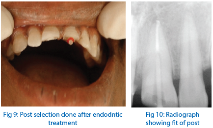
Following the healing endodontic treatment was performed for 11 and 21 . The teeth were cleaned and shaped using the ProTaper rotary system (Dentsply , Maillefer) upto the size F3 file. The teeth were obturated by the lateral condensation technique and temporized. A week after endodontic treatment it was decided to reattach the coronal fragment of 21 along with placement of a fiber post in both the teeth due to lack of sufficient coronal structure. The gutta percha from the canals were removed using Peeso reamer drills upto size 3 and a Para post ( Coltene Whaledent) drill no 2 was used to create space for the post (Fig. 9). Radiographs were taken to assess the fit of the post (Fig. 10) and the post was cemented using a dual cure resin cement RelyX U 200 (3M ESPE).Following the cementation the fractured tooth fragment was reattached.
A small hole was drilled in the cervical portion of the fragment to accommodate the head of the post and the fit of the fragment was reassessed. Once a satisfactory fit had been established the reattachment procedure was commenced. The tooth surface and post were etched with a 37% phosphoric acid gel, rinsed, and coated with an ethanol-based adhesive system (Adper Single BondPlus, 3M ESPE). The adhesive was not light cured at this point.
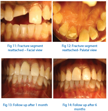
The fractured surface of the fragment was treated with 37% phosphoric acid gel for 30 seconds ,followed by delicate rinsing. The adhesive system was then applied to the etched surface. Composite resin (Filtek Z 350XT 3M ESPE) was applied to both fragment and tooth surfaces. The fractured segment was then accurately placed on the tooth, paying special attention to the fit between the segments. When the original position had been reestablished, excess resin was removed and the area was light cured for 40 seconds on each surface, making sure that no displacement of the fragment occurred before adhesive/resin polymerization was complete. The margins were properly finished with diamond burs and polished with a series of Sof-Lex disks (3M ESPE) and diamond polishing paste.(Figs 11 & 12) A porcelain fused to metal crown was fabricated for 11.
The occlusion was carefully checked and adjusted, and the patient was dismissed after receiving instructions to avoid exerting heavy function on this tooth and to follow regular home care procedures relative to oral hygiene. The patient was informed that the reattachment line might be visible, and, if necessary, this could be managed in future visits. The patient returned for 1- and 6-month follow-ups( Figs. 13 & 14) and it was observed that both endodontic and restorative treatments remained clinically acceptable for the entire time.
DISCUSSION
Inadequate emergency treatment of dental injuries with laceration of soft tissues are important problems that can contribute to increase complications such as infections and other disturbances.11 Usually a fractured or missed incisor does not pose a problem in diagnosis. However when there is also soft-tissue laceration, the possibility of tooth fragments should also be investigated. Occlusal, periapical and lateral radiographs help in the detection of any tooth fragments in the oral regions, especially if the laceration and bleeding make the clinical examination difficult.5
We located the tooth fragment by placing the film between the lower teeth and the lip.This radiographic assessment is extremely important, since tooth fragments may become foreign bodies in soft tissues causing pain , difficulties in speech or eating and oedemas. Also antibiotics are important to prevent systemic and local infection. Anti inflammatory drugs are indicated to reduce edema and provide increased comfort to the patient.12
The most conservative treatment option for a crown facture when a fragment is encountered, is the bonding of the fragment to the fractured tooth. The technique produces good, lasting esthetic results, as it maintains the original anatomic shape, colour and texture of the tooth.5 It is important to maintain the fragment in an aqueous mediums such as normal saline 0.5% until the bonding procedure.12 In the current case we recuperated the fragment and it was successfully bonded.
Although lip tissue is an unusual medium to preserve a tooth fragment, the coronal fragment embedded in the lip of our patient for 3 weeks maintained the conditions and colour of the fractured incisor crown. Moreover according to Tosun et al., 13 the reattachment of tooth fragments can successfully benefit periodontal health, esthetic needs and natural functioning.
Restoration with a post following endodontic treatment retains a core to support coronal restoration, particularly when tooth loss is extensive. In recent years, various types of fiber post have been introduced and excellent long-term clinical performance of pulpless teeth reported. It has been stated that fiber-reinforced resin, post that is bonded into the root canal of the incisor, provides increased retention of the crown’s fractured segment and is less subject to root fracture due to a combination of adhesive and elastic properties.13 However, long-term follow up is recommended.
The techniques described in this case report is reasonably simple, while restoring function and esthetics with a very conservative approach. However, the professional has to keep in mind that a dry and clean working field and the proper use of bonding protocol and materials is the key for achieving success in adhesive dentistry. Reports and clinical experience indicate that the reattachment of fractured coronal fragments results in successful short- and medium-term outcomes.10After the completion of treatment, the patient was recommended to wear a mouth guard if he engaged in sports activities to prevent new dental injuries and conserve the restoration.
It should be remembered that dento- alveolar fractures do adversely affect the patient’s individual quality of life and interferes with day to day activities. But we as dental professionals with the advances in dental technology at our disposal can help reverse the situation. With the materials available today, in conjunction with an appropriate technique, esthetic results can be achieved with predictable outcomes. Thus, the reattachment of a tooth fragment is a viable technique that restores function and esthetics with a very conservative approach, and it should be considered when treating patients with coronal fractures of the anterior teeth, especially younger patients.
REFERENCES
- O'Neil DW, Clark MV, Lowe JW, Harrington MS. Oral trauma in children: a hospital survey. Oral Surg Oral Med Oral Pathol. 1989 Dec;68(6):691-6.
- Galea H. An investigation of dental injuries treated in an acute care general hospital. J Am Dent Assoc. 1984 Sep;109(3):434-8
- Humter ML et al. Traumatic injuries to maxillary incisor teeth in Swedish children. Endod Dent Traumatology 1990;6: 260-4
- Andreason JO, Andreason FM. Textbook and colour atlas of traumatic injuries to anterior teeth, 3rd edn. Saint Louis C. V. Mosby; 1997. P 170-88
- da Silva AC, de Moraes M, Bastos EG, Moreira RW, Passeri LA. Tooth fragment embedded in the lower lip after dental trauma: case reports. Dent Traumatol. 2005 Apr;21(2):115-20.
- Tennery N.T.: The fractured tooth reunited using the acid etch bonding technique. Tex Dent J 1988; 96: 16 - 17.
- Burke F.J.T.: Reattachment of a fractured central incisor tooth fragment. Br Dent J 1991; 170: 223 - 225.
- Badami A., Dunnes, Scheer B.: As in vitro investigation into shear bond strengths of two dentine bonding agents used in the reattachment of incisal edge fragments. Endo Dent Traumat 1995; 11: 129 - 135.
- Baratieri L.N., Monteiro S.: Tooth fragment reattachment: Fundamentals of the technique and the case report. Quint Int 2003; 34: 99- 107
- Baratieri L.N., Monteiro S.: Tooth fracture reattachment: Case reports. Quint Int 1990; 21: 261 - 270.
- Al-Jundi SH. The importance of soft tissue examination in traumatic dental injuries: a case report. Dent Traumatol. 2010 Dec;26(6):509-11.
- Lips A, da Silva LP, Tannure PN, Farinhas JA, Primo LG, de Araújo Castro GF. Autogenous bonding of tooth fragment retained in lower lip after trauma. Contemp Clin Dent. 2012 Oct;3(4):481-3.
- Tosun G, Yildiz E, Elbay M, Sener Y. Reattachment of fractured maxillary incisors using fiber-reinforced post: Two case reports. Eur J Dent. 2012 Apr;6(2):227-33.
Disclaimer from Guident
The views and opinions expressed in articles published in Guident are solely those of the authors and do not necessarily reflect the views of the editors, the editorial board or the publisher, Ivory India. Guident does not endorse any product, technique or treatment described. Clinical content is intended for qualified dental and medical professionals and is not a substitute for individual clinical judgement. Guident, its editors and the publisher accept no liability for any consequence arising from the use of information published in the journal.
How to cite this article
Dr.Kartik Pendharkar, Dr. Pratim Talukdar, Dr.Mukut Seal. Reattachment of Tooth Fragment Embedded in Lower Lip Following Dental Trauma Using Fiber Post. Guident. 2014 Nov;7(12). Available from: https://guident.in/articles/prosthodontics/reattachment-of-tooth-fragment-embedded-in-lower-lip-following-dental-trauma-using-fiber-post.html