Endodontic Perforation Repair with Bioceramic an Endosequence Root Repair Material
Department of Conservative Dentistry & Endodontics D.J College Of Dental Sciences And Research, Modinagar, U.P
Abstract
Introduction:Perforation repair is a fairly common endodontic procedure. The aim of this article was to present a case of perforation repair with endosequence root repair material a six months recall period.
Methods:A nonsurgical endodontic case was treated and repaired with BIOCERAMIC an endosequence root repair material and restored. Recalls upto six months are presented.
Results : Perforation repair was shown to be successful in this case report.
Conclusions:If managed properly, perforation repairs can result in long-term clinical success.
Perforations are unfortunate complications that occur in the course of endodontic treatment1, 2. How their management can make the difference between retention and the loss of a tooth.
Perforation is a mechanical or pathological communication between the root canal/pulp space and the supporting apparatus of the tooth, which leads to a compromise on the health of the periradicular tissue. Perforation can be of two types, one that results from a resorptive process and the other, that is iatrogenically produced, which can occur during the bio mechanical preparation of the root canal or during a post endodontic procedure. Fuss and Trope3 developed a classification of perforations as a predictor of prognosis. They described perforations that are small, within bone, and repaired immediately as having the best prognosis.
Bioceramic a new repair material is, in fact, the EndoSequence Root Repair material, which comes either premixed in a syringe (just like BC Sealer) or as a premixed putty.5,6.This is a tremendous help not just in terms of assuring a proper mix but also in terms of ease of use. We now have a root repair material with an easy and efficient delivery system.7,8,9.
Factors of significance to prognosis for treatment are time, size, and shape of the perforation as well as its location impacts the potentials to control infection at the perforation site.
The aim of this article was to present a case of perforation repair with endosequence root repair material a six months recall period.
CASE REPORT
A 36 year old male patient was referred to the Department of conservative dentistry and endodontics at DJ College of dental sciences and research, modinagar, with chief complaint of pain in the right lower back tooth region since 2-3 months. On clinical examination 46 had a carious lesion on the mesial aspect of the tooth and a radiolucency was present in the furcation area.[Fig. 1 A]. On removal of the same, an ovoid shaped perforation was noticed in the furcation area and mesial side of the mesiobuccal and mesiolingual canals. On probing the area, bleeding was noticed from the perforation site.[Fig. 1 B].
Intra oral periapical radiographic analysis of 46 showed a large radiolucency extending into the furcation area and mesially. The root canals were negotiated, biomechanical preparation completed, calcium hydroxide saline paste was used as the intra canal medicament in between appointments and the root canal obturatedusing AH plus sealer (Dentsply DeTrey GmbH) and Guttapercha cones (DentsplyMaillefer), using a monocone technique.[Fig. 1 C, D]. The site is cleansed with physiological solution. The perforation site was subsequently sealed using bioceramicand finally, radiographic evidence identifies the perforations’ sealing. [Fig. 1 E]. After one month, a control X-ray is taken. [Fig.2 A]. After three months the tooth is restored, with proper bone regeneration in the furcation zone.[Fig. 2 B]. After six months, radiographically, the repair of the defect is almost complete.[Fig. 2 C].

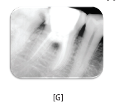
FIGURE 1: [A] Preopoerative Radiograph showing carious lesion on the mesial aspect of the tooth and a radiolucency in the furcation area;[B]On removal of the same, an ovoid shaped perforation was noticed in the furcation area and mesial side of the mesiobuccal and mesiolingual canals. On probing the area, bleeding was noticed from the perforation site,Intra oral periapical radiographic analysis of 46 showed a large radiolucency extending into the furcation area and mesially;[C] Photographic view of the perforation site;[D]The root canals were negotiated, biomechanical preparation completed, calcium hydroxide saline paste was used as the intra canal medicament in between appointments ;[E] The root canalobturated using AH plus sealer (Dentsply DeTrey GmbH) and Guttapercha cones (Dentsply Maillefer), using a monocone technique. The perforation site was subsequently sealed using bioceramic showing the perforations’ sealing; [F] Photographic view showing the sealing of perforation site with bioceramic.
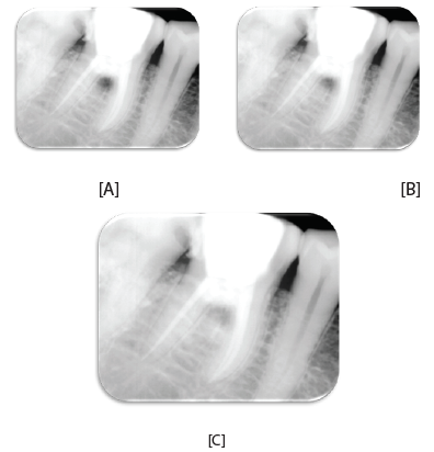
FIGURE 2. [A] One month, a control X-ray; [B] Three months X ray with proper bone regeneration in the furcation zone; [C] Six months, radiographically, the repair of the defect is almost complete
DISCUSSION
Maintaining the integrity of the natural dentition is essential for function and esthetics. Endodontic therapy can play a vital role in achieving this goal. Occasionally technical problems do occur during endodontic treatment i.e. perforating a wall or floor of the pulp chamber or root canal during caries removal, during access cavity preparation, locating of canals and mechanical debridement. This can significantly impair the long-term prognosis of a tooth (Breault et al, 2000).
Prognosis for a perforated tooth depends on the location of the perforation, the time in which the perforation allows entry of contamination, the possibility of sealing it and the accessibility of the main canal.15
Different materials have been used for endodontic perforation repair and the search for an ideal perforation repair material is a challenge. A repair material has to be placed in intimate contact with hard tissues of the tooth and soft tissues of the periodontium. These materials may pose a threat to endodontic treatment outcome by causing local or systemic adverse effects, either through direct contactwith or leaching of chemical components into the periodontal tissues and alveolar bone (Breault et al, 2000).
In this case bioceramic- an endosequence root repair material was choosen to repair the perforation.This material is biocompatible and hydrophilic. It also has a high pH and a strength of 70-90 MPa . It promotescementogenesis.It has high resistance to compression, low water permeability , low porosity and rapid setting reaction which are desired characteristics for reparative purposes.
REFERENCES
- Ng YL, Mann V, Gulabivala K. A prospective study of the factors affecting outcomes of nonsurgical root canal treatment: part 1: periapical health. IntEndod J 2011;44: 583–609.
- de Chevigny C, Dao TT, Basrani BR, et al. Treatment outcome in endodontics: the Toronto study—phases 3 and 4: orthograde retreatment. J Endod 2008;34:131–7.
- Fuss Z, Trope M. Root perforations: classification and treatment choices based on prognostic factors. Endod Dent Traumatol 1996;12:255–64.
- Best S.M., Porter A.E., Thian E.S., Huang J., Bioceramics: Past, Present and for the Future, Journal of the European Ceramic Society 28 (2008) 1319–1913.
- Hench L., Bioceramics: From Concept to Clinic, Journal Amer. Ceram. Soc., 74(7) 1487–1510 (1991).
- Koch K., Brave D., Bioceramic technology-the game changer in endodontics. Endodontic Practice US.2009;12:7–11.
- Koch KA, Brave DG, Nasseh AA, Bioceramic Technology: closing the endo-restorative circle, Part I, Dent. Today, 2010;29:100–105.
- Ghoneim Ag, Lutfy RA,Sabet NE, et al. Resistance to fracture of roots obturated with novel canal-filling system. J Endod,2011;37:1590–1592.
- Koch, K., Brave D., Bioceramic technology — the game changer in endodontics. Endodontic Practice US.2009;12:7–11.
- Seltzer S, Bender IB, Smith J, Freedland I, Nazemov H. Endodontic failures: an analysis based on clinical radiographic and histologic findings. Oral Surg Oral Med Oral Pathol1967;23:500-30.
Disclaimer from Guident
The views and opinions expressed in articles published in Guident are solely those of the authors and do not necessarily reflect the views of the editors, the editorial board or the publisher, Ivory India. Guident does not endorse any product, technique or treatment described. Clinical content is intended for qualified dental and medical professionals and is not a substitute for individual clinical judgement. Guident, its editors and the publisher accept no liability for any consequence arising from the use of information published in the journal.
How to cite this article
Dr. Anil Dhingra, Dr. Saurabh, Dr. H K Bhullar. Endodontic Perforation Repair with Bioceramic an Endosequence Root Repair Material. Guident. 2014 Nov;7(12). Available from: https://guident.in/articles/endodontics/endodontic-perforation-repair-with-bioceramic-an-endosequence-root-repair-material.html