Muscular Control and Complete Denture Stability
Department of Prosthodontics, A. J. Institute of Dental Sciences, Mangalore
INTRODUCTION
The musculature is directly involved during important phases of complete denture treatment. Denture wearers learn to effectively control the dentures with muscles of the lips, cheeks and tongue. Good muscular control and coordination are essential in complete denture stability especially the mandibular denture.
THE DENTURE SPACE
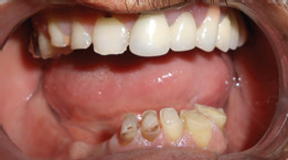
Denture space is that space in the edentulous mouth vacated by the natural dentition and supporting tissues and bounded by tongue medially and the lips and cheeks laterally1 (Figure 1). It is that space in the oral cavity which is bordered by the lips anteriorly, the cheeks laterally and the tongue inferiorly, the maxillary ridge and the palatal vault above, the mandibular ridge below, the lingual tissue attachment at the borders of the floor of the mouth and the palate and by continuation of the oral cavity posteriorly2

Figure 1: Denture Space
Structures in the oral cavity undergo considerable changes following loss of teeth. The lips and cheeks become unsupported by the teeth and the bone. The tongue enlarges and its lateral border comes in contact with the buccal mucosa. This has been termed as proptosis lingualis3. As resorption progresses muscle attachments lie close to the ridge crest.
MUSCULAR CONTROL OF THE DENTURE
As Fauchard stated, “We must consider the form and the curvature that the outside and the inside surfaces must have to avoid discomfort of the tongue, the gingivae and the inside of both cheeks4.
Factors affecting the muscular control of the denture
- Design of the dentures
- Ability of the patient to acquire the necessary skill
Design of the dentures
A properly contoured polished surface of the denture aids in denture stability by active and passive muscle function.
ACTIVE MUSCLE FIXATION
During various phases of mastication, muscles of the cheeks, lips and tongue press against the polished surface of the denture to seat them. When the powerful tongue pressure against a mandibular denture is not counteracted by equal force from the antagonist muscles of the cheeks and lips denture will be dislocated.
Antagonistic activity of muscles can be used to advantage in stabilizing the dentures3.- Right side of the tongue and the right buccinator muscle acting in opposite directions at the same time will hold the denture in place.
- The functioning genioglossus and orbicularis oris muscles will stabilize a mandibular denture by opposing forces in the anterior section.
- When the right and left buccinator muscles contract at the same time an active muscular fixation can be established even with an inactive tongue.
Passive muscle fixation
When the muscles are at rest, the relaxed tissues sit on the dentures, thereby maintaining dentures in position. The mass and weight of the structures and the pressure exerted by muscle tonus will stabilize the denture in passive muscle fixation3.
Two factors are important for the passive muscle fixation i.e inclination and position of the polished surfaces of the dentures in the zone of equilibrium.
Inclination of Polished surfaces
The labial, lingual and palatal parts of the denture base together with nonarticulating buccal and lingual parts of prosthetic teeth surfaces form the polished surface of the denture.6 The polished surface must be modelled into series of inclined planes each of which presents itself to the muscle with which it comes into contact at such an angle that when the muscle moves it pushes the denture into place.7 The general cross sectional shape of the polished surface through the residual ridge area should be triangular. The correct faciolingual positioning of the artificial teeth establishes accurate inclination of the polished surface.
Inclination of the maxillary denture
Buccal surface of the maxillary denture should look outwards and downwards to direct a lateral force from the contracting Buccinator Muscle in a superior direction and thus stabilize the denture. The alveolar palatal surface of the maxillary denture should be concave permitting the tongue to stabilize the denture.
INCLINATION OF THE MANDIBULAR DENTURE
Stability of the mandibular denture depends on the following
- The available tongue space
- The anterior lingual plane held by the anterior third of the tongue
- The flanges of lower dentures should extend under the fold of the Buccinator Muscle and under the tongue to act as handles to hold the denture in place.
- The lower denture must be narrow in the biscuspid region and the posterior teeth must not encroach on the tongue space2.
The flanges should be concave, to face upwards and outwards permitting the cheek to cradle in against it and give the desired inferior component of forces. The lingual flange of the lower denture should be concave and face inwards and upwards. The flange height in the anterior lingual region should be shallow and thicker. In the first molar region the polished surface should be more nearly vertical to provide room for the tongue. In the distolingual region, the flanges turn lingually under the lateral surface of the tongue forming a shallow concavity to permit the tongue to direct forces inferiorly against the flange.
POSITION OF POLISHED SURFACE
Neutral zone
The neutral zone concept was put forward by Sir Wil-Ford Fish8. The neutral zone has been defined as that area in the potential denture space where the forces of tongue pressing outwards are neutralized by the forces of the cheeks and lips pressing inwards9. Positioning the teeth in the neutral zone aids in denture stability. The functional muscular forces vary in magnitude and direction in different individuals10. Neutral zone is also known as dead space11, the stable zone2, and the zone of minimal conflict.12
Patient skill
The patient’s ability to accept and learn the skills required to control the new dentures determines the complete denture success. During the initial learning period patients may take some time to get adapted and habituated. For example, when a patient incises, the forces tend to tip the maxillary denture, causing the posterior border to drop. This movement is counteracted by the dorsum of the tongue which presses against the denture and stabilizes it.
FUNCTIONAL ANATOMY OF MUSCLE IN RELATION TO DENTURE
The musculature of the denture space is divided into two groups
- Dislocating muscles: Those muscles which primarily dislocate the denture during function (Figure 2).
- Fixing muscles: Those muscles that fix the denture by muscular pressure on its secondary supporting surfaces (Figure 3).
- Cagna DR, Massad JJ, Schiesser FJ. The neutral zone revisited: From historical concepts to modern application. J Prosthet Dent 2009; 101:405-412.
- Tuckfield WJ. Problem of the mandibular denture. J Prosthet Dent 1953;3:8-28.
- Brill N, Tryde G, Canter R. The dynamic nature of the lower denture space. J Prosthet Dent 1965; 15:401-418.
- Lott F, Levin B. Flange technique: An anatomic and physiologic approach to increased retention, function, comfort and appearance of dentures. J Prosthet Dent. 1966;16:394-413.
- Brodie AG .Facial patterns. Angle Orthodontics. 1946; 16:75-87.
- Zarb Hobkirk, Eckert, Jacob. Prosthodontic treatment for edentulous patients. In: Mosby ed. 13th edn. Complete dentures Implant supported Prosthesis. St Louis. Missouri: Elsevier Inc., 2013: 180.
- Fish EW. Principles of full denture Prosthesis. In: Staple Press ed, London. 5th edition. 1952. 34,39.
- Fish EW. An analysis of stabilizing factors in complete dentures. Br Dent J 1931; 52:559-570.
- Beresin VE, Schiesser FJ. The neutral zone in complete dentures. J Prosthet Dent. 2006; 95:93-100.
- Beresin VE, Schiesser FJ. Neutral zone in complete dentures. J Prosthet Dent 1976; 36: 356-367.
- Fish EW. Using the muscles to stabilize the full lower denture. J Am Dent Assoc 1933; 20:2163-2169.
- Matthews E. The polished surfaces. Br Dent J 1961; 111:407-411.
- Shannon JL: The mentalis muscle in relation to dentulous mandible. J Prosthet Dent 1972; 27:477-484.
- Last RJ, The muscles of the Head and Neck- A review. Internat Dent J 1955; 5:338-354.
- Lawson WA. Influence of sublingual fold on retention of complete dentures. J Prosthet Dent 1961; 11:1038-1044.

Figure 2: Dislocating muscles

Figure 3: Fixing muscles
VESTIBULAR DISLOCATING MUSCLES
Masseter Muscle
The masseter muscle originates from the zygomatic arch to insert outer surface of the mandible. It is a powerful elevator of the mandible. The contour of the distobuccal region of the mandibular denture is determined by the action of the masseter muscle. The fibres of buccinator muscle are at right angles to the fibres of the masseter muscle. When the masseter is activated, it pushes inward against the Buccinator muscle and produces a bulge into the mouth. When properly molded the distobuccal border of the impression must converge superiorly and medially forming a groove called the masseteric groove.
Mentalis muscle
The mentalis muscle originates in the incisal or canine fossa and inserts into the skin of the chin13 (Figure 4). The origin of the muscle extends closer to the crest of the residual ridge than the mucosal reflection in the labial vestibule. Therefore, when the mentalis muscle contracts, the depth and available space in the labial vestibule gets reduced. The contraction of this muscle is capable of dislodging a mandibular denture particularly when there is severe ridge resorption.
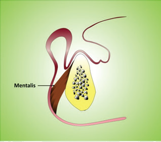
Figure 4: Origin and insertion of Mentalis
Incisive labii inferioris muscle
This muscle originates from the lower canine jugum and extends anteriorly and laterally towards the oral angle. The incisive labii inferioris muscle originates near the crest of the residual ridge. Therefore, during contraction it can destabilize the denture.
LINGUAL DISLOCATING MUSCLES
Internal pterygoid muscle
The internal pterygoid muscle originates from the medial surface of the lateral pterygoid plate and a superficial head arises from the maxillary tuberosity. The muscle inserts on the medial surface of the angle of the mandible. Contraction of the medial pterygoid muscle causes a bulge in the wall of the retromylohyoid curtain. It determines the distolingual extension of the mandibular denture.
Palatoglossus muscle
The muscle descends from the soft palate onto the lateral margin of the tongue. Right and left side muscle fibres meet each other at the midline of the tongue to create a sphincteric function when closing the posterior portion of the oral cavity14. The distolingual part of the denture space is influenced by this muscle. When the muscle contracts the terminating part of the alveololingual sulcus will be included in this movement.
Styloglossus muscle
Styloglossus is attached to the styloid process and the tongue. It acts in the same way as the palatoglossus muscle.
Mylohyoid muscle
The mylohyoid muscle forms the floor of the mouth. The bilateral muscle originates from the whole length of the mylohyoid ridge of the mandible4. Medially, the fibres join those from the mylohyoid muscle of the opposite side and posteriorly they continue to the hyoid bone. Thus, it forms a berthlike structure in which the tongue rests (Figure 5). The anterior fibres of the mylohyoid muscle run horizontally whereas the posterior fibres run in a vertical direction.

Figure 5: Origin and insertion of Mylohyoid
The depth and contour of the lingual flange of the mandibular denture is determined by the position of the mylohyoid muscle. Its action elevates hyoid bone, the tongue and the floor of the mouth during swallowing. When making impression, it is easy to carry the impression material into the undercut below the mylohyoid ridge because it is a thin sheet of fibre in a relaxed state and will not resist the impression material. Extension of the lingual flange under the mylohyoid ridge will interfere with the action of the mylohyoid muscle when it contracts and this will displace the denture, causing soreness.
In the distolingual region, the lingual flange can go beyond the mylohyoid muscle attachment. The impression may depart from the lingual surface of the ridge to be suspended under the tongue in soft tissue on both sides of the mouth to create a border seal.
VESTIBULAR FIXING MUSCLES
Buccinator muscle
The buccinator is the principal muscle of the cheek. The upper and lower fibres originate from the base of the maxillary and mandibular alveolar process in the molar region respectively. They join the middle fibres which arise posteriorly on the pterygomandibular raphe. All the fibres converge in the modiolus at the angle of the mouth. The upper and lower fibres are continuous with the lower and upper fibres of the orbicularis oris muscle respectively2. The fibres of buccinator muscle run anteroposteriorly parallel to the occlusal plane and allow some lateral encroachment by the buccal flange of the denture without causing dislodgement.
When the buccinator muscle contracts, the corner of the mouth is pulled laterally and posteriorly. Its main function is to keep the cheeks taut otherwise when the jaws close the cheeks would collapse and be caught between the teeth.
The buccinator muscle also assists in positioning food between the teeth and returning food that has escaped into the vestibular sulcus to the occlusal table. The lower fibres of the buccinators are relatively flaccid and the middle fibres of the buccinators are active in holding food in position during masticatory effort and which stabilize the dentures. When the muscles contract to hold the food between the teeth they do not reduce the depth of the sulcus in the molar region as they do in the premolar region. They form a pouch in which a piece of food which has been cut off a large bolus can be stored or held while other food is being further triturated6. Therefore, buccinator is also known as the accessory muscle of mastication.
Orbicularis oris muscle
Orbicularis oris is the anterior sphinter muscle of the oral cavity and forms the greater part of the bulk of the lips. The orbicularis oris muscle is attached to the maxillae by the incisive labii superioris muscle and to the mandible by the incisive labii inferioris muscle. It has no bony attachment. The muscle is active when the lips are pressed against the teeth and the alveolar processes.Modiolus
Modiolus is a point at the corner of the mouth where eight muscles of facial expressions meet (Figure 6). The term modiolus is derived from Latin, meaning nave of a wheel because the eight muscles radiate from it like the spokes of a wheel from the hub.7 These Convergences of the muscles form a muscular knot of considerable strength with a wide versatility of movement up, down, forward and backward. Modiolus is located in a strategic position to unseat mandibular dentures and sometimes maxillary dentures as well.4
If the buccinator and orbicularis muscles were the only ones to be considered with, everytime the buccinators contract, the corner of the mouth will be pulled backwards, but a series of muscles cross and intercept the buccinator and orbicularis oris muscles at the angle of the mouth. They are the caninus which arises from the canine eminence, the zygomaticus arising from the zygoma and the triangularis which originates from the lower border of the mandible.2
Narrowing of the mandibular denture base in premolar region avoids denture displacement, otherwise modiolus will not be fixed leading to destabilization of the denture.

Figure 6: Modiolus
Lingual fixing muscles
The tongue consists of the extrinsic and intrinsic group of muscles. The extrinsic muscles have their origin outside the body of the tongue but their course terminates within it.
The extrinsic muscles of the tongue produce upward, downward, forward and backward movements. The genioglossus muscle forms the greater part of the tongue and arises from the superior genial tubercles on the inner surface of the mandible (Figure 7). The anterior group of fibres insert into the tip of the tongue and posterior fibres reach the base of the tongue.13 Anterior fibres are responsible for the protrusion of the tongue and the posterior fibres for the retracting the tip of the tongue.15

Figure 7: Intrinsic muscles of tongue
The intrinsic muscles (Figure 7) lie completely within the tongue and their activities affects the tongue size and form. It has a pair of superior and inferior longitudinalis muscles that can shorten the tongue and turn the tip and sides up or down, a transverse pair of muscles that can narrow and elongate it and a vertical pair of muscles that can flatten and broaden it. These muscles run in three planes and at right angles to each other.4
CONCLUSION
Failure to identify the fundamental importance of tooth position and contour of polished surface often results in dentures which are unstable and unsatisfactory, even though they were skillfully designed and constructed. The coordination of complete dentures with neuromuscular function is the basis of stable dentures.
REFERENCES
Disclaimer from Guident
The views and opinions expressed in articles published in Guident are solely those of the authors and do not necessarily reflect the views of the editors, the editorial board or the publisher, Ivory India. Guident does not endorse any product, technique or treatment described. Clinical content is intended for qualified dental and medical professionals and is not a substitute for individual clinical judgement. Guident, its editors and the publisher accept no liability for any consequence arising from the use of information published in the journal.
How to cite this article
Dr. Vidya K Shenoy, Dr. Mithun Upadhya, Dr. Glynis Miranda. Muscular Control and Complete Denture Stability. Guident. 2020 Jan;13(2). Available from: https://guident.in/articles/prosthodontics/muscular-control-and-complete-denture-stability.html