Minimizing Radiation: A Prosthodontic Approach
Department of Prosthodontics Thai Moogambigai Dental College And Hospital, Chennai
INTRODUCTION
Radiotherapy has advantage of treating a tumor in situ avoiding removal of the tissue or organ. Radiation can be given to a tissue by using an implant (brachytherapy) or an external beam therapy external beam should be given in such a way that does not involve unaffected area. Radiation dose to the eye, optic chiasm or spinal cord, salivary glands, alveolar bone, and soft tissue can be limited through shielding and by moving the uninvolved tissue out of the field.
Laskar and yathiraj pointed out mucositis are seen in almost 100% of patients during head and neck radiotherapy and despite tremendous research, very little has been done regarding the prevention and management of the same. Protecting normal tissues from radiation injury as important as target diseased tissues with radiation.
Radio therapy has good advantage of preserving function and aesthetics of structures which could have been compromised by of surgical management of head and neck cancer. Early examples of preventive prosthodontic appliances used during radio therapy dates back to 1920’s and 1930’s. Radium needle carrier used devices made from simple materials such as wax and modelling compound.
There are many oral complications during radio therapy of head and neck cancer. National institute of health estimated 400000 patients develop oral complication as result of treatment of cancer1. These complications increase agony and creates a painful regime of treatment for patients who are already distressed2. Early management and prevention are the most futile ways of avoiding these complications. Radiation as a treatment very less concern is taken of normal tissues and inadvertent side effects of the therapy. Protecting normal tissues in the radio therapy is as much as important as targeting the diseased tissue.
The line of treatment for cancer patients destroys normal oral architectures such as muscles, salivary glands, teeth and bone, severely compromising the quality of life in these patients so the preventive prosthodontic steps should be taken to avoid/minimize certain unexpected complications. Therefore, increasing the quality of life and encouraging patients for taking treatment and ensuring the quality of care given to avoid destruction of normal tissues by using certain stents and appliances. Therefore, these article outlines the bright side different types of appliances which can be used for patients undergoing radiation for head and neck cancer.
CLASSIFICATION OF RADIO THERAPY PROSTHESIS:
i) According to Santiago 3
a number of prostheses used by dentists help the radiotherapists in treating patients with malignancies of oral and para-oral tissues.
1. The carrier prosthesis holds radium or cesium-137 securely in a planned site during every treatment
2. The prosthesis for radiation beam therapy holds the intraoral cone in a definite position and also places the tongue away from the field of irradiation
3. Another is the prosthesis to displace movable tissues. The shape varies according to the direction in which the tissues are to be displaced. The lips, tongue cheeks, and soft palate may be displaced away from or toward the source of radiation.
4. A prosthesis to displace the tongue varies in shape depending on the direction in which the tongue is to be displaced. The shape also depends on whether the tongue is to be displaced away from the source of radiation or included with the tissues to be irradiated
5. A protector of the tongue is a wall made of a specific thickness of plastic that displaces the tongue. Lead can be added to this wall when additional protection is desired for the tongue
6. Two types of prostheses are used in conjunction with radium needle implants when treating the tongue. One prevents the patient from biting the tongue and the implants by preventing contact of the posterior teeth, the other prosthesis is used after the needle implant treatment is finished. It has a shield to prevent the tongue from coming between the teeth and allows the patient to bring his teeth together.
7. A prosthesis for measuring radiation may be needed. Lithium fluoride capsules attached to the prosthesis enable the radiotherapist to measure radiation received, by tissues in contact with the capsules.
ii) according to japanese academy of maxillofacial prosthetic, they are classified into 3 broad categories
- Shields
- Carriers
- Holders
At Tokyo medical dental college university total of 434 cases of patients with head and neck cancer were treated by radiographic stents. All the stents were fabricated by prosthodontics on the request of radio therapist. Types of stents and early number of stents were fabricated is shown in figure-1[1]. In this case 80% of the stents were spacers. This was because from 1988 onwards rule was employed to fabricate spacers for all the tongue cancer patients. Spacers is not the only appliance used in radio therapy. Recent years many appliances were developed such as shields, carriers etc. An increasing trend is moving towards extra oral spacers and stents using 3d printers.
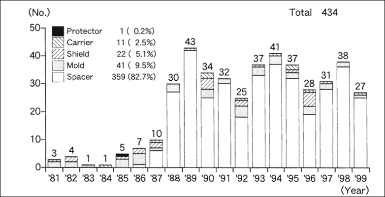
Fig.-1 year by year case count of radiotherapy prosthesis.
(courtesy -Taniguchi H. Radiotherapy prostheses4)
Johannes h et. Al demonstrated 5 cases with the use of dental prosthetic devices to protect normal tissues which showed reduction in radiation induced mucositis and improving radiation distribution.5
In 2013 dosimetry study was conducted by camargosaopaulo, brazil. In a patient with squamous cell carcinoma, it was a comparative study with and without use of intraoral scan and concluded that major structures such as maxillary teeth, hard palate, parotid glands and sub mandibular glands will be preserved from radiation therapy6.
Another study conducted by Juliana Rocha Verome et al. They wanted to determine the effectiveness of intra oral stent during intensity modulated radiotherapy in 33 patients with cancer on the floor of mouth and tongue. And research/results where mean dose to maxilla was 20.0 gy when compared to 35.8 gy in patients without stents. In mean dose to parotid gland was 35 compared to 41.8 gy in patients without stents7.
The study was conducted by Goel et. Al which was the first prospective trial to find the efficiency of stent. In a study group of 352 patients divided into study group and control group. The control group showed much more impact of radiation than study group.
Another study was done by Breno. A. Rocha et. Al in 2017, study of 6 patients with lip cancer were treated with personalized intraoral stent and analyzed for complications. Out of 6 only 1 patient showed oral dryness.8
REQUIREMENT OF A RADIOTHERAPY PROSTHESIS
Adaptability
A radiation prosthesis apart from thickness should extend till the border covering all the area required should adapt properly to the tissue, ill-fitting prosthesis may cause ulceration which may complicate the treatment. Proper impression method to be used to get maximum adaptability without any irritation to underlying tissue, customized tray is advocated in case of presence of tumor or any other defect. Clear acrylic gives the proper view on adaptability of prosthesis to underlying structure. Poor adaptability may result in improper positioning of device which will alter the requirement of prosthesis.
Retention and stability
Proper retention must be provided depends on duration of treatment, Interstitial treatment may require long term retention. When clasp is not advised extra oral retention should be advocated such as chin cap. Patient or radiotherapist should be able to remove and insert on their own, over retention should be avoided. Poor retention can cause movement of denture and may lead to unnecessary trauma to tissue hence proper retention should be provided in prosthesis.
Structure and shape
Structure of prosthesis must be strong enough to resist fracture and shape should be simple, can be molded in to required form and extent. Protruding and sharp edges to be avoided it must be able to fabricate in limited time and avoiding complicated structures and parts.
MATERIALS USED TO FABRICATE RADIATION STENTS
heat cure acrylic resin most common material used for the fabrication of radiographic stents. Common alloys us for shielding are Cerroband, and Lipowitz. Cerrobend composed of bismuth- 50% lead-26.7%, tin-13.3%, cadmium-10%. Cerrobend is mostly preferred than lead as its melting temperature is 140f and hence it can be melted and poured into the cavity prepared, lead has high melting point (600f) and hence cannot be in molten state. Used mainly when patient receives unilateral dose of radiation e.g.-buccal mucosa, skin, alveolar ridge. 1cm thickness of Lipowitz easily reduce an 18mev electron beam by 95%. The walls of reservoir should be 0.5% thick to prevent back scatter of radiation.
Prosthetic device used in radiation therapy are
- Stents
- Shields
- Positioners
- Carriers
- Splints

RADIATION CARRIERS
Radiation carrier are used to carry the material to the site. Tube, needle, beads, capsules are commonly used with radioactive material such as such as radium226, iridium192 or cesium13.most commonly used for carcinoma of salivary gland, buccal mucosa, lip. Basic requirement of this prosthesis is that it should be easily loaded and unloaded, maintain radiation source without any movement in desired place, can be fabricated according to the sources desired or prescribed by the radiotherapist and should be easily removed and placed back in same location. There are are two types carrier used they are pre-loaded and afterlaoding carriers.
Pre loading carriers
In this type of carrier radioactive material is sealed and placed inside the carrier in desired location9, the carrier is coated with Cerrobend to avoid leakage. Disadvantage is that unnecessary exposure to patient and radiotherapist during placement of carrier fig -2.

Fig-2 Pre loading carriers
AFTER LOADING CARRIER
Preferred more than preloading because the radioactive element is placed after thecarrier is in desired location avoiding unnecessary exposure. This type of carrier can be temporary or permanent type. Temporary type the device is removed after the therapy and permanent type the nylon tubing is left inside the tumor after the treatment fig-3.
Glenn E. Minsley and Steven Rothenberg, Use of the afterlaoding technique for intraoral radiation carriers, Maxillofacial Prosthetics L Dental Implants, May 1985 Volume 53 Number 5
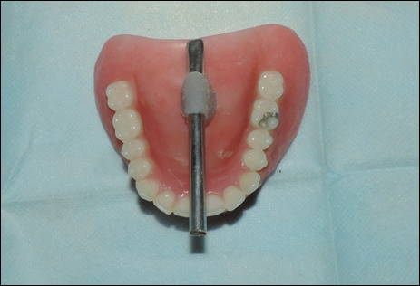
Fig-3 After loading carriers
PERIORAL CONE POSITIONING STENT
This type of stent directs the beam in desired location avoiding any movement in fixed repeatable position thus avoiding unnecessary exposure during the procedure. Usually used in carcinoma of plate sinus, glottis, tonsils, nasopharynx. Perioral cone position stent size is the actual size of the cone used in the treatment, fabricated by 5 to 6mm acrylic ring is fabricated around it. Then the ring is attached to the bases in edentulous case and should be placed directly over the desired location for ration treatment.
DOSIMETER POSITIONING STENTS
Dosimeter is a device used for measuring the radiation exposure. So this device can also be used to calculate amount of radiation exposed to targeted lesion and calculating the required amount of radiation required. Dosimeter stents holds the dosimeter in a required position, lithium flourished capsules are most commonly used. Capsule is embedded in the stent and a wire is attached to punch the capsule in position by the radiotherapist.
INTRA ORAL POSITIONING APPLIANCE FOR STEREOTACTIC RADIOTHERAPY
It is a type of therapy where alternative factional radiation dose is delivered from different positions. Several different positions maximize target doss and minimizing dose to the normal tissue. It is important to have accuracy for treatment and to maintain the head in reproducible manner within the stereotactic space. The head ring is positioned by the means of a screw in the cranial cortical bone this causes unwanted exposure to intraoral tissue to minimize this tungsten spheres of 3mm diameter embedded into maxillary occlusal splint. The metal spheres embedded in the appliance serve as the reference points to verify the location and orientation of the head within the stereotactic space
SHIELDING STENTS
This type of stents is fabricated to protect the vital structures near to the affected tissue from radiation exposure thus minimizing the complication, without any protective measure has led to mucositis and xerostomia. Shields can be made to protect the tongue, salivary glands and the opposite side of the mandible10. These stents consist of a radio-opaque material placed in a position that it shields the vital structure from radiation exposure, material used commonly are Cerroband, pb-bi-sn, and Lipowitz are used as shielding materials fig-4.

Fig-4 Sheilding device
TONGUE DEPRESSING STENT
the major use of this stent is to depress the tongue and preventing it from unwanted radiation exposure. Protects the parotid gland by depressing the mandible during treatment. It has an interocclusal stent extending lingual with a flat plate of acrylic resin which serves to depress the tongue11. Indicated in carcinoma of tongue, head and neck cancersfig-5 and6.

Fig-5 and 6 Tongue depressing stent
DISPLACING STENTS
Used to displace the vital structures from the radiation field. Commonly used in the treatment of tumor involving the alveolus of the mandible, posterolateral borders of the tongue and the buccal mucosa. It separates the maxilla from mandible, which prevents maxilla from unwanted radiation exposure fig-7.
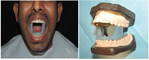
Fig-7 Displacing stents
BOLUS COMPENSATORS
bolus compensators help in the treatment of with irregular contours. Due to irregularities areas within the field may be untreated, while others may develop isolated hotspots. It is a tissue substitute material which fills the irregular spaces thus creating homogenous layer. Commonly used materials for bolus are tissue conditioners, water, saline, waxes and acrylic resin.
TISSUE RECONTOURING STENTS
Required in treating lesions of lips. Curvature of the lip low dose of radiation is delivered at the corners of the mouth and high dose at the center. This stent serves to flatten the lip maintaining in same plane. Fabricated by modeling plastic/wax which are later processed in acrylic resin fig-8a and 8b.
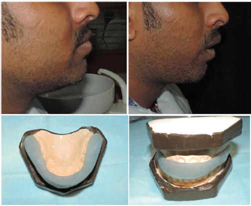
Fig-8a and 8b Tissue recontouring stents
CONCLUSION
Radiotherapy is noninvasive method of treating head and neck, oral cancers and also used adjuvant with chemotherapy and surgical resection. Radiation beam mostly involves normal tissue and organs in the line of beam or near to the targeted site, thus this article depicts various stents to minimize or avoid unwanted damage the normal tissue indirectly.
Case should be properly discussed with radiotherapist and fabricated upon his requirements and request keeping in mind radiotherapy site as primary goal, Quotes go back to “prevention is better than cause” Different stents have shown benefit to the patient during the therapy and should be implicated whenever required to increase the quality of treatment provided to the patients.
With increase in cancer cases of head and neck cancer preventive measure in preventing radiation dose to normal tissue during the treatment procedure will overcome or decrease the fatality rate from it and with the recent advancement like 3D printing the feasibility in fabrication of such stent will become easy.
REFERENCES
- National Institute of Health Consensus Development Conference. Oral complications of cancer therapies: Diagnosis, prevention, and treatment. J Am Dent Assoc 1989;119: 179-83
- Rosenthal LE, Wilkie B. The effects of radiotherapy on oral tissues. The Journal of prosthetic dentistry. 1965 Jan 1;15(1):153-6.
- Santiago A. The role of the dentist in radiotherapy. The Journal of prosthetic dentistry. 1973 Aug 1;30(2):196-201.
- Taniguchi H. Radiotherapy prostheses. Journal of medical and dental sciences. 2000;47(1):12-26.
- Kaanders JH, Fleming TJ, Ang KK, Maor MH, Peters LJ. Devices valuable in head and neck radiotherapy. International Journal of Radiation Oncology* Biology* Physics. 1992 Jan 1;23(3):639-45.
- Verrone JR, Alves FD, Prado JD, Boccaletti KW, Sereno MP, Silva ML, Jaguar GC. Impact of intraoral stent on the side effects of radiotherapy for oral cancer. Head & neck. 2013 Jul 1;35(7):E213-7.
- Verrone JR, Alves FA, Prado JD, das DoresMarcicano A, de Assis Pellizzon AC, Damascena AS, Jaguar GC. Benefits of an intraoral stent in decreasing the irradiation dose to oral healthy tissue: dosimetric and clinical features. Oral surgery, oral medicine, oral pathology and oral radiology. 2014 Nov 30;118(5):573-8.
- Rocha BA, Lima LM, Paranaíba LM, Martinez AD, Pires MB, de Freitas EM, Boas CV, de MeloFilho MR. Intraoral stents in preventing adverse radiotherapeutic effects in lip cancer patients. Reports of Practical Oncology & Radiotherapy. 2017 Nov 1;22(6):450-4.
- Sela M, Taicher S. Intraoral radiation carrier for edentulous patients. The Journal of prosthetic dentistry. 1983 Dec 1;50(6):815-7.
- Fleming TJ, Rambach SC. A tongue-shielding radiation stent. The Journal of prosthetic dentistry. 1983 Mar 1;49(3):389-92.
- Wang RR, Olmsted LW. A direct method for fabricating tongueshieldingstent. J Prosthet Dent 1995;74:171–173.
Disclaimer from Guident
The views and opinions expressed in articles published in Guident are solely those of the authors and do not necessarily reflect the views of the editors, the editorial board or the publisher, Ivory India. Guident does not endorse any product, technique or treatment described. Clinical content is intended for qualified dental and medical professionals and is not a substitute for individual clinical judgement. Guident, its editors and the publisher accept no liability for any consequence arising from the use of information published in the journal.
How to cite this article
Dr. Rathika Rai, Dr. Rahul B.. Minimizing Radiation: A Prosthodontic Approach. Guident. 2018 Dec;12(1). Available from: https://guident.in/articles/prosthodontics/minimizing-radiation-a-prosthodontic-approach.html