Management of Flabby Ridges
Raichur, Karnataka, Maharashtra
ABSTRACT
A fibrous or flabby ridge is a superficial area of movable soft tissue affecting the maxillary or mandibular alveolar ridges.Masticatory forces can displace this mobile denture-bearing tissue, leading to altered denture positioning and loss of peripheral seal. Denture fabrication on flabby tissue is a challenging task for the clinician.The use of conventional impression techniques may lead to an unstable and unsatisfactory denture. This article presents two case reports for rehabilitation of patients with flabby ridges with different impression techniques.
INTRODUCTION
The performance of a complete denture prosthesis during function is a reflection of its support & retention.1 An accurate secondary impression of the denture bearing and limiting structures will ensure maximum support, stability and retention of the prosthesis during its use.2 The task of ensuring adequate function increases manifold when the dentinst confronts denture bearing areas which are movable and displaceable.
A fibrous or flabby ridge is a superficial area of displacable soft tissue affecting maxillary or mandibular alveolar ridges. It develops when hyperplastic soft tissue replaces the alveolar bone and is a common finding particularly in the maxillary anterior region of long term denture wearers. The reported prevalence has varied, but has been demonstrated in upto 24% of edentulous maxillae and 5% edentulous mandibles. Masticatory forces can displace this mobile denture-bearing tissue leading to loss of peripheral seal. Forces exerted during impression making can result in distortion of the mobile tissue.3
Flabby ridge has also been believed to occur in cases with a maxillary complete denture opposing mandibular anterior natural teeth, without proper posterior occlusal support.4 Another reason for flabby tissue is unplanned and uncontrolled dental extraction.5
Flabby tissues get compressed during conventional impression making and later tend to recoil and dislodge the overlying denture.6 The objective of this article is to describe impression techniques for rehabilitation of patients with flabby ridges.
CASE REPORT 1
A 45 year old male patient reported to the Department of Prosthodontics , Crown and Bridge, AME’s Dental College, Raichur, with the chief complaint of completely edentulous maxillary and mandibular arches since one year and required a new set of complete dentures . On examination, flabby tissue in the maxillary anterior region extending from right first premolar to left canine region was found [Fig-1]. Tissue blanching was noticed on pressure application.
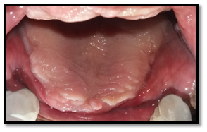
Fig- 1: Flabby Tissue In The Maxillary Anterior Region
Fabrication of new complete dentures was planned for the patient with recording of flabby tissue in undisplaced condition using Hobkirk technique.7 The maxillary preliminary impression was made using irreversible hydrocolloid (Zelgan Alginate Impression Material, Dentsply India Pvt Ltd.) in perforated edentulous tray and the primary cast was poured [Fig-2]. Special tray was fabricated using double spacer over the flabby tissue area and in the region of mid palatine raphe. After checking the proper tray extensions, border molding was done in conventional manner using green stick impression compound (DPI Pinnacle Tracing Sticks). [Fig-3]

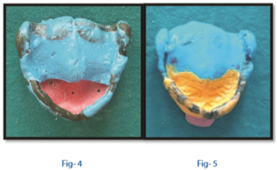
Spacer wax was removed and impression was made with medium body elastomeric impression material (Reprosil Medium bodied silicone, Dentsply Intl Inc.,Milford, USA). The tray was then removed from the mouth and impression material was removed in the region of flabby tissue using a scalpel [Fig-4]. Relief holes were made and tray was loaded in this region with light body elastomeric impression material (Reprosil light bodied silicone, Dentsply Intl Inc.,Milford, USA) to record flabby tissue. [Fig-5]
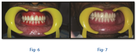
Beading and boxing of the final impression was done and master cast was poured in Type 3 gypsum product. A tentative jaw relation was recorded and wax try in done [Fig-6]. The dentures were then processed, trimmed, finished and polished. It had good retention and stability with proper recording of flabby tissue. [Fig-7]
CASE REPORT 2
A 52 year old male patient named Mallikarjun reported to the Dept of Prosthodontics, Crown and Bridge and Implantology , Ame’s Dental College, Raichur, with a chief complaint of ill fitting dentures since six months. The patient had been a denture wearer since 4 years.
On examination, flabby tissue in the maxillary anterior region extending from canine to canine was found. Treatment plan was formulated for the fabrication of a new set of complete dentures.
Window impression technique8 was planned for this patient. The maxillary preliminary impression was made using irreversible hydrocolloid (Zelgan Alginate Impression Material, Dentsply India Pvt. Ltd) in perforated edentulous tray and primary cast was poured. Spacer wax was adapted over the primary cast except in the region of flabby tissue. Special tray was fabricated providing a window in the region of flabby tissue [Fig-8]. Border molding was done using green stick compound (DPI Pinnacle Tracing Sticks ). Spacer wax was removed, tray adhesive was applied around the window and impression was made with zinc oxide eugenol impression material. With the zinc oxide eugenol impression (DPI Impression Paste) in the mouth, light body silicone impression was injected over the region of flabby tissue [Fig-9]. Silicone impression material was allowed to set and tray was removed from the mouth. Beading and boxing of the impression was done and master cast was poured. The denture was fabricated in which flabby tissue was properly recorded and given adequate relief. [Fig-10]

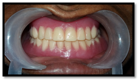
Fig- 10
DISCUSSION
Unstable occlusal conditions resulting in excessive forces on the edentulous arches can lead to the formation of flabby ridges which inturn lead to poor support for the maxillary denture. This is caused by the replacement of bone by fibrous tissue and is associated with marked fibrous inflammation.9 This condition was first described by Watson in 1970. Later in 1972, Ellsworth Kelly and his colleagues first described 'combination syndrome' caused by the presence of opposing natural teeth to an edentulous area. His observations included alveolar bone resorption in the anterior maxilla, enlargement of the tuberosities and bone resorption underneath the mandibular denture bases.10
Different impression techniques have been proposed so that the flabby ridge can be recorded with minimal displacement of the fibrous tissue. However there is controversy as to whether a mucodisplacive technique which compresses the mobile tissue aiming to achieve maximum support from it or whether a mucostatic technique with the aim of achieving maximum retention should be employed.11 Liddlelow in 1964 described a technique whereby two separate impression materials were used in a custom tray (using 'plaster of Paris' over the flabby tissues and zinc oxide eugenol over the normal tissues). In 1964, Osborne described a technique where two separate impression trays and materials were used to separately record the 'flabby' and 'normal' tissues and then related intra-orally.12
Impression making plays a very important role in complete denture fabrication. Problems of support and retention of the upper denture are encountered if flabby tissue is present on an otherwise 'normal’ denture bearing area. An impression technique is required which will compress the non flabby tissues to obtain optimal support and at the same time, will not displace the flabby tissues.
The current article describes simple techniques to record flabby tissues in their undisplaced state using readily available clinical materials like polyvinylsiloxanes. The choice of treatment is made based on the requirement that the stability and retention of prosthesis must be balanced along with the preservation of health of the oral tissues.
CONCLUSION
Managing a patient with flabby maxillary ridge can be a challenging task to the clinician. The two impression techniques described in this article require the same amount of appointments as required for the fabrication of the conventional complete denture and the materials are also readily available. The treatment of flabby ridges is controversial and no one treatment stands out against the rest, so it is often the method favoured by the clinician that is used which ensures both accurate fit and optimal functioning of the prosthesis.
REFERENCES
- Fenlon MR, Sherriff M, Walter JD. Comparison of patients' appreciation of 500 complete dentures and clinical assessment of quality. Eur Journal of prosthodont Rest Dent 1999 ;7(1):11-4.
- British Society for the Study of Prosthetic Dentistry. Guidelines in prosthetic and implant dentistry. London: Quintessence. 1996;1.
- Crawford RW, Walmsley AD. A review of prosthodontic management of fibrous ridges. Br Dent J 2005;199:715-9
- Naik R, Madhukiran, lttagi J. Impression Technique for Flabby Ridges.J Dent Sci Res 2012;5(1):7-9
- Q. Xie, T. O. Närhi, J. M. Nevalainen, J. Wolf, and A. Ainamo, “Oral status and prosthetic factors related to residual ridge resorption in elderly subjects,” Acta Odontologica Scandinavica, vol. 55, no. 5, pp. 306–313, 1997
- Basker R M, Davenport J C. Prosthetic treatment of the edentulous patient. 4th edn. Oxford: Blackwell, 2002
- Hobkirk JA. Complete Dentures: A Dental Practitioner Hand book. Bristol: Wright; 1986:44-5.
- Khan Z, Jaggers J, Shay J. Impressions of unsupported movable tissues. J Amer DentAssoc 1981;103:590-92
- Sajani R, Rankumari A. Impression techniques for Effective management of flabby ridge-An overview. J of Scientific Dentistry. 2012;2(2):34-8.
- Kelly E. Changes caused by a mandibular removable partial denture opposing a maxillary complete denture. J Prosthet Dent 1972;27:140-50.
- Pai UY, Reddy VS, Hosi RN. A single step impression technique of flabby ridges using monophase polyvinylsiloxane material: a case report. Case reports in dentistry. 2014 Apr 27;2014.
- Lynch CD, Allen PF. Management of the flabby ridge: Using contemporary materials to solve an old problem. Br Dent J 2006;200:258-61.
- Kelly E. Changes caused by a mandibular removable partial denture opposing a maxillary complete denture. J. Prosthet Dent. 1972;27(2):140-50.
- Bansal R, Kumar M, Garg R, Saini R, Kaushala S. Prosthodontic rehabilitation of patient with flabby ridges with different impression techniques. Indian Journal of Dentistry. 2014 Apr;5(2):110.
Disclaimer from Guident
The views and opinions expressed in articles published in Guident are solely those of the authors and do not necessarily reflect the views of the editors, the editorial board or the publisher, Ivory India. Guident does not endorse any product, technique or treatment described. Clinical content is intended for qualified dental and medical professionals and is not a substitute for individual clinical judgement. Guident, its editors and the publisher accept no liability for any consequence arising from the use of information published in the journal.
How to cite this article
Dr.Anupama-NeelakantanDr.Chandrashekhar Sajjan Department of Prosthodontics AME Dental College & Hospital. Management of Flabby Ridges. Guident. 2017 Nov;10(12). Available from: https://guident.in/articles/prosthodontics/management-of-flabby-ridges.html