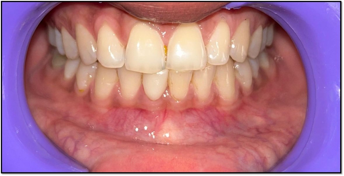
Hemisection: A Ray of Hope for Freezing Tooth : A Case Report
Department of Prosthodontics and Crown & Bridge, Subbaiah Institute of Dental Sciences, Karnataka India
ABSTRACT
Modern advances in dentistry have provided the opportunity for patients to maintain a functional dentition for lifetime. Hemisection is a conservative way of preserving tooth.1 It is most likely to be performed on mandibular molars.The purpose of this study was to report a case of hemisection management in teeth with bifurcation lesion. A 49-year-old female patient complained of pain and discomfort arising from the right mandibular first molar during mastication. The patient underwent endodontic treatment. Based on the clinical examination, a deep cavity was found in 46. It revealed oblique radiolucent line extending from cervical 3rd of mesial root to furcation area with widening of the pdl space and intact lamina dura of mesial root. A hemisection procedure was performed in the mesial root. The remaining root restored as an individual tooth, restoring individual roots with premolar morphology using the adjacent tooth as an abutment in a full coverage fixed partial denture. Hemisection can prove to be a reliable treatment option in saving molar teeth once considered non restorable.2
Keywords: Crown, Bifurcation, Hemisection
INTRODUCTION
Loss of the posterior teeth is eventful and undesirable often leading to teeth drifting, loss of masticatory function and loss of arch length, which requires prevention and maintenance measure. Advances in dentistry have provided an opportunity for patients to maintain their functional dentition for a lifetime. Nevertheless, treatment strategy to retain such teeth involves periodontal, prosthodontic and endodontic assessment for appropriate selection to allow for stronger survival.1
Hemisection is a conservative way of preserving tooth. Hemisection is a surgical procedure which involves resection and subsequent extraction of a compromised root up to the level of furcation1.The main goal of endodontic surgery is to eliminate the disease, prevent the recurrence of the disease, and facilitate healing on the side of the involved tooth tissue so that it can be restored according to its function.
The success or prognosis of the surgical endo depends on diagnosis factors, case selection, the accuracy of indications, contraindications, treatment procedures, periodic evaluations, statistical analysis and criteria of success itself.2
The Endodontic indications of hemisection are:
- Prosthetic failure of abutments within a splint.
- Endodontic failure: perforation through the floor of the pulp chamber, or pulp canal of one of the roots which cannot be instrumented.
- Vertical fracture of one root: vertical fracture traverses one root while the other roots are unaffected.
- Severe destructive process: furcation or subgingival caries, traumatic injury, and large root perforation during endodontic therapy.
The Periodontal Indications Of Hemisection are:
- Severe vertical bone loss involving only one root of multi-rooted teeth.
- Through and through furcation destruction.
- Unfavourable proximity of roots of adjacent teeth, preventing adequate hygiene maintenance in proximal areas. Severe root exposure due to dehiscence
The Contra-indications of hemisection are:
- Strong adjacent teeth available for bridge abutments as alternatives to hemi-section which could act as an abutment to a prosthesis, the remaining root may be inoperable for root canal treatment.
- Inoperable canals in root to be retained.
- Root fusion-making separation impossible.
Hemisection and root resection have now been established as successful treatment modalities. Yuh et al. assessed survival rates of a large number of root-resected molars retrospectively and reported interesting findings with respect to demographic variables. The overall survival rate for root-resected molars was found to be 91.1%.Carnevale et al. reported a survival rate of about 93% over a 10 year follow up among patients who received hemisection as the management of furcated molars.
CASE REPORT
A 49 year old female was referred to the department of conservative dentistry and endodontics with a chief complaint of pain in the lower right back tooth region since 15 days. Pain was dull aching, non-radiating and intermittent in nature. Aggravated on chewing and on intake of cold and hot food and relieved on its own. Patients visited the dental office visited 10 months ago for extraction of 31,41 and 36. Patient is a known areca nut chewer since 20 years.
On intraoral examination Occlusal Caries irt 14 and 24 ,Occlusal Pit irt 15 ,Tender on percussion irt 46 and Missing tooth irt 31,41 and 36. On probing, a periodontal pocket of 6 mm was found on the distal aspect. However, no mobility was observed in the affected tooth. On radiographic examination It revealed oblique radiolucent line extending from cervical 3rdof mesial root to furcation area with widening of the pdl space and intact laminadura of mesial root (figure 1).
On the basis of history, clinical and radiographic examination, a diagnosis of chronic apical periodontitis was made with respect to tooth #46.,class 1 caries-14,15,24 and kennedys class III modification I. Since the extent of decay rendered the tooth non restorable, the patient was explained about the condition and prognosis of tooth with feasible treatment options including extraction and placement of dental implant. However, she opted for hemisection followed by fixed dental prosthesis over other treatment options. The periodontal prognosis of the distal root was fair with good bone support. After completion of endodontic treatment that also included removal of all carious tooth structures , hemisection of distal root was performed under local anesthesia.
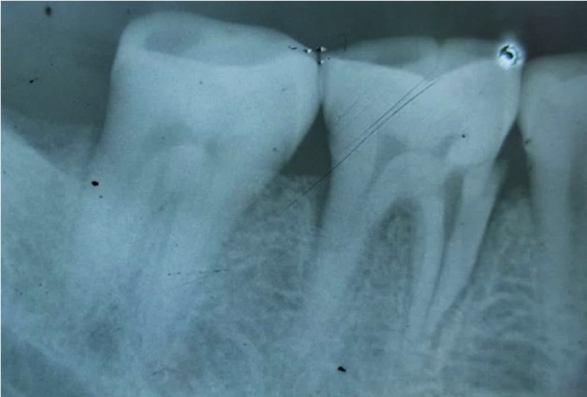
Figure 1
The treatment plan was decided into 3 phases.
- Root canal treatment in distal root of 46
- Hemisection surgical procedure
- Fixed partial denture irt distal root of 46 and 45.Removable partial denture irt 31,41 and 36
Phase 1:
Access opening was done irt 46 and the distal canal was negotiated (figure 2). The patency of the canal was established using no 10 K-file Working length of 18 mm was determined. Biomechanical preparation was done with 21 mm 4% taper upto F2 rotary file. F2 master cone was selected (figure 3) and the obturation (figure 4) was done with Zinc Oxide Eugenol sealer. Post endo restoration was done with Glass Ionomer Cement (figure 5).
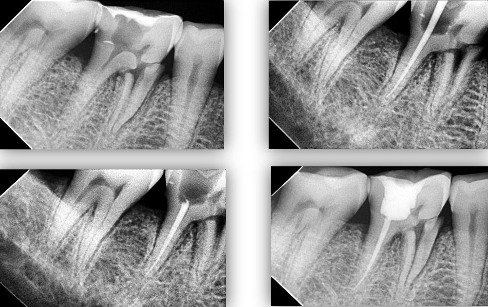
Figure 2, Figure 3, Figure 4, Figure 5
Phase 2:
Blood investigation was advised. Under the normal confinements of blood report, patient was prepared was the surgical procedure. Inferior Alveolar nerve block, Lingual nerve block and Long Buccal nerve block was administered (figure 6). Tooth number 46 was split buccolingually upto the furcation area with long shank carbide bur (HP 701) (figure 7). Straight elevator and Coupland elevator were used to luxate the mesial half of 46 and the fragments were extracted (figure 8), socket was compressed and copiously irrigated with saline and betadine. Two simple interrupted sutures (figure 9) were placed and post operative instructions were given. Immediate post operative x ray was taken (figure 10).

Figure 6, Figure 7, Figure 8, Figure 9, Figure 10
Phase 3:
Diagnostic impressions were made with irreversible hydrocolloid impression material and diagnostic casts were obtained. Tooth preparation of the mesial portion of the second permanent molar and first permanent molar was performed followed by metal prosthesis. Tooth preparation was performed using principles of tooth preparation and full metal fixed restoration.with pre molar anatomy. Hemisected abutment was given a taper greater than 6–10 degree to have a path of insertion Occlusal table is reduced in size in order to decrease the forces on the retained hemisected root. Cuspal inclines are made less steep to reduce laterally directed forces and eliminate the non-working contacts. Final impression was made using putty-reline technique and master cast was obtained. Mandibular master cast was mounted using interocclusal record. Wax pattern was fabricated, sprued, and invested. Casting procedure was carried out using standard techniques. Metal framework was tried in the patient’s mouth. Final prosthesis was cemented using glass ionomer cement.Post cementation instructions regarding periodontal maintenance were given. Recall was done periodically to assure the healing and success of the restoration.
Radiographs at 3 months and 9 months suggested progressive formation of bone in the extraction socket along with resolution of radiolucency around the mesial root of #46.
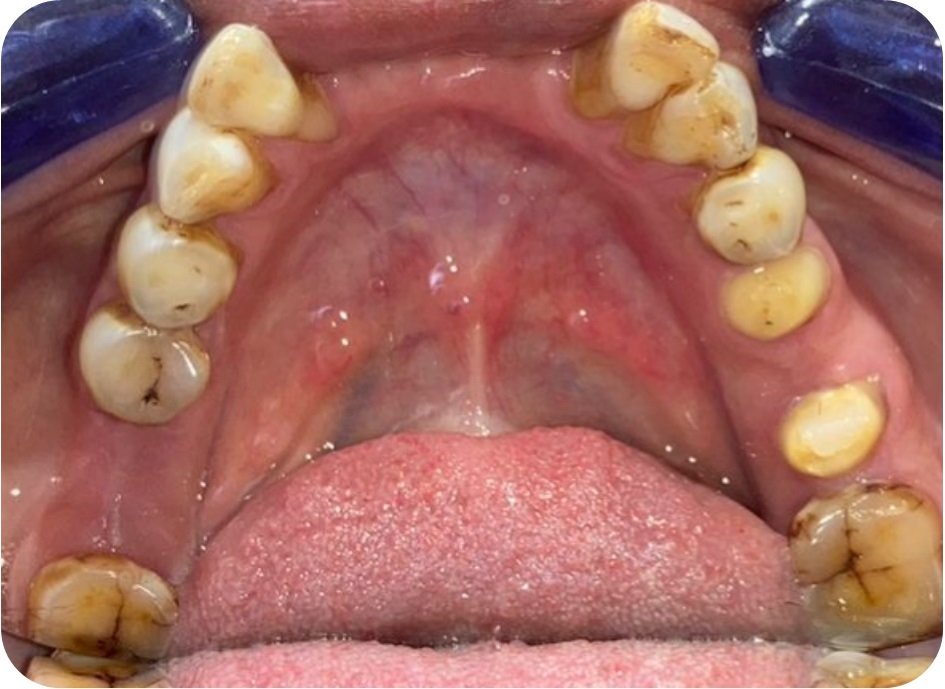
Figure 11
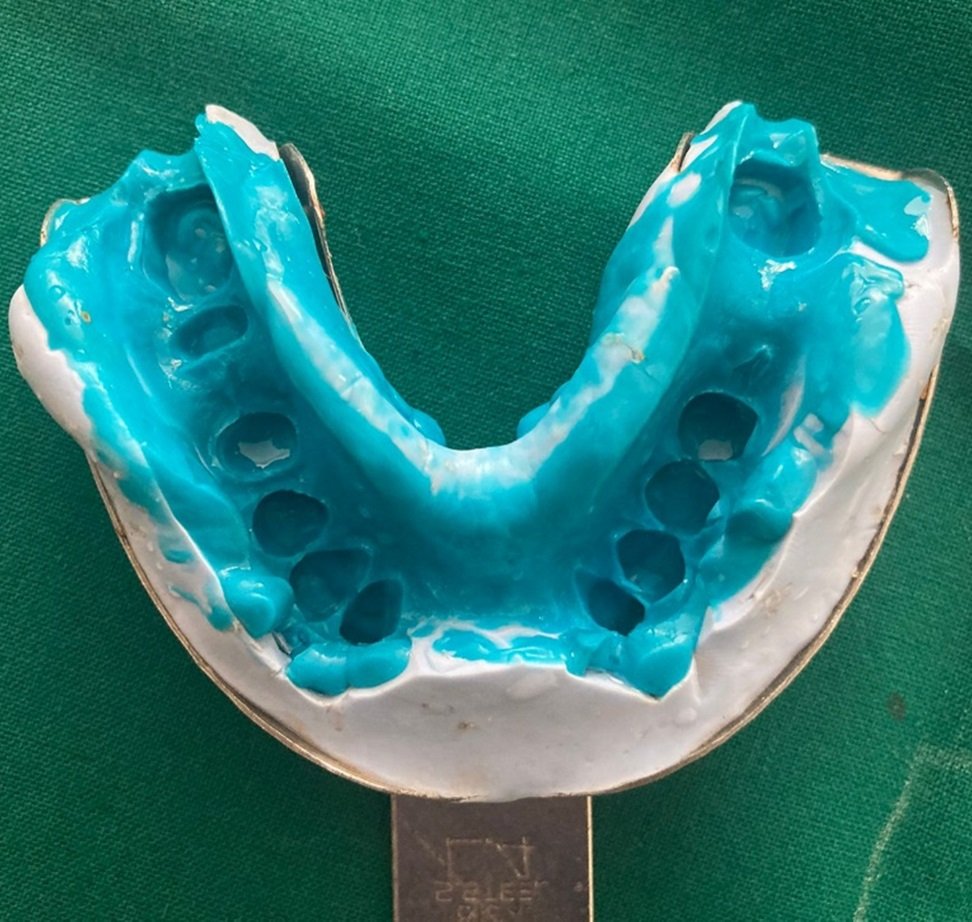
Figure 12

Figure 13

Figure 14

Figure 15

Figure 16
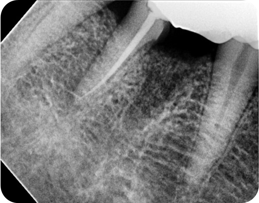
Figure 17

Figure 18
DISCUSSION
A useful alternative method to save multi-rooted teeth that have been recommended for extraction is hemisection, often known as root amputation. If the right cases are chosen, it is a reasonably straightforward, conservative, affordable treatment with good prospects of success. The clinician's decision to select a case for hemisection is influenced by a variety of circumstance.
Hemisection has been used successfully to retain teeth with furcation involvement. However, there are a few disadvantages associated with it. It can cause pain and anxiety; more susceptibility to caries especially at the site of hemisection, an improperly shaped occlusal contact area, may convert acceptable forces into destructive forces and predispose the tooth to trauma from occlusion.
It is important to consider the following factors while restoring a hemisected molar:
- Advanced bone loss around one root with an acceptable level of bone around the remaining roots.
- Angulations and position of the tooth in the arch. A molar that is buccally, lingually, mesially or distally titled cannot be resected.
- Length and curvature of roots
Hemisection is a dependable treatment choice for molars with a dubious prognosis since it will keep the teeth without a noticeable bone loss for a protracted length of time if the patient maintains excellent dental hygiene. Additionally, it is important to evaluate the root's accessibility for simple separation and the root's remaining bone support. The overall survival rate of a large number of root-resected molars by Yuh et al. in a retrospective study was found to be 91.1%. A survival rate of nearly 93% over a 10-year follow-up was reported in another study by Carnevale et al. in cases where hemisection was performed for the management of furcated molars.When subgingival caries, residual roots, or ledges were present in 21 (30%) of the resections, those procedures were deemed flawed. Maxillary molar failure rates were higher (33.3%) than mandibular molar failure rates (22.7%).
In 2010, Anthony and his associates demonstrated an alternative treatment for extraction of a whole tooth and salvation of a healthy tooth structure where the patient did not wish to have the tooth removed. In this case, a hemisected molar was restored with individual premolar morphology on the molar and adjacent premolar. Anshuman Kharbanda et al , in which a hemisected mandibular first molar was restored with a molar morphology and an inlay type of restoration on the second molar. In the present case, good prognosis was observed with proper occlusion, absence of mobility, and healthy periodontal condition.
Thus, conservative management of extensive carious molar tooth in young patients can not only preserve the tooth but also reduce the financial burden, psychological trauma and occlusal dysfunction.
CONCLUSION
Conservative management of grossly carious multirooted teeth in young patients not only preserves the dentition butalso reduces the financial burden, psychological trauma, and occlusal dysfunction associated with tooth loss. Hemisection seems to be a reliable treatment option for saving a nonrestorable molar which otherwise needs to be extracted.
REFERENCES
- Karimah F, Hutami ER, Nugraheni T, Mulyawati E. Hemisection as an Alternative Management for Mandibular First Molar With Bifurcation Lesion and Root Fracture: A Case Report. In4th International Conference on Sustainable Innovation 2020–Health Science and Nursing (ICoSIHSN 2020) 2021 Jan 16 (pp. 209-212). Atlantis Press.
- Naveen YG, Patel JR, Parikh P, Shah K. Alternatives for restoration of a hemisected mandibular molar. Case Reports. 2014 Jul 30;2014:bcr2013010275.
- Jain A, Bahuguna R, Agarwal V. Hemisection as an alternative treatment for resorbed multirooted tooth-A case report. Asian J Oral Health Allied Sci. 2011 Jan;1:45.
- Behl AB. Hemisection of a Multirooted Tooth-A Case Report. 1: 336. doi: 10.4172/scientificreports. 336 Page 2 of 3 Volume 1• Issue 6• 2012 access cavity was restored with IRM and the patient was reviewed after 15 days and hemisection procedure was carried out. Figures 4a and 4b) Decision was made to extract the distal root. Post endodontic restoration was done with composite resin and a three unit fixed partial denture involving. 2012;35:36.
- Sharma S, Sharma R, Ahad A, Gupta ND, Mishra SK. Hemisection as a conservative management of grossly carious permanent mandibular first molar. Journal of natural science, biology, and medicine. 2018 Jan;9(1):97.
- Mishra P, Sharma A, Mishra SK. Hemisection: A conservative approach of tooth preservation. Journal of Current Research in Scientific Medicine. 2016 Jan 1;2(1):46-8.
- Mohammad Khalid Shafiq[13] hemisected the tooth and mesial root was removed along with the dislodged part. A three unit bridge combining the hemisected root and adjacent second premolar was inserted which is successfully in service for more than a year. He concluded that the retention of a part of a tooth seems to extend the life of prosthesis; the patient certainly deserves the option of hemisection or root amputation rather than extraction.
- Shafiq MK, Javaid A, Asaad S. Hemisection: An option to treat apically fractured & dislodged part of a mesial root of a molar. Jpda. 2011 Jul;20(3):183-6.
- Khetan Jr RR, Mahapatra J. Hemisection: A Boon for the Hopeless Tooth. Cureus. 2024 May;16(5).
- Thakur V, Sharma V, Minocha A. Hemisection: Split to save a compromised tooth-A case report. UNIVERSITY JOURNAL OF DENTAL SCIENCES. 2024 Jun 12;10(2).
Disclaimer from Guident
The views and opinions expressed in articles published in Guident are solely those of the authors and do not necessarily reflect the views of the editors, the editorial board or the publisher, Ivory India. Guident does not endorse any product, technique or treatment described. Clinical content is intended for qualified dental and medical professionals and is not a substitute for individual clinical judgement. Guident, its editors and the publisher accept no liability for any consequence arising from the use of information published in the journal.
How to cite this article
Dr. Chaitra SL, Dr. Naveen YG, Dr. Deeksha Tomar. Hemisection: A Ray of Hope for Freezing Tooth : A Case Report. Guident. 2025 Sep;18(10). Available from: https://guident.in/articles/prosthodontics/hemisection-a-ray-of-hope-for-freezing-tooth-a-case-report.html