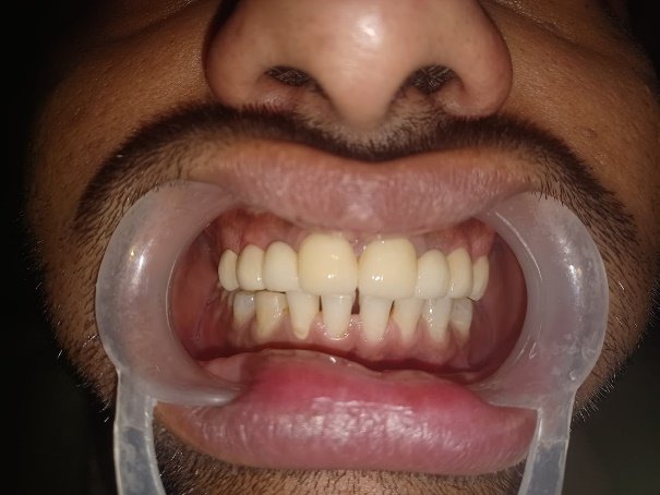
Endocrowns: An Effective Solution for Restoring Severely Damaged Molars Post-Endodontic Treatment: A Case Report
Department of Prosthodontics and Crown & Bridge K. D. Dental College and Hospital, Mathura 281006, Uttar Pradesh, India
ABSTRACT
Introduction- Endocrowns are designed for teeth that have undergone root canal treatment and have significant loss of structure. They provide structural support, protect the tooth, and offer better retention and stability compared to traditional crowns. Endocrowns distribute occlusal forces evenly, reduce fracture risk, and preserve more natural tooth structure, ensuring both functionality and aesthetics. This study describes the procedure to restore severely damaged molar.
Case description- A 25-year-old male patient presented to the Department of Prosthodontics Crown and Bridge with a root canal-treated lower right first molar (#47) that had sustained significant coronal damage and required a crown. He exhibited good oral hygiene and a favourable bite. Following the removal of the previous restoration, an endocrown was recommended due to the extent of the remaining tooth structure.
Discussion- Literature describes restoration of severely damaged molar by Endocrowns. These comprehensive studies and reports highlight the effectiveness of endocrowns in providing structural support and aesthetic appeal for teeth that have undergone significant damage. By covering the entire occlusal surface and extending into the pulp chamber, endocrowns offer a reliable solution for restoring functionality and preventing further deterioration. This method is particularly beneficial for molars that have suffered extensive coronal damage, ensuring a durable and lasting restoration.
Introduction
Many dentists face a significant challenge when considering the restoration of endodontically treated teeth with extensive coronal damage. The biomechanical principles of retention and resistance are compromised. Changes in biomechanics due to root canal therapy and the extent of dental tissue loss influence restorative treatment planning. For teeth severely damaged by decay or fractures, a treatment involving a full crown supported by a cast metal core has been recommended. However, this approach can lead to root perforation and thinning of the root canal walls due to excessive preparation for intraradicular posts .
Additionally, the use of intraradicular posts is limited by factors such as calcified root canals, narrow canals, or instrument fractures, prompting dentists to consider alternative solutions.
Endocrowns can be manufactured using either heat pressing or computer-aided design/computer-aided manufacturing (CAD/CAM) methods. A variety of materials are used in endocrown fabrication, with lithium disilicate glass-ceramic being highly recommended due to its excellent mechanical properties, aesthetic appeal, and strong bond with tooth structure.
Case Report
A 25-year-old male patient has come on department of Prosthodontics Crown and Bridge with root canal treated lower right molar 47, which had experienced significant coronal damage and required prosthesis (crown) for his first molar. The tooth underwent endodontic treatment. The patient demonstrated satisfactory oral hygiene and a favorable bite. Following removal of the previous restoration, an endocrown restoration was advised due to the extent of remaining tooth structure. After removing the restoration, an endocrown restoration was recommended. The decision was made to restore tooth #47 with an endocrown made of lithium disilicate ceramic, considering the remaining tooth structure and wall thickness Unlike traditional full crowns, the preparation for an endocrown involves specific techniques tailored to its monolithic, ceramic adhesive nature, addressing unique biomechanical requirements.
Due to less crown height, bone filing was undergone around 47 (buccally and lingually) to get an appropriate crown height for occlusal reduction (figure 1 and figure 2).
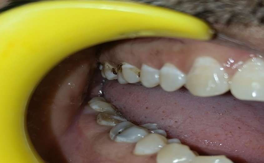
Figure 1
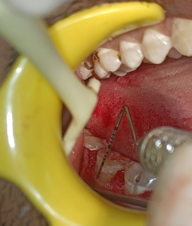
Figure 2
The goal is to reduce the overall height of the occlusal surface by at least 2 mm in the axial direction and create a cervical margin, often referred to as a "cervical sidewalk," resembling a butt joint. This margin should be positioned above the gum line (supragingival), and any enamel walls less than 2 mm thick need to be removed. To avoid a step-like appearance caused by differences in levels across the cervical margin, slopes connecting these parts should not exceed 60°. Our approach involved using a cylindrical-conical diamond bur aligned parallel to the occlusal plane to reduce the occlusal surface. Subsequently, a diamond wheel bur was employed to ensure controlled reduction orientation and achieve a flat surface due to its specific shape.
A cylindrical-conical diamond bur was used for occlusal convergence of 7° to establish a transition from the coronal pulp chamber to the endodontic access cavity. The bur was aligned parallel to the long axis of the tooth, and the preparation was performed with gentle pressure, ensuring that the pulpal floor was not touched. Excessive removal of tissue from the pulp chamber walls will decrease their thickness and the enamel strip width. The cavity depth should be a minimum of 3 mm.
Occlusal preparation was done to the depth of not more than 2 mm to utilize the saddle-like anatomy of the cavity floor. Non-abrasive instruments were used to preserve the integrity of the canal entrance, and no dentin drilling was performed. The preparation concluded with lining the root canal entrances using glass ionomer cement to safeguard the canal orifice (Figure 3 and figure 4).
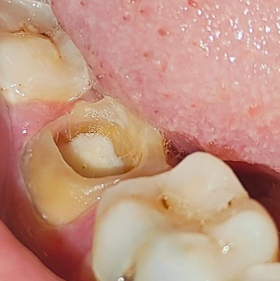
Figure 3
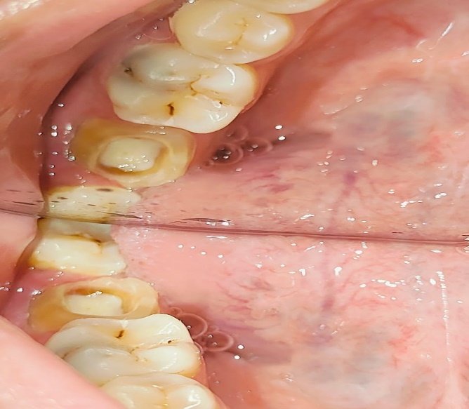
Figure 4
After evaluating the entire preparation, impression of the tooth was made by technique using addition silicone. selection of the ceramic shade was done and sent with the impression to laboratory. A provisional acrylic resin restoration was made by using and cemented with eugenol-free temporary cement (Figure 5 and Figure 6).
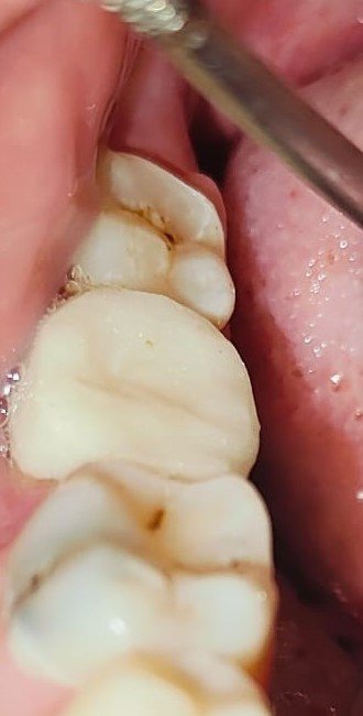
Figure 5

Figure 6
Then try-in of the endocrown was done and tested occlusion, internal, and proximal adjustments (Figure 7 and figure 8). In the following session, the internal surface of the endocrown was etched with hydrofluoric acid, rinsed with water, and dried with an air syringe. Next, a coat of a silane coupling agent was applied for 1 minute and dried. phosphoric acid was applied onto the tooth surface for15 sec on dentin and 30 sec on enamel, then abundantly washed and dried, applied with adhesive, and polymerized for 20 sec with light curing. A thin layer of a dual polymerizing resin was applied to the prosthetic endocrown and then was inserted into the tooth and polymerized at intervals of 5 seconds, making it easy to remove cement excesses. After that, it was polymerized for 60 seconds on all surfaces. The restoration was examined for any occlusal interference using ceramic finishing instruments (Figure 9 and Figure 10).
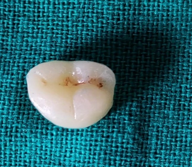
Figure 7
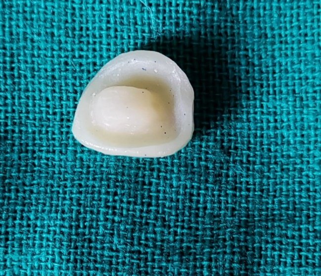
Figure 8
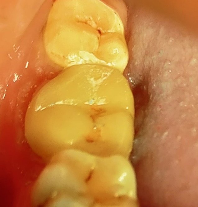
Figure 9
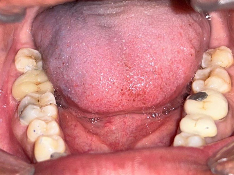
Figure 10
CONCLUSION
The endocrown is particularly suitable for all molars, especially those with clinically low crowns, calcified root canals, or narrow canals. However, it is not recommended in situations where adhesion cannot be guaranteed, if the pulpal chamber is less than 3 mm deep, or if the cervical margin is less than 2 mm wide around most of its circumference. This technique is advantageous because it simplifies the procedure, makes impression taking easier, and protects the periodontium. Additionally, ceramic materials offer benefits such as biocompatibility and biomimicry, and their wear coefficient is similar to that of natural teeth. Moreover, the single interface of a one-piece restoration enhances cohesion.
REFERENCES
- Dogui H, Abdelmalek F, Amor A, Douki N. Endocrown: An Alternative Approach for Restoring Endodontically Treated Molars with Large Coronal Destruction. Case Rep Dent. 2018 Aug.
- Assif D, Gorfil C. Biomechanical considerations in restoring endodontically treated teeth. J Prosthet Dent1984; 71:565–7.
- Lander E, Dietschi D. Endocrown: a clinical report. Quintessence Int 2008; 39:99–106.
- Valentina V, Aleksandar T, Dejan L, et al. Restoring endodontically treated teeth with all-ceramic endo-crowns—case report. Serbian Dent J 2008; 55:54–64.
Disclaimer from Guident
The views and opinions expressed in articles published in Guident are solely those of the authors and do not necessarily reflect the views of the editors, the editorial board or the publisher, Ivory India. Guident does not endorse any product, technique or treatment described. Clinical content is intended for qualified dental and medical professionals and is not a substitute for individual clinical judgement. Guident, its editors and the publisher accept no liability for any consequence arising from the use of information published in the journal.
How to cite this article
Dr. Sonali Chugh, Dr. Chiranjeev Gogoi, Dr. Abhinav Agarwal. Endocrowns: An Effective Solution for Restoring Severely Damaged Molars Post-Endodontic Treatment: A Case Report. Guident. 2024 Aug;17(9). Available from: https://guident.in/articles/prosthodontics/endocrowns-an-effective-solution-for-restoring-severely-damaged-molars-post-endodontic-treatment-a-case-report.html