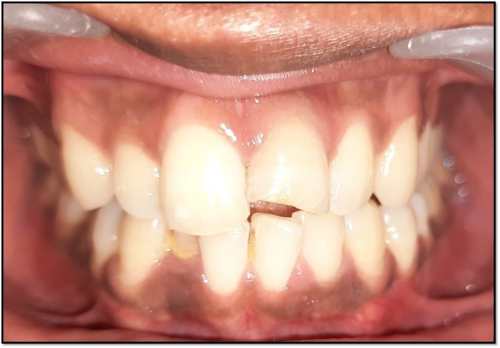
Crafting Smiles: The ART of Precision - Complete Denture Fabrication with Balanced Occlusion Using Extra-Oral Tracers: A Clinical Report
Department of Prosthodontics and Crown & Bridge, K.D. Dental College & Hospital, Mathura, UP, India.
INTRODUCTION
Recording accurate maxillo-mandibular determinants of occlusion is crucial to enhancing esthetics, function, and phonetics during the fabrication of complete dentures. Proficient clinical skills are necessary to establish a precise, verifiable, and reproducible vertical dimension of occlusion (VDO), recognized as the comfort zone, ensuring optimal harmony among musculoskeletal determinants. Additionally, achieving an accurate centric relation (CR), the unstrained position of the mandible allowing various movements, is essential. This position serves as a clinically valuable and repeatable reference point. Balanced occlusion, involving simultaneous contact of maxillary and mandibular teeth in both centric and eccentric positions, is designed to minimize tipping or rotating of denture bases in relation to supporting structures in the posterior and anterior occlusal areas on both the right and left sides.
Any errors or shortcomings in capturing the maxillo-mandibular jaw relation record can lead to uncomfortable and inappropriate dentures. Utilizing a tracing device, such as the gothic arch tracer, which functions as a central bearing device, is a demonstrably effective method for determining centric relation. Gothic arch tracing is categorized into two types—namely, intraoral and extraoral—based on the positioning of the stylus and tracing board, whether intraorally or extraorally, respectively. While extraoral gothic arch tracing is commonly employed in prosthodontics, intraoral tracing offers several advantages over it. Various methods exist for recording centric relation, including graphic methods such as intraoral and extraoral gothic arch tracing, as well as physiological methods.
CLINICAL REPORT
A 55-year-old female was referred to the Department of Prosthodontics at K.D. Dental College & Hospital, Mathura. The patient presented with a complete absence of teeth in the upper and lower anterior and posterior regions. Upon general examination, the patient appeared conscious, well-built, and had no significant medical history. (Fig 1,2)
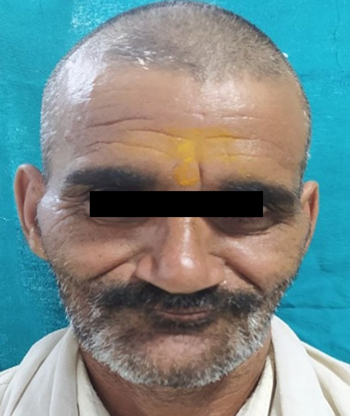
Fig 1

Fig 2
Intraoral examination revealed complete edentulism in both the maxilla and mandible, with no apparent abnormalities. (Fig 3,4) Treatment options were discussed, and it was decided to proceed with the fabrication of complete dentures with balanced occlusion. For extraoral tracing, the Height tracers was chosen.

Fig 3 & Fig 4
Thorough examinations were conducted, and a muco-compressive primary impression was taken using high-fusing impression compound (Fig 5,6), leading to the fabrication of a primary cast (Fig 7,8). Spacer wax was adapted, and a special tray was fabricated over it.

Fig 5 & Fig 6
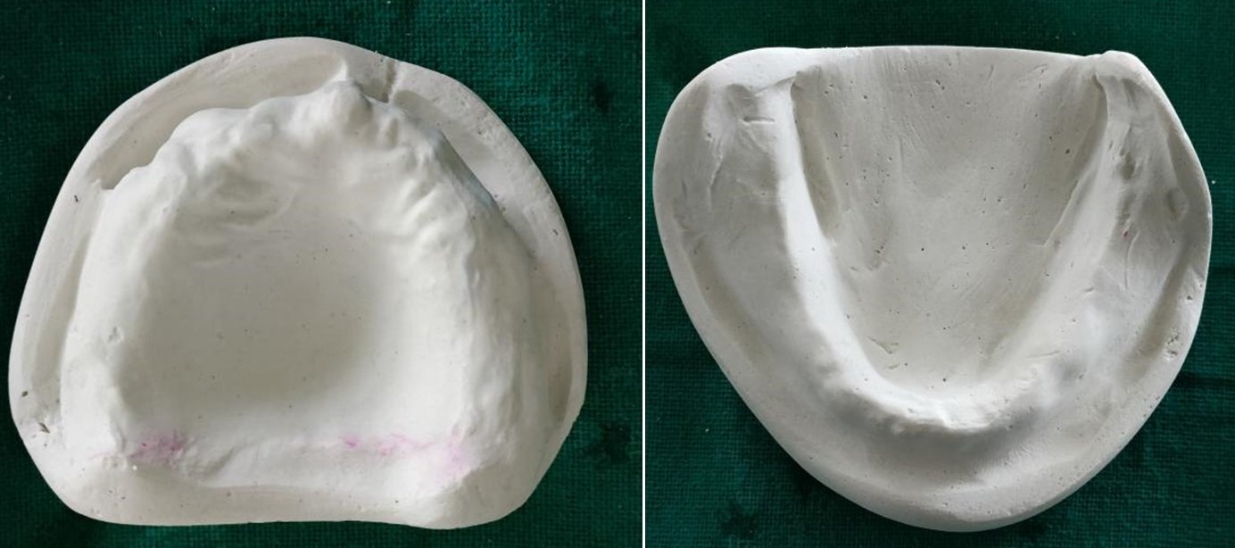
Fig 7 & Fig 8
Border molding was performed with green stick compound (Fig 9,10), and a secondary impression was made using zinc oxide eugenol impression material.(Fig 11,12)

Fig 9 & Fig 10

Fig 11 & Fig 12
A master cast was then fabricated using Type III dental stone & a split cast was also made for both casts. (Fig 13,14,15)

Fig 13 & Fig 14

Fig 15
Subsequently, denture bases and occlusal rims were constructed (Fig 16)

Fig 16
Tentative centric recording was performed first. The tentative jaw relation allows for an initial assessment of the relationship between the upper and lower jaws. It provides an opportunity to evaluate the tentative centric relation and make any necessary adjustments before proceeding to the orientation jaw relation. This step ensures that the preliminary position is satisfactory before finalizing the details. It serves as an intermediate step to ensure the accuracy and stability of the centric relation before finalizing the orientation jaw relation in the complete denture fabrication process. It allows for adjustments, patient adaptation, and verification of recording accuracy, contributing to the overall success of the denture construction. Then Orientation jaw relation was performed by attaching bitefork on the maxillary rim, 3mm above and parallel to the occlusal plane, so as not to hamper the occlusal plane and secured with the help of sticky wax. (Fig 17) The bitefork should be centered over the midline on the rim and its stem should be parallel to the Camper’s plane. (Fig 18)

Fig 17 & Fig 18
Facebow transfer was executed (Fig 19) wherein U shaped frame represents plane of cranium, bite fork represents plane of maxilla & locking device locks the two without altering their plane. U shaped frame is attached to transfer rod and moved up and down till orbital pointer is aligned with patient’s orbitale and it is locked by screw no 1. Screw no 2 aligns the midline of facebow to that of patient. Check from above of patient the correct alignment before locking. Screw no 3 tells confirms orientation by ask the patient if denture is seating properly or not and then tighten screw no 3. All the screws were checked and tighened and facebow record was transferred to the Hanau articulator. (Fig 20,21)

Fig 19

Fig 20 & Fig 21
The maxillary cast along with upper split cast was mounted onto the articulator. Next, vertical jaw relation was recorded and the mandibular cast along with split cast was also articulated. (Fig 22, 23) The assembly was secured with tight rubber band to prevent expansion during setting of plaster.

Fig 22 & Fig 23
The height tracer comprises several components, including central bearing device and extraoral tracers. To accommodate the central bearing plates & point, the height of the mandibular occlusal rims was reduced by 3mm. Central bearing plate was attached to the maxillary occlusal rim, and the central bearing point was affixed to the position was confirmed by placing a carbon paper between the point and plate and closing the articulator. (Fig 24,25,26,27) There should be a flush contact between the two. Indices were made on both the rims to help in positioning the interocclusal records. The tracing devices were then attached securely to the rims. The recording plate was attached to the mandibular occlusal rim such that it was at the midline. The stylus was attached to the maxillary occlusal rim. (Fig 28,29)

Fig 24 & Fig 25
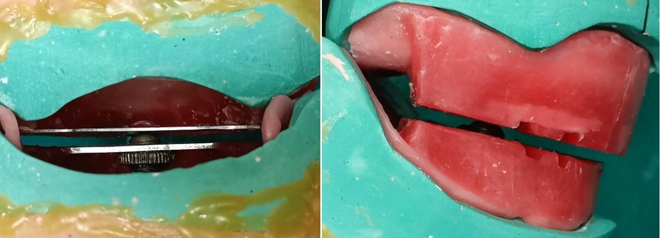
Fig 26 & Fig 27

Fig 28 & Fig 29
The patient was seated with his head upright and in a comfortable position. The record bases were inserted and inspected for stability. After confirming that there were no interferences between the occlusion rims when the mandible was moved in any direction, the stylus was lowered to the recording plate to determine that it contacted the recording plate during mandibular movements.The patient was instructed to move the mandible forward and backward in the median line, revealing the path of a straight protrusion. Following this, the patient retruded the mandible to its fullest extent and slid it to the right and left sides. After completing all excursive movements, the resulting tracing took the shape of an arrowhead. (Fig 30)

Fig 30
A layer of thin radiographic film was used to mark centic position and protusive position approximately at 6mm distance. (Fig 31)
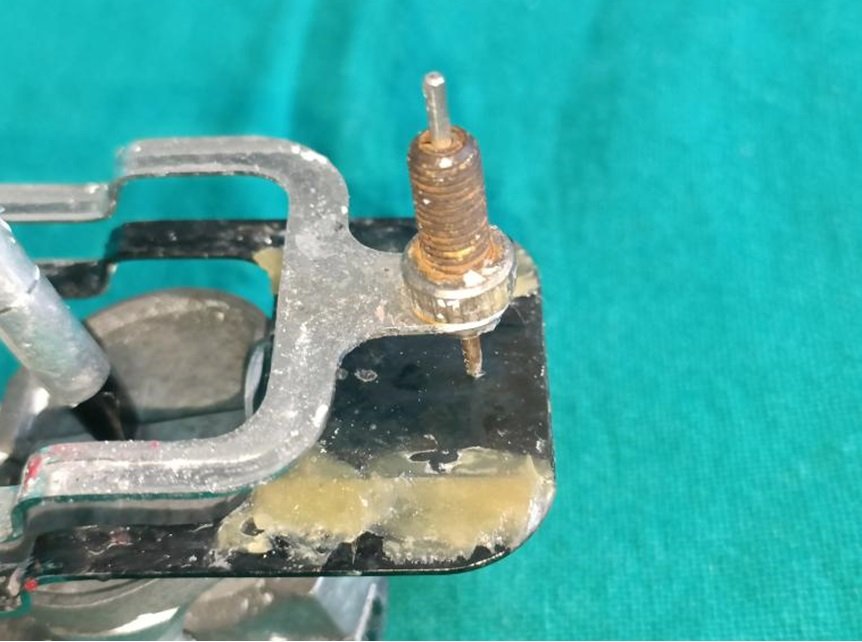
Fig 31
Perforations in film were made at these positions to help stabilize the position and an interocclusal record was made at centric relation and at protrusive relation using impression plaster to verify jaw relation. The setting was fastened by adding some salt to it. The centric record obtained should have a central perforation and the protrusive record should be near to perforation as the central bearing point contacts the central bearing plate at centric but as the mandible moves forward, the slight change in vertical dimension might create a minute space between them. (Fig 32)

Fig 32
The Centric record was used to verify centric relation by placing it between the maxillary & mandibular rim. The record should seat completely into the indiced surfaces of the rim, the stylus should be at the hole corresponding to centric relation position and there should be no space between rims, record or split cast and maxillary cast. (Fig 33)

Fig 33
For programming the articulator, two holes, 1mm wide and 5mm deep were drilled on the art portion of maxillary and mindbular casts such that they were anterior and posterior to the central bearing point and in line with each other. Die pins were placed into these holes and luted with cyanoacrylate. Now, the incisal pin was raised from incisal guide and cenric locks & thumbnuts for condylar guidance were unlocked. The protrusive record was placed between the rims. The maxillay cast, maxillary rim, protrusive inter-occlusal record, mandibular rim and madibular cast were all united into one by tying the die pins together with thread. The split cast and mounting plaster represent second unit. The condylar guidance was slowly rotated such that there was no space between the contacting surfaces of the two units i.e. the split cast and mounting surface of maxillary cast. (Fig 34,35) Using Hanau formula, Bennett angle was calculated and incorporated.

Fig 34 & Fig 35
Teeth selection was done using Berry’s biometric index, patient’s facial form, facial profile & complexion. Anterior teeth setting was done to develop an anterior guidance that is in harmony with patient’s esthetics and phonetics. During anterior try in for balanced dentures, the crucial step is to determine a labiolingual positioning of lower anteriors such that on protrusion, the occlusal rims posterior to them do not undergo marked seperation. (Fig 36) This step would make balancing easier as well as help to estabilish correct anterior guidance. Once anterior guidance was set intra-orally, the incisal guidance was set on the articulator and lateral wings were raised such that chisel end of incisal pin made contact with incisal guide on excursive movements of canine.

Fig 36
Later, posterior teeth were arranged in balanced occlusion. It is easier to gain balance as each tooth is being placed rather than arrange all teeth and then obtain balance. In balanced occlusion, there should be the simultaneous contact of the maxillary and mandibular teeth on the right and left and in the posterior and anterior occlusal areas in centric and eccentric (Protrusive & Lateral excursive) positions. Posterior try in was performed to determine the fit,aesthetics, balancing. (Fig 37,38, 39,40)
Centric Relation

Fig 37 & Fig 38
Then the trial dentures were secured on their respective casts and carefully demounted from their split casts, flasked, dewaxed, packed, cured and deflasked. The dentures were secured mounted on their split casts and checked for any occlusal interferenes and selective grinding was performed for any inaccuracy. The dentures were finished & polished and inserted.
Centric Relation

Fig 39, Fig 40, Fig 41
Protrusive Relation
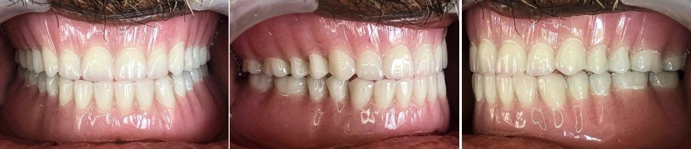
Fig 42, Fig 43, Fig 44
Right Lateral Relation

Fig 45, Fig 46, Fig 47
Left Lateral Relation
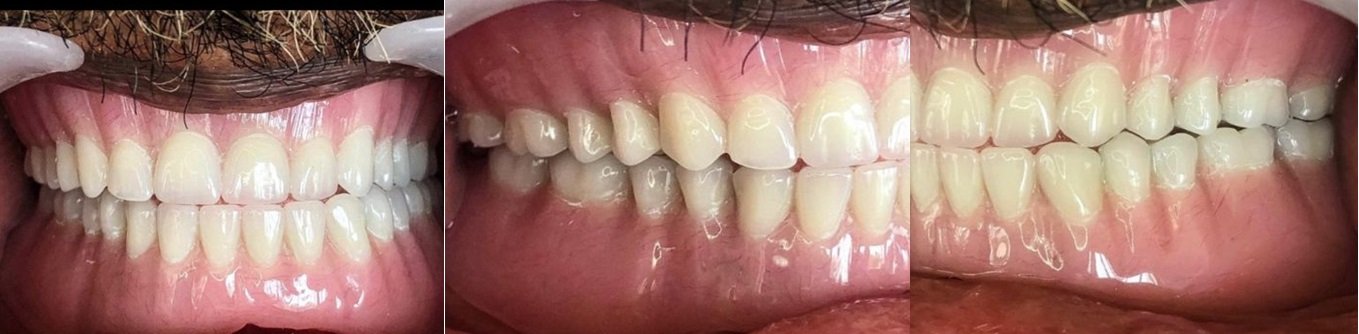
Fig 48, Fig 49, Fig 50
Patient was given instructions to wear them only during day time and store them in box at night, to clean the denture daily with soap water and denture brush and not use toothpaste as the abrasives in it can cause wearing away of acrylic. Gentle tissue cleansing and massaging of oral tissues should also be done. Patient might have speech problems initially and should do reading practice while at home. The problem of speaking will disappear and the tongue will adapt within a few weeks. Patient was instructed to eat non sticky, crispy, soft food initially and try to eat from both sides. During initial stages, patient should avoid sticky and hard food. Once comfortable, patient could start having solid food. While sneezing / coughing, patient should keep mouth closed to avoid denture's accidental falling off. Excess saliva production after new denture is normal and can last upto one week. This problem will disappear with time. A regular recall should be followed at 24hrs & 7 days after insertion with a maintenance at 3months, 6 months and 1 year interval.
DISCUSSION
Balanced occlusion dentures are considered superior to conventional dentures due to their ability to provide enhanced stability, comfort, and functionality for denture wearers. The fundamental principle of balanced occlusion involves ensuring simultaneous and harmonious contact of upper and lower teeth during various oral functions, such as chewing and speaking. This approach minimizes denture movements, reducing the risk of dislodgment and enhancing overall retention. The even distribution of occlusal forces across the dental arch improves chewing efficiency and reduces the strain on specific areas of the denture, contributing to increased longevity and durability. Balanced occlusion also aims to establish a stable centric relation, promoting comfort and minimizing the risk of temporomandibular joint issues associated with occlusal imbalances. Patients with dentures featuring balanced occlusion often report a more natural feeling, improved speech articulation, and higher overall satisfaction, highlighting the clinical advantages of this approach in providing comprehensive and patient-centered prosthodontic care.
Extraoral Gothic arch tracing is often preferred over intraoral tracing in certain cases due to its distinct advantages. This method involves recording the movements of the mandible from outside the mouth, typically using a Gothic arch tracing device. One key benefit is enhanced accessibility, as the tracing device is positioned extraorally, eliminating any interference within the oral cavity. This is particularly advantageous in patients with limited mouth opening or those who may find intraoral procedures uncomfortable. Additionally, extraoral tracing allows for a broader range of mandibular movements, providing a comprehensive understanding of the jaw's functional dynamics. The simplicity of the procedure and reduced reliance on patient cooperation contribute to the reliability and reproducibility of results. While both intraoral and extraoral Gothic arch tracing methods have their merits, extraoral tracing stands out in situations where patient comfort, accessibility, and a thorough assessment of mandibular movements are paramount considerations.
The creation of a 6mm protrusive record holds significant importance in the meticulous process of complete denture fabrication. This specific measurement, referring to the anterior limit of mandibular protrusion, plays a crucial role in achieving functional precision and patient satisfaction. The 6mm protrusive record serves as a guide for prosthodontists in replicating the natural range of mandibular movement during protrusion, allowing for the establishment of balanced occlusion. By capturing this functional dimension, the dental professional can optimize the positioning of anterior teeth, ensuring smooth articulation and coordinated movements during essential functions like speaking and swallowing. This record also aids in identifying and addressing potential interferences in the occlusal scheme, contributing to the denture's stability and the prevention of discomfort or dislodgment during protrusive activities. Ultimately, the 6mm protrusive record is a key element in the comprehensive design of complete dentures, promoting both functional efficacy and patient comfort.
CONCLUSION
Fabricating dentures for geriatric patients is crucial for enhancing their overall quality of life by addressing functional and aesthetic challenges resulting from tooth loss. Dentures restore the ability to chew, speak, and smile, improving nutritional intake and fostering social interactions. Beyond function, dentures contribute to psychological well-being by alleviating the loss of natural teeth, boosting self-esteem, and enabling comfortable social engagement. Proper denture fabrication is vital for maintaining oral health, as poorly fitted dentures can lead to issues like irritation and sore spots, potentially contributing to systemic health problems. Regular dental check-ups are essential for adjustments and evaluations to prevent complications and ensure ongoing comfort and functionality. Well-fabricated dentures also help prevent adverse effects of edentulism, such as changes in facial structure, preserving oral health and preventing further complications.
In summary, the fabrication of dentures for geriatric completely edentulous patients goes beyond the restoration of oral function; it plays a pivotal role in promoting overall well-being, psychological health, and the maintenance of oral and systemic health in the aging population. A thoughtful and precise approach to denture fabrication is vital to ensuring that geriatric individuals can enjoy a fulfilling and comfortable lifestyle despite the challenges posed by complete edentulism.
Disclaimer from Guident
The views and opinions expressed in articles published in Guident are solely those of the authors and do not necessarily reflect the views of the editors, the editorial board or the publisher, Ivory India. Guident does not endorse any product, technique or treatment described. Clinical content is intended for qualified dental and medical professionals and is not a substitute for individual clinical judgement. Guident, its editors and the publisher accept no liability for any consequence arising from the use of information published in the journal.
How to cite this article
Dr. Meher Yazdani, Dr. Srishti Mishra, Dr. Manesh Lahori. Crafting Smiles: The ART of Precision - Complete Denture Fabrication with Balanced Occlusion Using Extra-Oral Tracers: A Clinical Report. Guident. 2023 Nov;16(12). Available from: https://guident.in/articles/prosthodontics/crafting-smiles-the-art-of-precision-complete-denture-fabrication-with-balanced-occlusion-using-extra-oral.html