Combating the Compromised Edentulism
Department of Prosthodontics Dr. D.Y. Patil Dental College and Hospital, Nerul, Navi Mumbai
ABSTRACT
The vibrant phase of Life - ‘Youth’, brings with it immense vitality, vigor and a consciousness about aesthetics. However, nothing in life is permanent. Time leaves its imprint on every living being and ushers in an inevitable phase - ‘Aging’. Just as every human tissue undergoes changes of wear and tear throughout life, so does the oral cavity.
Gerodontics, a specialized prosthodontic practice extensively deals with the diagnosis, treatment planning and prognosis of these age related issues.
This article, is a review that shall highlight some of the optimum rehabilitative solutions and effective prostheses which have been fabricated to restore the form and function of our stomatognathic tissues, thus improving the quality of life of the so called ‘Dental Cripples’ - The Geriatric patients.
KEYWORDS : Hollow, severely atrophic ridges, sectional, constricted mouth opening, saliva reservoir, xerostomia
INTRODUCTION
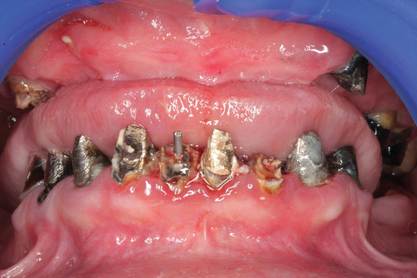
With growing urbanization, rapidly changing lifestyles and ‘stress’ laying its heavy burden on each one of us in every sphere of life, what other factors can complicate the retaining potential of dentures in the oral cavity?. What adds to the growing dilemma of an aging edentulous patient regarding his oral health?. To answer these questions, we can classify geriatric patients into 3 categories : 1) Physiologic, 2) Psychologic, 3) Pathologic ; owing to the clinical signs and symptoms evaluated at chair side and evidence based research. Physiologic refers to the state of oral tissues battered by aging, showing characteristic features of thinning of tissues with depleted repair potential, loss of healthy oral mucosa resiliency and firmness, residual ridge resorption leading to loosening of dentures, loss of vertical dimension, loss of normal facial contours, impaired neuromuscular control. Psychological refers to patients withstanding the above mentioned features with depression, anxiety, lack of self esteem, shying from socializing and stress. The Pathologic category refers to patients targeted with systemic disorders like diabetes, hypertension, nutritional deficiencies, ill effects on tissues due to tobacco chewing related habits, congenital deformities, terminally ill and institutionalized patients, patients on medications, conditions like xerostomia, trismus, microstomia, loss of proprioception, to name a few. These characteristic changes drastically complicate the fabrication and retaining capacity of dentures for the patients. This should bring us to another question – does one type of conventional complete denture suit all?1,2,3
Here are enlisted different treatment modalities that we can avail of in constructing optimally fitting oral prostheses to counteract a few of these debilitating conditions, thus improving the quality of life of our aging patients.
RESORBED RIDGES
Extreme resorption of the maxillary and mandibular denture-bearing areas result in narrower, more constricted residual ridges as resorption progresses, decreased supporting tissues, and a resultant large restorative space between the maxillary and mandibular alveolar ridges. Restoring such a situation will lead to increased volume of the effective dentures that will challenge the poor denture-bearing ability of the tissues and lead to decreased retention and resistance. We can solve these problems by fabricating hollow dentures to optimize functioning.
What are Hollow Dentures ?
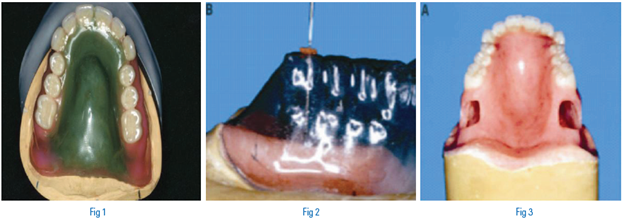
It is a modified complete denture prosthesis which is formed with a cavity form that ensures the appropriate dimensions for structural integrity of the denture base and optimal weight reduction. (FIG 1-3)6
Prime Indication
Fabricated in situations where the severely atrophic maxilla and mandible and increased inter-arch distance pose clinical challenges for fabrication of successful complete dentures
PROSTHESES
- During fabrication of the denture, acrylic thickness may be estimated using endodontic file and rubber stop.
- In the processed maxillary denture, openings are prepared to facilitate removal of putty. Heat polymerized clear acrylic resin windows provide for adequate sealing of the denture at its terminal ends
Advantages
- Light weight
- Can be used in conjunction with an obturator to obliterate an associated orofacial defect (Eg: Hollow bulb obturator)
Disadvantages
- Junctions between two polymerized portions increase risk of seepage of fluids
- Cavity void cannot act as a shock absorber
LIMITED ORAL OPENING
Limited oral opening, may result from surgical treatment of orofacial cancers, cleft lips, trauma, burns, Plummer-Vinson syndrome, or scleroderma. Scleroderma is a disease that causes fibrosis by affecting the connective tissue and the blood vessels; which is reportedly believed to stem from a disorder of the immune system. Characterized by the skin becoming tight and the wrinkles disappearing, giving the face a mask-like appearance. Hardening of the skin around the mouth causes the oral opening to become limited.
A maximal oral opening that is smaller than the size of a complete denture can make prosthetic treatment challenging. Different designs for the fabrication of maxillary and mandibular sectional trays, foldable complete dentures can aid in resolving these issues.8,9
What are Sectional Dentures ?
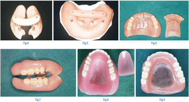
A modified complete or partial denture prosthesis which enables insertion, removal and optimum functioning in the oral cavity with compromised and limited opening. Also called as ‘split’ and ‘collapsible’ dentures. (FIG 4-7)
Prime Indication
Constricted mouth opening inhibiting range of functional movement within normal physiological limits.
Prostheses
- The sectioned maxillary and mandibular halves of the tray are fixed by means of metal pins and an acrylic block in the horizontal plane for proper orientation
- Various customized metal attachments, magnetic attachments, press button components can be used to aid in aligning and fixing tray and denture parts
Advantages
- Two rigid components have different path of insertion, thus fill opposing undercuts without necessity of clasps
- Restores vertical dimension in patients with compromised oral opening
Disadvantages
- Technique sensitive
- Tedious casting and manufacturing of metal components
DRY MOUTH
Xerostomia, dryness of mouth; is a clinical condition caused by a decrease in production of saliva flow which presents itself as a local symptom of Sjogren’s syndrome, diabetes, alcoholism, side effects of medication and therapeutic radiation to treat cancers of head and neck regions.
Several saliva substitutes are available for the rehabilitation of patients with xerostomia. However, saliva substitutes must be placed in the mouth by sucking, dripping, or spraying saliva from a container. These methods of applying artificial saliva seem tedious to cope up with over a period of time. To overcome this, techniques were applied to construct complete dentures that would permit a steady flow of artificial saliva to keep the oral tissues lubricated.
What are artificial saliva reservoir dentures ?
Dentures incorporating a reservoir filled with artificial saliva which is released at a controlled pace to moisten the oral tissues. (FIG 8-9)
Prostheses:
- The completed prosthetic assembly consists of the lid which was permanently attached to the denture with the help of autopolymerising acrylic resin and was smoothened and given a final polish, so as to facilitate communication between reservoir and the oral cavity
- Two saliva releasing openings of 0.1-0.2 mm are drilled in the palatal lid of the final processed denture to load (using a syringe)artificial saliva in the prepared reservoir
At recall visits (twice a week initially and then every 3 months) the prostheses can be thoroughly
cleaned by the dentist. The reservoirs are filled with an artificial saliva that contains mucin to allow adequate lubrication of oral tissues and prevent drying.
Research indicates that the period of continuous wetting of the oral cavity after filling the such constructed reservoirs was at least 2 hours. The release holes were small (0.1 to 0.2 mm) to prevent early release of the artificial saliva from the reservoir. If the holes were blocked by food particles, they could be cleared with a needle. For optimal physiologic activity and protection of the oral cavity, continuous coating of the mucous membranes with natural or artificial saliva is necessary. In patients with xerostomia this may involve application of artificial saliva with a squeeze bottle or atomizer.
Because retention of a saliva substitute in the mouth is short, this method of application does not provide a continuous flow of saliva. Artificial saliva administered by an intraoral reservoir guarantees continuous flow into the mouth. A reservoir is not suitable for all patients. The large size of the maxillary and mandibular reservoirs is required to provide storage of an adequate volume of saliva. Therefore, an adequate reservoir cannot be constructed for patients who have only a slight resorption of the mandibular alveolar process or a shallow palatal vault.11,13,15
Advantages
- Maintains a steady flow of artificial saliva to wet oral mucosa
- More suitable than topical application of saliva substitutes
- Additionally, it also enables the patient to clearly visualize the levels of saliva substitute within the chamber.
- The ready access to the reservoirs, both by the patient and by the dentist.
Disadvantages
- Fabrication is technique sensitive and increased laboratory steps
- Design might accumulate slight debris, but complete access for thorough cleansing is present
CONCLUSION
A Prosthodontist should critically analyze the clinical situation he/she is confronted with, apply the acumen and skill in creating prostheses that will aid in solving the plight of aging patients and relieving their symptoms to the highest level possible. Accurate diagnosis and subsequent clinically effective treatment planning will alleviate the fore mentioned symptoms.
Our motto lies in promoting oral rehabilitative care by fabricating patient sensitive prostheses and give pleasing end results to our geriatric patients, so that they can embrace aging and age gracefully.
REFERENCES
- Silverman Sol. Geriatrics and tissue changes- Problem of the aging denture patient. J Prosthet Dent 1958;8(5):734-739.
- W.O. Ramsay. Nutritional problems of the aged. J Prosthet Dent 1983;49(1):16-19.
- Carlson Gunner. Clinical morbidity and sequelae of treatment with complete dentures. J Prosthet Dent 1998;79(1):17-23.
- Hal G. Pickett, Ralph G , Margaret Osborn. Changes in the dentures supported tissues associated with the aging process. J Prosthet Dent 1972;27(3):257-262.
- Storer R. Geriatric Dentistry. Br Dent J 1966;121:547-552.
- Michael O’Sullivan, Nancy Hansen, Robert J. Cronin and David R. Cagna. The hollow maxillary complete denture: A modified technique. J Prosthet Dent 2004;91(6):591-594.
- LaDeane Fattore, Louis Fine and Qavid C. Edmonds. The hollow denture: An alternative treatment for atrophic maxillae. J Prosthet Dent 1988;59(4):514-516.
- Cenk Cura, Serdar Cotert and Atilla User. Fabrication of a sectional impression tray and sectional complete denture for a patient with microstomia and trismus: A clinical report. J Prosthet Dent 2003;89(6):540-543.
- Ikuya Watanabe, Yasuhiro Tanaka, Chikahiro Ohkubo and Amp W. Miller. Application of cast magnetic attachments to sectional complete dentures for a patient with microstomia: A clinical report. J Prosthet Dent 2002;88(6):573-577.
- Laxman Singh Kaira. Prosthodontic Management of Xerostomic Patient with Reservoir Denture - A Case Report. www.journalofdentofacialsciences.com Vol 1. Issue 1. Accessed 10 August 2013.
- A. Vissink et al. Artificial saliva reservoirs. J Prosthet Dent 1984;52(5):710-715.
- GS Chandu, MN Homesh. Management of xerostomia and hyposalivation in complete denture patients. Indian J Stomatol 2011;2(4): 263-266.
- A. Vissink, M. C. Huisman and E. J.‘s-Gravenmade. Coristruction of an artificial saliva reservoir in an existing maxillary denture. J Prosthet Dent 1986;56(1):70-74.
- Ana M. Diaz-Arnold and Cindy A. Marek. The impact of saliva on patient care: A literature review. J Prosthet Dent 2002;88(3):337-343.
- Joseph A. Toljanic and Terry G. Zucuskie. Use of a palatal reservoir in denture patients with xerostomia. J Prosthet Dent 1984;52(4):540-544.
Disclaimer from Guident
The views and opinions expressed in articles published in Guident are solely those of the authors and do not necessarily reflect the views of the editors, the editorial board or the publisher, Ivory India. Guident does not endorse any product, technique or treatment described. Clinical content is intended for qualified dental and medical professionals and is not a substitute for individual clinical judgement. Guident, its editors and the publisher accept no liability for any consequence arising from the use of information published in the journal.
How to cite this article
Dr. Asha M. Rathod, Dr. Omkar K. Shetty, Dr. Aditya M. Motwani. Combating the Compromised Edentulism. Guident. 2014 May;7(6). Available from: https://guident.in/articles/prosthodontics/combating-the-compromised-edentulism.html